. 2015 Feb;89(4):2209-19.
doi: 10.1128/JVI.03073-14. Epub 2014 Dec 3.

Structure-function analysis of vaccinia virus H7 protein reveals a novel phosphoinositide binding fold essential for poxvirus replication

Affiliations

Structure-function analysis of vaccinia virus H7 protein reveals a novel phosphoinositide binding fold essential for poxvirus replication

Swapna Kolli et al. J Virol. 2015 Feb.

Abstract

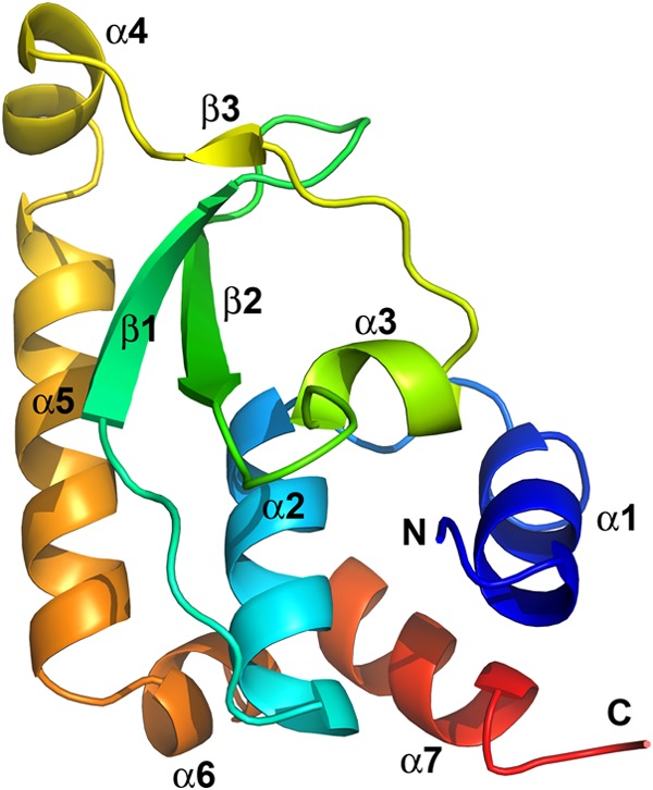
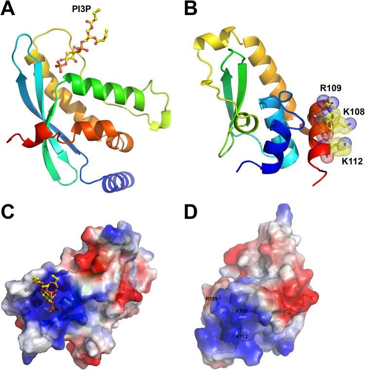
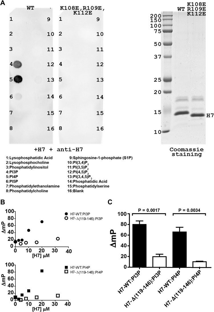
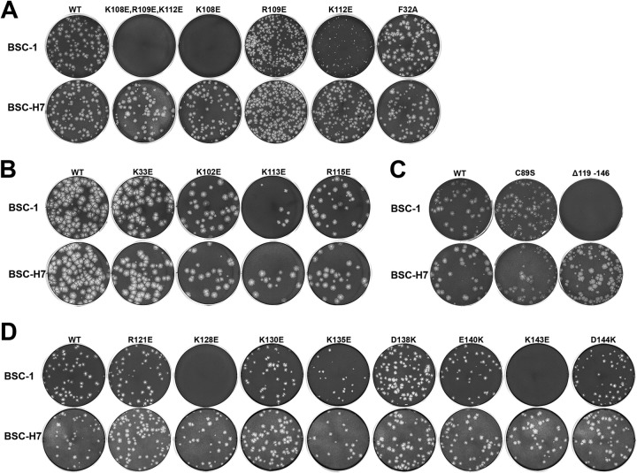
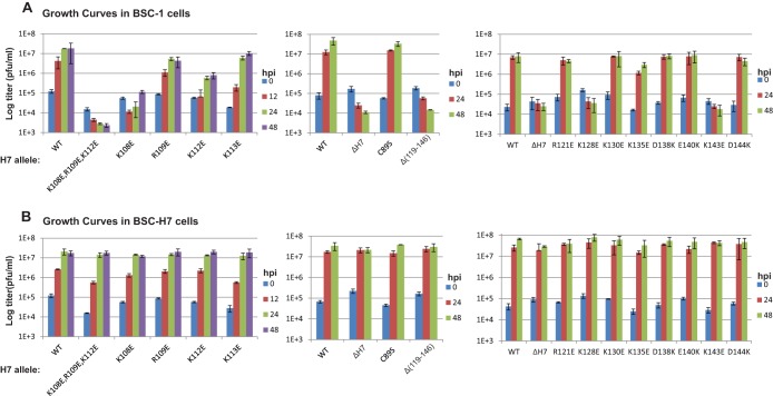
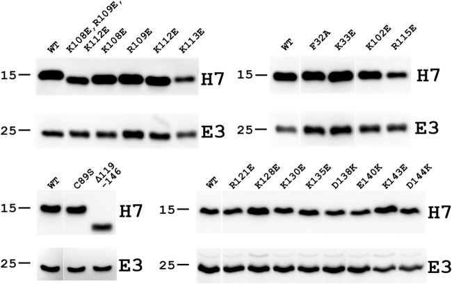
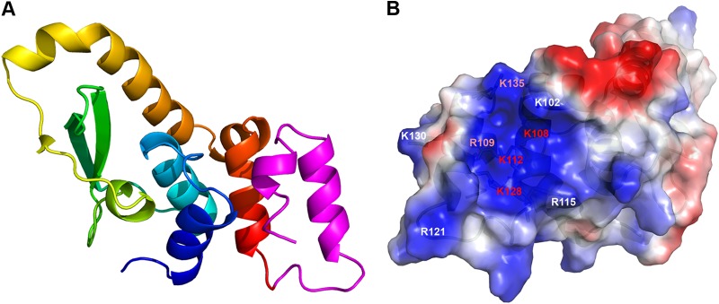
Phosphoinositides and phosphoinositide binding proteins play a critical role in membrane and protein trafficking in eukaryotes. Their critical role in replication of cytoplasmic viruses has just begun to be understood. Poxviruses, a family of large cytoplasmic DNA viruses, rely on the intracellular membranes to develop their envelope, and poxvirus morphogenesis requires enzymes from the cellular phosphoinositide metabolic pathway. However, the role of phosphoinositides in poxvirus replication remains unclear, and no poxvirus proteins show any homology to eukaryotic phosphoinositide binding domains. Recently, a group of poxvirus proteins, termed viral membrane assembly proteins (VMAPs), were identified as essential for poxvirus membrane biogenesis. A key component of VMAPs is the H7 protein. Here we report the crystal structure of the H7 protein from vaccinia virus. The H7 structure displays a novel fold comprised of seven α-helices and a highly curved three-stranded antiparallel β-sheet. We identified a phosphoinositide binding site in H7, comprised of basic residues on a surface patch and the flexible C-terminal tail. These residues were found to be essential for viral replication and for binding of H7 to phosphatidylinositol-3-phosphate (PI3P) and phosphatidylinositol-4-phosphate (PI4P). Our studies suggest that phosphoinositide binding by H7 plays an essential role in poxvirus membrane biogenesis.

Importance: Poxvirus viral membrane assembly proteins (VMAPs) were recently shown to be essential for poxvirus membrane biogenesis. One of the key components of VMAPs is the H7 protein. However, no known structural motifs could be identified from its sequence, and there are no homologs of H7 outside the poxvirus family to suggest a biochemical function. We have determined the crystal structure of the vaccinia virus (VACV) H7 protein. The structure displays a novel fold with a distinct and positively charged surface. Our data demonstrate that H7 binds phosphatidylinositol-3-phosphate and phosphatidylinositol-4-phosphate and that the basic surface patch is indeed required for phosphoinositide binding. In addition, mutation of positively charged residues required for lipid binding disrupted VACV replication. Phosphoinositides and phosphoinositide binding proteins play critical roles in membrane and protein trafficking in eukaryotes. Our study demonstrates that VACV H7 displays a novel fold for phosphoinositide binding, which is essential for poxvirus replication.


Figures

FIG 1
FIG 1
The structure of vaccinia virus H7 protein displays a novel fold. The secondary structures are shown as ribbon models and were colored in rainbow colors and labeled. The N and C termini of H7 are indicated. The C-terminal 29 residues (aa 118 to 146) are disordered and are not visible in this structure.
FIG 2
FIG 2
Comparison of the structures of H7 and the PX domain. (A) The structure of the PX domain of the human p40 subunit of NADPH oxidase (PDB entry 1H6H) is shown as a ribbon. The bound PI3P is shown as a ball-and-stick model. (B) H7 is shown as a ribbon, and the basic surface residues (K108, R109, and K112) on helix α7 are shown as sticks, with dotted envelopes indicating the van der Waals radius. The color scheme is the same as that for Fig. 1. (C and D) Electropotential surfaces of the PX domain (PDB entry 1H6H) (C) and H7 (D). The PX domain uses a large basic pocket for lipid binding. Similarly, H7 also displays a prominent basic surface patch centered on a cluster of positively charged residues, including K108, R109, and K112.
FIG 3
FIG 3
H7 binds PI3P and PI4P. (A) Lipid overlay assay. Nitrocellulose strips containing 100-pmol spots of 15 different lipids were incubated with 1 μg/ml of the purified H7 protein, and the bound protein was detected by Western blotting with anti-H7 antibody. The numbers on the blots mark the positions of the lipids, and their names are given below the blots. The H7 proteins (WT or K108E/R109E/K112E mutant) used are indicated above the blots. Coomassie staining of the purified H7 protein used for lipid overlay is shown on the right. The sizes of molecular mass markers are indicated in kilodaltons. (B) Fluorescence polarization assay. A sample containing 0.2 nM BODIPY-labeled PI3P (top) or PI4P (bottom) was mixed with increasing concentrations of either H7-WT or H7-Δ(119-146). Binding was determined by measuring the change in millipolarization (ΔmP). (C) Binding experiments were conducted at a fixed concentration (20 μM) of either H7-WT or H7-Δ(119-146). Error bars represent standard errors of the means. Significance was found for differences between H7-WT and H7-Δ(119-146) (P < 0.01 by the two-tailed t test).
FIG 4
FIG 4
Plaque morphologies of H7 mutant VACVs. BSC-1 (top rows) and BSC-H7 (bottom rows) cells in 12-well plates were infected with the indicated H7R mutant VACVs in semisolid medium for 48 h. The cells were then stained with crystal violet to reveal plaques. For each mutant, the same amount of virus was used to infect BSC-1 and BSC-H7 cells. The mutants were studied in groups in four separate experiments (A to D), each with a WT control. Thus, the plaque sizes of different mutants can be compared within groups but not between groups.
FIG 5
FIG 5
Growth curves of H7 mutant VACVs. BSC-1 (A) and BSC-H7 (B) cells in 12-well plates were infected with the indicated viruses at a multiplicity of infection (MOI) of 5 PFU/cell. Viral titers at 0, 24, and 48 h postinfection (hpi) were determined by plaque assay on BSC-H7 cells. The mutants were studied in groups in three separate experiments, each with a WT control.
FIG 6
FIG 6
Western blots of viral proteins expressed by H7 mutant VACVs. BSC-H7 cells were infected with the indicated viruses at an MOI of 5 PFU/cell. The levels of H7 and E3 proteins at 8 hpi were determined by Western blotting with antibodies against H7 and E3, respectively. E3 is a viral protein that is unrelated to H7 and served as a control for infectivity and gel loading. The sizes of molecular mass markers are shown in kilodaltons. The mutants were studied in groups in four separate experiments, each with a WT control.
FIG 7
FIG 7
Modeled structure of full-length H7. The structure of full-length H7 was modeled with the I-TASSER server (43) by using the crystal structure of H7(1-118) as the template. The model with the highest confidence score (C score) as suggested by I-TASSER was selected. (A) The modeled full-length H7 structure is shown as a ribbon. The part of H7 observed from the crystal structure is colored as in Fig. 2B. The modeled C terminus (residues 119 to 146) is shown in magenta. (B) Electropotential surface of the modeled full-length H7 protein. The basic residues at the putative lipid binding pocket are indicated and labeled in color according to their importance for H7 function, as determined by the experiments shown in Fig. 5 and 6. Red, white, and salmon represent essential, nonessential, and modestly important regions, respectively.

References

    1. Di Paolo G, De Camilli P. 2006. Phosphoinositides in cell regulation and membrane dynamics. Nature 443:651–657. doi:10.1038/nature05185. - DOI - PubMed
    1. Balla T. 2013. Phosphoinositides: tiny lipids with giant impact on cell regulation. Physiol Rev 93:1019–1137. doi:10.1152/physrev.00028.2012. - DOI - PMC - PubMed
    1. Lemmon MA. 2008. Membrane recognition by phospholipid-binding domains. Nat Rev Mol Cell Biol 9:99–111. doi:10.1038/nrm2328. - DOI - PubMed
    1. Kutateladze TG. 2010. Translation of the phosphoinositide code by PI effectors. Nat Chem Biol 6:507–513. doi:10.1038/nchembio.390. - DOI - PMC - PubMed
    1. Teasdale RD, Collins BM. 2012. Insights into the PX (phox-homology) domain and SNX (sorting nexin) protein families: structures, functions and roles in disease. Biochem J 441:39–59. doi:10.1042/BJ20111226. - DOI - PubMed

Publication types

Associated data

LinkOut - more resources