Comparative Study
. 2015 Mar;63(3):181-9.
doi: 10.1369/0022155414564219. Epub 2014 Dec 3.

Human saliva-derived exosomes: comparing methods of isolation

Affiliations
Comparative Study

Human saliva-derived exosomes: comparing methods of isolation

Ayelet Zlotogorski-Hurvitz et al. J Histochem Cytochem. 2015 Mar.

Abstract

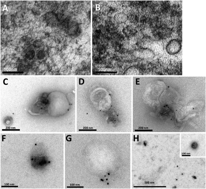
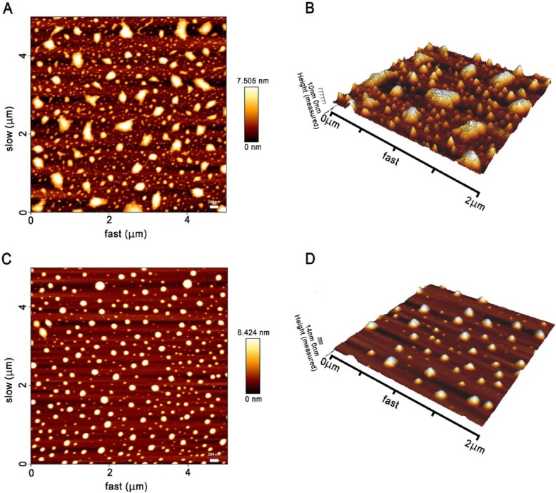
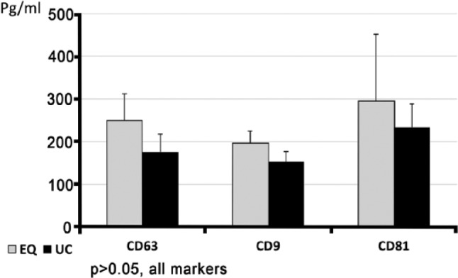
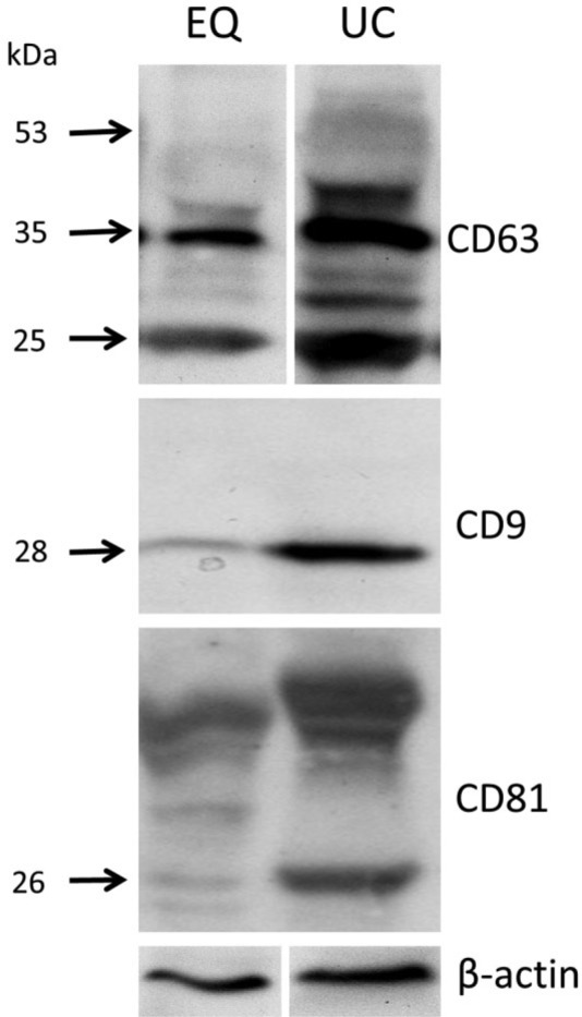
ExoQuick-TC(TM) (EQ), a chemical-based agent designed to precipitate exosomes, was calibrated for use on saliva collected from healthy individuals. The morphological and molecular features of the precipitations were compared with those obtained using the classical, physical-based method of ultracentrifugation (UC). Electron microscopy and immunoelectron microscopy with anti-CD63 showed vesicular nanoparticles surrounded by bi-layered membrane, compatible with exosomes in EQ, similar to that observed with UC. Atomic force microscopy highlighted larger, irregularly shaped/aggregated EQ nanoparticles that contrasted with the single, round-shaped UC nanoparticles. ELISA (performed on 0.5 ml of saliva) revealed a tendency for a higher expression of the specific exosomal markers (CD63, CD9, CD81) in EQ than in UC (p>0.05). ELISA for epithelial growth factor receptor, a non-exosomal-related marker, showed a significantly higher concentration in EQ than in UC (p=0.04). Western blotting of equal total-protein concentrations revealed bands of CD63, CD9 and CD81 in both types of preparations, although they were less pronounced in EQ compared with UC. This may be related to a higher fraction of non-exosomal proteins in EQ. In conclusion, EQ is suitable and efficient for precipitation of salivary exosomes from small volumes of saliva; however, EQ tends to be associated with considerably more biological impurities (non-exosomal-related proteins/microvesicles) as compared with UC.

Keywords: ExoQuick; exosomes; extracellular vesicles; isolation; saliva; ultracentrifugation.


Conflict of interest statement

Declaration of Conflicting Interests: The authors declared no potential conflicts of interest with respect to the research, authorship, and/or publication of this article.

Figures

Figure 1.
Figure 1.
Exosomal pellets. (A) Pellet obtained using the ExoQuick-TCTM (EQ) method from 0.5 ml of saliva is shown at the bottom of a 15 ml tube. (B) Pellet obtained using the ultracentrifugation (UC) method from 0.5 ml of saliva is shown in a close-up view of the bottom of a 1-ml ultracentrifugation tube. It was in the form of a very thin, barely visible grayish film (in between the arrows).
Figure 2.
Figure 2.
Characterization of salivary exosomes isolated by two methods. Electron micrographs of exosomes isolated from saliva by ExoQuick-TCTM (EQ) (A) and the classical ultracentrifugation (UC) method (B) demonstrating small vesicles of different sizes (ranging between 30–120 nm) with lipid bilayer membranes. Electron micrographs stained with 10-nm gold-conjugated anti-CD63 antibody and uranyl acetate counterstaining in EQ (C–E) and UC (F–H) preparations. Aggregate formation was a feature of the EQ preparations. With the UC method, exosomes were usually isolated as individual structures. (G) Note that the gold-stain method distinguished between a positively stained exosome (~50 nm) and a large (~250 nm), non-stained vesicular structure that was incompatible with an exosome.
Figure 3.
Figure 3.
Topography of salivary exosomes from healthy individuals observed under tapping mode atomic force microscopy (AFM). Exosomes on a mica surface precipitated by ExoQuick-TCTM (EQ) revealed heterogeneity in size and shape in both 2-dimensional (2D) (A) and 3D (B) images. In contrast, 2D (C) and 3D (D) images of the ultracentrifugation (UC) pellets showed individual, regular, fairly rounded structures.
Figure 4.
Figure 4.
Exosomal marker identification, as analyzed by ELISA. Equal volumes of 0.5 ml of whole saliva showed a trend toward higher concentrations in the expression of exosomal markers in pellets obtained using ExoQuick-TCTM (EQ) method as compared with that obtained using ultracentrifugation (UC).
Figure 5.
Figure 5.
Identification of exosomal markers by western blotting. Sixty µg of total protein derived from salivary exosomal pellet lysates isolated using ExoQuick-TCTM (EQ) or ultracentrifugation (UC) were separated by 12% SDS-PAGE (under reducing conditions), electro-transferred and incubated with antibodies against exosomal markers (CD63, CD9, CD81, 1:1000 each) and β-actin (1:500). The molecular weight standards (kDa) are marked on the left. The three markers were identified (arrows) in the exosomal pellets isolated by both methods. UC bands of the isolated proteins had consistently more intense and specific signals as compared to the EQ bands.

References

    1. Burns G, Brooks K, Wildung M, Navakanitworakul R, Christenson LK, Spencer TE. (2014). Extracellular vesicles in luminal fluid of the ovine uterus. PLoS One 9:e90913. - PMC - PubMed
    1. Caradec J, Kharmate G, Hosseini-Beheshti E, Adomat H, Gleave M, Guns E. (2014). Reproducibility and efficiency of serum-derived exosome extraction methods. Clin Biochem pii:S0009-9120(14)00405-6. - PubMed
    1. Chernousov MA, Stahl RC, Carey DJ. (2013). Tetraspanins are involved in Schwann cell-axon interaction. J Neurosci Res 91:1419-1428. - PubMed
    1. Davidovich E, Aframian DJ, Shapira J, Peretz B. (2010). A comparison of the sialochemistry, oral pH, and oral health status of Down syndrome children to healthy children. Int J Paediatr Dent 20:235-241. - PubMed
    1. Fabbri M, Paone A, Calore F, Galli R, Gaudio E, Santhanam R, Lovat F, Fadda P, Mao C, Nuovo GJ, Zanesi N, Crawford M, Ozer GH, Wernicke D, Alder H, Caligiuri MA, Nana-Sinkam P, Perrotti D, Croce CM. (2012). MicroRNAs bind to Toll-like receptors to induce prometastatic inflammatory response. Proc Natl Acad Sci U S A 31:E2110-E2116. - PMC - PubMed

Publication types