Review
. 2014 Nov 21:7:421-35.
doi: 10.2147/IJNRD.S42924. eCollection 2014.

Blood oxygenation level-dependent MRI for assessment of renal oxygenation

Affiliations
Review

Blood oxygenation level-dependent MRI for assessment of renal oxygenation

Joel Neugarten et al. Int J Nephrol Renovasc Dis. .

Abstract

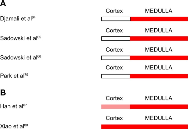
Blood oxygen level-dependent magnetic resonance imaging (BOLD MRI) has recently emerged as an important noninvasive technique to assess intrarenal oxygenation under physiologic and pathophysiologic conditions. Although this tool represents a major addition to our armamentarium of methodologies to investigate the role of hypoxia in the pathogenesis of acute kidney injury and progressive chronic kidney disease, numerous technical limitations confound interpretation of data derived from this approach. BOLD MRI has been utilized to assess intrarenal oxygenation in numerous experimental models of kidney disease and in human subjects with diabetic and nondiabetic chronic kidney disease, acute kidney injury, renal allograft rejection, contrast-associated nephropathy, and obstructive uropathy. However, confidence in conclusions based on data derived from BOLD MRI measurements will require continuing advances and technical refinements in the use of this technique.

Keywords: BOLD MRI; acute kidney injury; chronic kidney disease; contrast-associated nephropathy; diabetes mellitus; hypoxia; kidney; oxygenation.


Figures

Figure 1
Figure 1
Intrarenal oxygenation in human subjects with chronic kidney disease studied by blood oxygen level-dependent magnetic resonance imaging. Note: White indicates no change from control. Blue indicates reduced oxygenation.
Figure 2
Figure 2
Intrarenal oxygenation in animal models of diabetes mellitus studied by blood oxygen level-dependent magnetic resonance imaging. Note: White indicates no change from control. Blue indicates reduced oxygenation. Abbreviations: IS, inner stripe of the outer medulla; OS, outer stripe of the outer medulla.
Figure 3
Figure 3
(A and B) Intrarenal oxygenation in human subjects with diabetes mellitus studied by blood oxygen level-dependent magnetic resonance imaging. Note: White indicates no change from control. Blue indicates reduced oxygenation. The lighter shade of blue indicates a lesser degree of hypoxia. Abbreviation: GFR, glomerular filtration rate.
Figure 4
Figure 4
Selection of regions of interest. C 1–3 identify cortical regions of interest. O 1–3 identify outer medullary regions of interest. Notes: Adapted from Pohlmann A, Hentschel J, Fechner M, et al. High temporal resolution parametric MRI monitoring of the initial ischemia/reperfusion phase in experimental acute kidney injury. PLoS One. 2013;8(2):e57411.
Figure 5
Figure 5
(A and B) Intrarenal oxygenation in human subjects with acute rejection of renal allografts studied by blood oxygen level-dependent magnetic resonance imaging. Notes: White indicates no change from control. Red indicates increased oxygenation. The lighter shade of red indicates a lesser degree of hyperoxia.

References

    1. Gloviczki ML, Glockner J, Gomez SI, et al. Comparison of 1.5 and 3 T BOLD MR to study oxygenation of kidney cortex and medulla in human renovascular disease. Invest Radiol. 2009;44(9):566–571. - PMC - PubMed
    1. dos Santos EA, Li LP, Ji L, Prasad PV. Early changes with diabetes in renal medullary hemodynamics as evaluated by fiberoptic probes and BOLD magnetic resonance imaging. Invest Radiol. 2007;42(3):157–162. - PMC - PubMed
    1. Pedersen M, Dissing TH, Mørkenborg J, et al. Validation of quantitative BOLD MRI measurements in kidney: application to unilateral ureteral obstruction. Kidney Int. 2005;67(6):2305–2312. - PubMed
    1. Pohlmann A, Arakelyan K, Hentschel J, et al. Detailing the relation between renal T2* and renal tissue pO2 using an integrated approach of parametric magnetic resonance imaging and invasive physiological measurements. Invest Radiol. 2014;49(8):547–560. - PubMed
    1. Zhang JL, Morrell G, Rusinek H, et al. Measurement of renal tissue oxygenation with blood oxygen level-dependent MRI and oxygen transit modeling. Am J Physiol Renal Physiol. 2014;306(6):F579–F587. - PMC - PubMed

LinkOut - more resources