Review
. 2014 Dec;44(4):325-32.
doi: 10.5624/isd.2014.44.4.325. Epub 2014 Nov 25.

Basic principles and applications of (18)F-FDG-PET/CT in oral and maxillofacial imaging: A pictorial essay

Affiliations
Review

Basic principles and applications of (18)F-FDG-PET/CT in oral and maxillofacial imaging: A pictorial essay

Galal Omami et al. Imaging Sci Dent. 2014 Dec.

Abstract

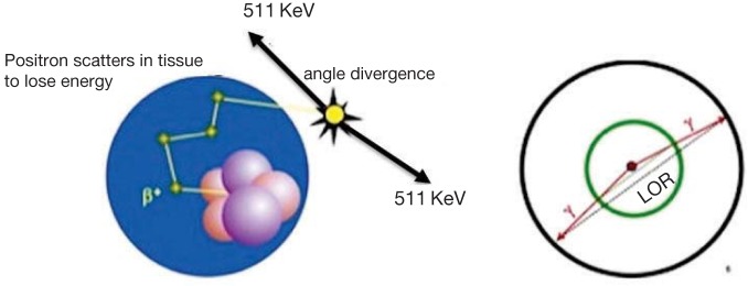
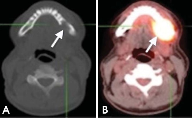
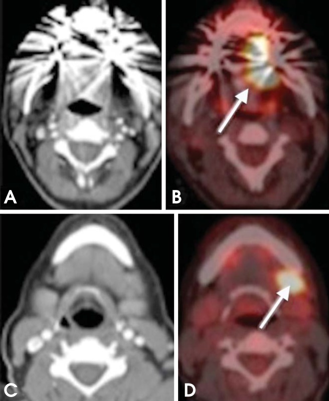
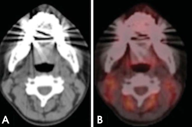
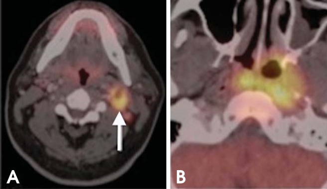
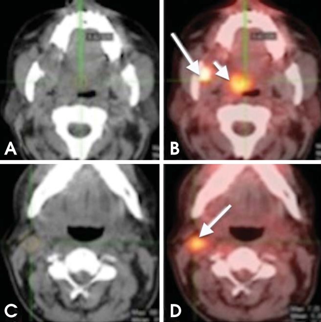
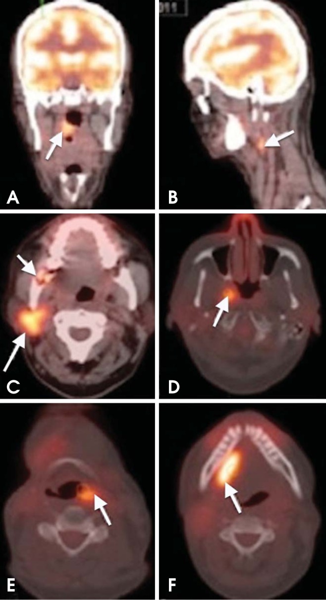
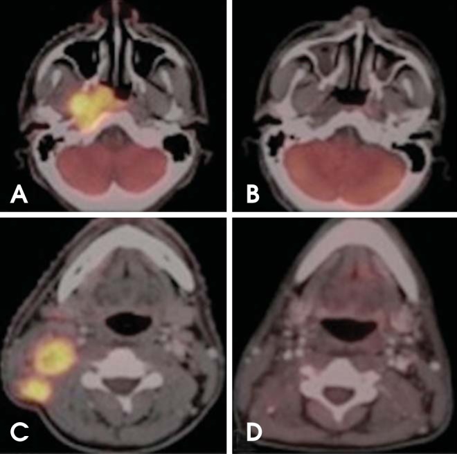
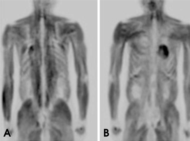
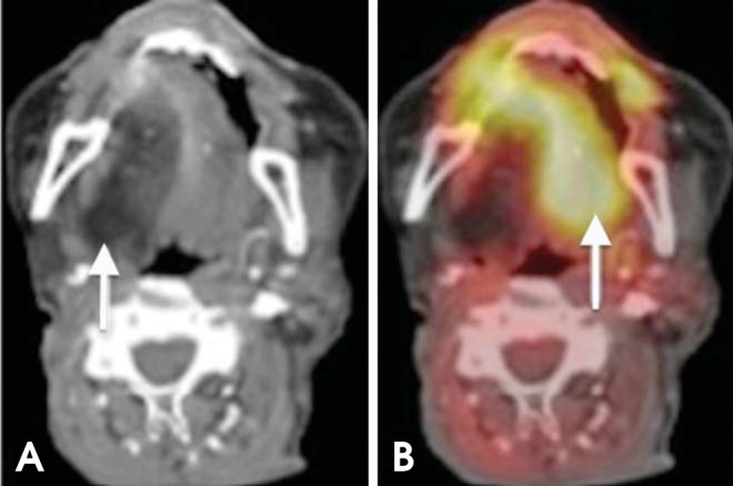
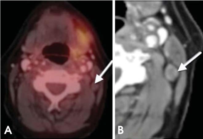
A combination of positron emission tomography (PET) with (18)F-labeled fluoro-2-deoxyglucose ((18)F-FDG) and computed tomography ((18)F-FDG-PET/CT) has increasingly become a widely used imaging modality for the diagnosis and management of head and neck cancer. On the basis of both recent literature and our professional experience, we present a set of principles with pictorial illustrations and clinical applications of FDG-PET/CT in the evaluation and management planning of squamous cell carcinoma of the oral cavity and oropharynx. We feel that this paper will be of interest and will aid the learning of oral and maxillofacial radiology trainees and practitioners.

Keywords: Head and Neck Neoplasms; Positron-Emission Tomography; Tomography, X-Ray Computed.


Figures

Fig. 1
Fig. 1
Annihilation coincidence detection (ACD). When a positron is emitted by a nuclear transformation, it scatters through matter losing energy and annihilates with an electron, resulting in two 511-keV photons that are emitted in nearly opposite directions (left). When two interactions are simultaneously detected within a ring of detectors surrounding the patient (right), it is assumed that annihilation occurred on the line connecting the interactions (line of response, LOR). ACD acts as a collimator for the positron emission tomography (PET) scanner (electronic collimation) by determining the path of the detected photons.
Fig. 2
Fig. 2
A 64-year-old man with T4 squamous cell carcinoma (SCC) of the lingual gingiva of the left posterior mandible. (A) Axial plain computed tomography (CT) image shows bone destruction of the left posterior mandible (arrow). (B) Axial PET/CT scan shows intense fluoro-2-deoxyglucose (FDG) uptake in the same area (arrow).
Fig. 3
Fig. 3
A 56-year-old man diagnosed with SCC of the lateral border of the tongue (left side). (A, B) Axial CT and PET/CT images reveal increased FDG uptake in the lateral border of the tongue (arrow). (C, D) Axial CT and PET/CT images at a lower level show increased uptake in a left level IB lymph node (arrow).
Fig. 4
Fig. 4
False-negative CT overlooks nodal disease. A 57-year-old man diagnosed with Hodgkin's lymphoma. (A) Non-enhanced CT scan showed normal study. The lymph nodes are normal according to radiographic criteria. However, the fused PET/CT images (B) show many pathologic nodes surrounding the paraspinal musculature.
Fig. 5
Fig. 5
Metastatic head and neck SCC (HNSCC). A 61-year-old male with T3 SCC of the right tosillar fossa. Whole-body PET shows a focus of increased uptake (arrow) in the sternum, which proved to be a distant metastasis. The CT component is still needed to adequately assess lesion location. PET/CT is useful for detecting distant metastasis in patients undergoing restaging or surveillance.
Fig. 6
Fig. 6
Unknown primary tumor. A 54-year-old patient presented with an enlarging left neck mass. (A) CT shows a level IIA lymph node (arrow); fine-needle aspiration biopsy showed metastatic squamous cell carcinoma. CT chest and panendoscopy failed to disclose the primary tumor. (B) PET/CT shows the patient's primary tumor in the nasopharynx.
Fig. 7
Fig. 7
Synchronous tumor. A 65-year-old man recently diagnosed with SCC of the right mandibular retromolar trigon. (A, B) Axial CT and PET/CT images show intense FDG uptake in the retromolar trigon; another focal area of increased uptake can be seen in the right soft palate (short arrow); further biopsy confirmed a tumor (metachronous). (C, D) Axial CT and PET/CT images show increased FDG uptake in a right level IIA lymph node (arrow).
Fig. 8
Fig. 8
Metachronous tumors. A 64-year-old woman diagnosed with SCC of the right tonsillar fossa. (A, B) Axial PET/CT images show increased FDG uptake in the right tonsillar fossa and ipsilateral level IIA level nodal metastasis. (C, D) Surveillance PET/CT scan performed 10 months after chemoradiation demonstrates new tumors in the right parotid gland and right lateral wall of the nasopharynx. The right retromolar trigon shows bone destruction with slightly increased metabolic activity of maximum standardized uptake value (SUVmax) of 5.68. This area represented radiation-induced osteomyelitis (short arrow in C). (E, F) The PET/CT scan performed 9 months later reveals additional metachronous tumors in the left piriform fossa and right floor of the mouth.
Fig. 9
Fig. 9
Monitoring therapy response. Nasopharyngeal carcinoma with ipsilateral level IIB nodal metastasis. (A, C) Axial PET/CT images show increased FDG uptake in the right nasopharynx and ipsilateral level IIB lymph nodes. (B, D) Six months after initiation of chemoradiation, complete anatomic and metabolic response to treatment.
Fig. 10
Fig. 10
Coronal PET images show diffuse whole-body muscle uptake of FDG. This condition can be caused by insulin, recent food intake, and strenuous exercise that involves many muscle groups.
Fig. 11
Fig. 11
XII nerve dysfunction. (A) Axial CT image shows fatty degeneration of the right half of the tongue. (B) PET/CT shows increased FDG avidity in the left (normal) half of the tongue (paradoxical finding).
Fig. 12
Fig. 12
A 51-year-old man with a 3-year history of nasopharyngeal carcinoma. The patient presented with new mucosal swelling in the left retromolar trigon; recurrence was suspected. PET-CT image shows increased metabolism in the corresponding area (arrow); however, further clinical examination and biopsy revealed a dental abscess.
Fig. 13
Fig. 13
False-negative PET/CT overlooks nodal metastasis. (A) On axial PET/CT, the lymph node (arrow) has low FDG uptake belying nodal disease. (B) On axial contrast-enhanced CT, the central necrosis and rounded configuration are clues to the nodal metastasis; confirmed with further biopsies. A PET/CT examination of such conditions may yield false-negative findings (metastatic thyroid carcinoma).

References

    1. Chandra R. Nuclear medicine physics: the basics. 6th ed. Philadelphia: Lippincott Williams & Wilkins; 2004.
    1. Blodgett TM, Fukui MB, Snyderman CH, Branstetter BF, 4th, McCook BM, Townsend DW, et al. Combined PET-CT in the head and neck: part 1. Physiologic, altered physiologic, and artifactual FDG uptake. Radiographics. 2005;25:897–912. - PubMed
    1. Jemal A, Siegel R, Ward E, Murray T, Xu J, Smigal C, et al. Cancer statistics, 2006. CA Cancer J Clin. 2006;56:106–130. - PubMed
    1. Forastiere A, Koch W, Trotti A, Sidransky D. Head and neck cancer. N Engl J Med. 2001;345:1890–1900. - PubMed
    1. Antoch G, Vogt FM, Freudenberg LS, Nazaradeh F, Goehde SC, Barkhausen J, et al. Whole-body dual-modality PET/CT and whole-body MRI for tumor staging in oncology. JAMA. 2003;290:3199–3206. - PubMed