. 2014 Dec 4;10(12):e1004804.
doi: 10.1371/journal.pgen.1004804. eCollection 2014 Dec.

Genetic analysis of circadian responses to low frequency electromagnetic fields in Drosophila melanogaster

Affiliations

Genetic analysis of circadian responses to low frequency electromagnetic fields in Drosophila melanogaster

Giorgio Fedele et al. PLoS Genet. .

Abstract

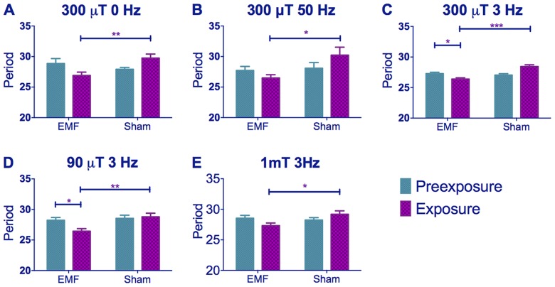
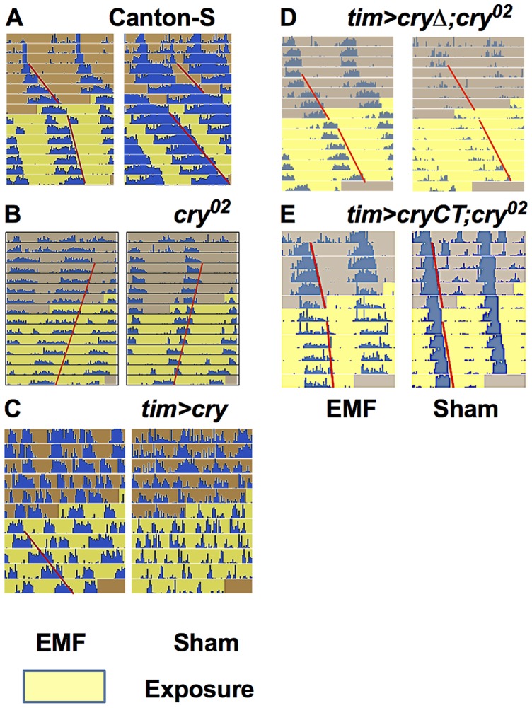
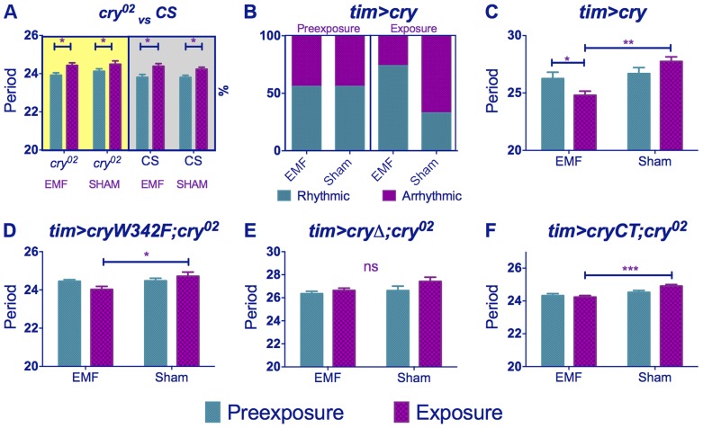
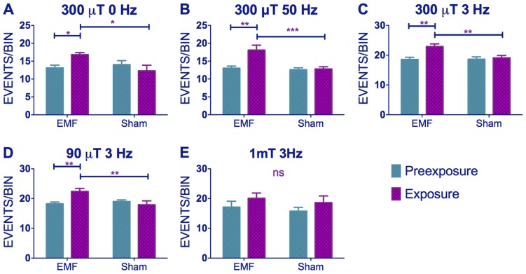
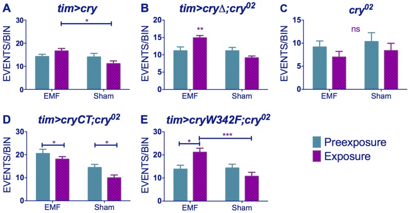
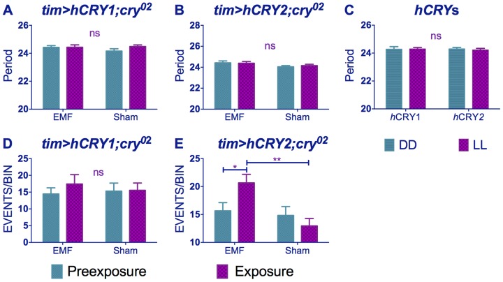
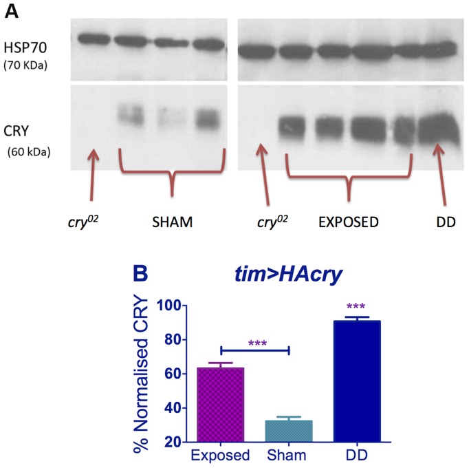
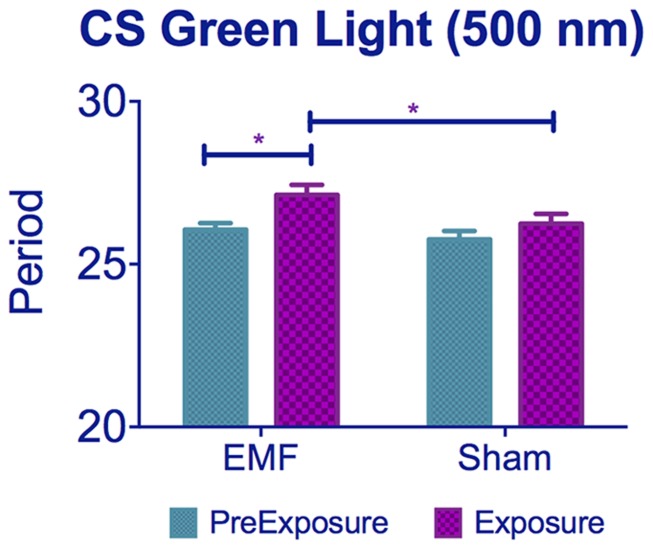
The blue-light sensitive photoreceptor cryptochrome (CRY) may act as a magneto-receptor through formation of radical pairs involving a triad of tryptophans. Previous genetic analyses of behavioral responses of Drosophila to electromagnetic fields using conditioning, circadian and geotaxis assays have lent some support to the radical pair model (RPM). Here, we describe a new method that generates consistent and reliable circadian responses to electromagnetic fields that differ substantially from those already reported. We used the Schuderer apparatus to isolate Drosophila from local environmental variables, and observe extremely low frequency (3 to 50 Hz) field-induced changes in two locomotor phenotypes, circadian period and activity levels. These field-induced phenotypes are CRY- and blue-light dependent, and are correlated with enhanced CRY stability. Mutational analysis of the terminal tryptophan of the triad hypothesised to be indispensable to the electron transfer required by the RPM reveals that this residue is not necessary for field responses. We observe that deletion of the CRY C-terminus dramatically attenuates the EMF-induced period changes, whereas the N-terminus underlies the hyperactivity. Most strikingly, an isolated CRY C-terminus that does not encode the Tryptophan triad nor the FAD binding domain is nevertheless able to mediate a modest EMF-induced period change. Finally, we observe that hCRY2, but not hCRY1, transformants can detect EMFs, suggesting that hCRY2 is blue light-responsive. In contrast, when we examined circadian molecular cycles in wild-type mouse suprachiasmatic nuclei slices under blue light, there was no field effect. Our results are therefore not consistent with the classical Trp triad-mediated RPM and suggest that CRYs act as blue-light/EMF sensors depending on trans-acting factors that are present in particular cellular environments.


Conflict of interest statement

The authors have declared that no competing interests exist.

Figures

Figure 1
Figure 1. EMF exposure shortens free-running circadian periods in dim blue light.
Mean circadian periods (h) +/− sem are shown for the EMF and sham-exposed groups. Note how periods are considerably longer than 24 h. (A–C) period changes in CS flies under static, 50 and 3 Hz field respectively at 300 µT (C–E) period changes in CS flies under 300, 90 and 1000 µT (1 mT) field respectively at 3 Hz. EMF-exposed flies show significant period shortening. For period and N see Table S1. (post-hoc *p<0.05, **p<0.01, ***p<0.001).
Figure 2
Figure 2. EMF exposure shortens circadian period.
Representative free-running locomotor rhythms in dim blue, constant LL before and during the exposure to EMF (300 µT, 3 Hz). A. Exposed Canton-S flies showed a significant period shortening compared to sham. B. cry02 flies did not show any EMF effect and maintain their free-run during the exposure period. C. Most exposed tim>cry flies showed arrhythmia before, but a well-defined period during the EMF exposure. D. tim>cryΔ;cry02 are not EMF sensitive. E. tim>cryCT;cry02 show an EMF effect with a slight period shortening compared to sham exposed flies. Each horizontal line show activity events (blue) double plotted for two successive 24 hour periods, day 1 and 2 on the top line, day 2 and 3 on the second line and so on. The red line outlines the activity offset.
Figure 3
Figure 3. cry variants alter normal circadian responses to EMFs.
Circadian periods (h) in dim blue LL are shown for EMF and sham-exposed groups. Mean periods ± sem. (A) cry02 flies exposed to EMF show only ageing effects on period (yellow shaded box). Wild-type flies kept in DD (grey shaded box) show similar ageing effects (B) tim>cry % rhythmic/arrhythmic flies during pre-exposure and exposure to EMF or sham. Exposure to EMF dramatically increases the proportion of rhythmic flies (χ2 (3) = 12.78, p<0.01). (C) tim>cry period for EMF exposed and sham flies before and during exposure (D) tim>cryW342F;cry02 (E) tim>cryΔ;cry02. (F) tim>GFPcryCT;cry02. (See Table S1, post-hoc *p<0.05, **p<0.01, ***p<0.001).
Figure 4
Figure 4. EMFs increase activity levels in wild-type flies.
(A–C) Hyperactivity in EMF-exposed CS under static, 50 and 3 Hz field respectively at 300 µT. (C–E) Hyperactivity in CS flies under 300, 90 and 1000 µT field respectively at 3 Hz. N's are the same as in Figure 1. Mean activity events per 30 min time bin (± sem). For average activity and N refer to Table S2 (post-hoc *p<0.05, **p<0.01, ***p<0.001).
Figure 5
Figure 5. EMF-induced hyperactivity in cry variants.
(A) tim>cry (B) tim>cryΔ;cry02 (C) cry02 (D) tim>cryCT;cry02 (E) tim>cryW342 F;cry02 N's are the same as in Figure 3. Mean ± sem. (see Table S2, post-hoc *p<0.05, **p<0.01, ***p<0.001).
Figure 6
Figure 6. hCRY2 but not hCRY1 reveals a sensitivity to EMFs.
(A) tim>hCRY1; cry02 or (B) tim>hCRY2; cry02 transformants do not show period shortening under EMF (pre-exposure*EMF/sham interaction hCRY1 F(1,48) = 1.41, p = 0.3 hCRY2 F(1,54) = 0.2, p = 0.63 (see Table S1). (C) hCRY1/2 flies do not show period increase in dim blue LL compared to DD (F(1, 82) = 0.125, p = 0.72) (D) hCRY1 are not hyperactive under EMF (F(1,48) = 0.33, p = 0.56). (E) hCRY2 are hyperactive under EMF exposure. Mean ± sem (see Table S2, post hoc * = p<0.05, ** = p<0.01).
Figure 7
Figure 7. EMF exposure increases CRY stability.
Top panel. Western blots for CRY using anti-dCRY in wild-type flies expose to EMF or sham in dim blue LL with cry02 and DD control. HSP is used as loading control. Bottom panel. Quantification based on 3 biological replicates each with 3 technical replicates (repeated measures ANOVA F(2,6) = 113.1, p<0.001, post hoc *** p<0.001). Mean ± sem.
Figure 8
Figure 8. Exposure to 500 nm green light lengthens circadian period under EMF.
CS flies kept under 500 nm show period lengthening when exposed to EMF compared to sham flies. See Table S1, post-hoc *p<0.05, ***p<0.001). Mean ± sem.

References

    1. Kato M (2006) In: Kato M, ed. Electromagnetics in Biology. Tokyo: Springer Japan.
    1. Johnsen S, Lohmann KJ (2008) Magnetoreception in animals. Phys Today 61: 29.
    1. Gould JL (2010) Magnetoreception. Curr Biol 20: R431–5. - PubMed
    1. Wiltschko R, Wiltschko W (2013) The magnetite-based receptors in the beak of birds and their role in avian navigation. J Comp Physiol A 198: 89–98. - PMC - PubMed
    1. Eder SHK, Cadiou H, Muhamad A, McNaughton P a, Kirschvink JL, et al. (2012) Magnetic characterization of isolated candidate vertebrate magnetoreceptor cells. Proc Natl Acad Sci U S A 109: 12022–12027. - PMC - PubMed

Publication types