Oral streptococci utilize a Siglec-like domain of serine-rich repeat adhesins to preferentially target platelet sialoglycans in human blood
- PMID: 25474103
- PMCID: PMC4256463
- DOI: 10.1371/journal.ppat.1004540
Oral streptococci utilize a Siglec-like domain of serine-rich repeat adhesins to preferentially target platelet sialoglycans in human blood
Abstract
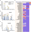
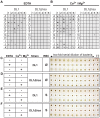
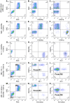
Damaged cardiac valves attract blood-borne bacteria, and infective endocarditis is often caused by viridans group streptococci. While such bacteria use multiple adhesins to maintain their normal oral commensal state, recognition of platelet sialoglycans provides an intermediary for binding to damaged valvular endocardium. We use a customized sialoglycan microarray to explore the varied binding properties of phylogenetically related serine-rich repeat adhesins, the GspB, Hsa, and SrpA homologs from Streptococcus gordonii and Streptococcus sanguinis species, which belong to a highly conserved family of glycoproteins that contribute to virulence for a broad range of Gram-positive pathogens. Binding profiles of recombinant soluble homologs containing novel sialic acid-recognizing Siglec-like domains correlate well with binding of corresponding whole bacteria to arrays. These bacteria show multiple modes of glycan, protein, or divalent cation-dependent binding to synthetic glycoconjugates and isolated glycoproteins in vitro. However, endogenous asialoglycan-recognizing clearance receptors are known to ensure that only fully sialylated glycans dominate in the endovascular system, wherein we find these particular streptococci become primarily dependent on their Siglec-like adhesins for glycan-mediated recognition events. Remarkably, despite an excess of alternate sialoglycan ligands in cellular and soluble blood components, these adhesins selectively target intact bacteria to sialylated ligands on platelets, within human whole blood. These preferred interactions are inhibited by corresponding recombinant soluble adhesins, which also preferentially recognize platelets. Our data indicate that circulating platelets may act as inadvertent Trojan horse carriers of oral streptococci to the site of damaged endocardium, and provide an explanation why it is that among innumerable microbes that gain occasional access to the bloodstream, certain viridans group streptococci have a selective advantage in colonizing damaged cardiac valves and cause infective endocarditis.
Conflict of interest statement
The authors have declared that no competing interests exist.
Figures


Similar articles
-
Streptococcal Siglec-like adhesins recognize different subsets of human plasma glycoproteins: implications for infective endocarditis.Glycobiology. 2018 Aug 1;28(8):601-611. doi: 10.1093/glycob/cwy052. Glycobiology. 2018. PMID: 29796594 Free PMC article.
-
Novel aspects of sialoglycan recognition by the Siglec-like domains of streptococcal SRR glycoproteins.Glycobiology. 2016 Nov;26(11):1222-1234. doi: 10.1093/glycob/cww042. Epub 2016 Apr 1. Glycobiology. 2016. PMID: 27037304 Free PMC article.
-
Recognition of specific sialoglycan structures by oral streptococci impacts the severity of endocardial infection.PLoS Pathog. 2019 Jun 24;15(6):e1007896. doi: 10.1371/journal.ppat.1007896. eCollection 2019 Jun. PLoS Pathog. 2019. PMID: 31233555 Free PMC article.
-
Platelet-streptococcal interactions in endocarditis.Crit Rev Oral Biol Med. 1996;7(3):222-36. doi: 10.1177/10454411960070030201. Crit Rev Oral Biol Med. 1996. PMID: 8909879 Review.
-
[Adhesins of oral streptococci].Nihon Saikingaku Zasshi. 2013;68(2):283-93. doi: 10.3412/jsb.68.283. Nihon Saikingaku Zasshi. 2013. PMID: 23727707 Review. Japanese.
Cited by
-
Role of Neuraminidase-Producing Bacteria in Exposing Cryptic Carbohydrate Receptors for Streptococcus gordonii Adherence.Infect Immun. 2018 Jun 21;86(7):e00068-18. doi: 10.1128/IAI.00068-18. Print 2018 Jul. Infect Immun. 2018. PMID: 29661931 Free PMC article.
-
Distinct Biological Potential of Streptococcus gordonii and Streptococcus sanguinis Revealed by Comparative Genome Analysis.Sci Rep. 2017 Jun 7;7(1):2949. doi: 10.1038/s41598-017-02399-4. Sci Rep. 2017. PMID: 28592797 Free PMC article.
-
Streptococcus abundance and oral site tropism in humans and non-human primates reflects host and lifestyle differences.NPJ Biofilms Microbiomes. 2025 Jan 17;11(1):19. doi: 10.1038/s41522-024-00642-1. NPJ Biofilms Microbiomes. 2025. PMID: 39824852 Free PMC article.
-
Structural basis for the role of serine-rich repeat proteins from Lactobacillus reuteri in gut microbe-host interactions.Proc Natl Acad Sci U S A. 2018 Mar 20;115(12):E2706-E2715. doi: 10.1073/pnas.1715016115. Epub 2018 Mar 5. Proc Natl Acad Sci U S A. 2018. PMID: 29507249 Free PMC article.
-
Serine-rich repeat proteins: well-known yet little-understood bacterial adhesins.J Bacteriol. 2024 Jan 25;206(1):e0024123. doi: 10.1128/jb.00241-23. Epub 2023 Nov 17. J Bacteriol. 2024. PMID: 37975670 Free PMC article. Review.
References
-
- Werdan K, Dietz S, Loffler B, Niemann S, Bushnaq H, et al. (2014) Mechanisms of infective endocarditis: pathogen-host interaction and risk states. Nat Rev Cardiol 11: 35–50. - PubMed
-
- Baddour LM, Wilson WR, Bayer AS, Fowler VGJ, Bolger AF, et al. (2005) Infective endocarditis: diagnosis, antimicrobial therapy, and management of complications: a statement for healthcare professionals from the Committee on Rheumatic Fever, Endocarditis, and Kawasaki Disease, Council on Cardiovascular Disease in the Young, and the Councils on Clinical Cardiology, Stroke, and Cardiovascular Surgery and Anesthesia, American Heart Association: endorsed by the Infectious Diseases Society of America. Circulation 111: e394–e434. - PubMed
-
- Hoen B, Duval X (2013) Clinical practice. Infective endocarditis. N Engl J Med 368: 1425–1433. - PubMed
-
- Duval X, Leport C (2008) Prophylaxis of infective endocarditis: current tendencies, continuing controversies. Lancet Infect Dis 8: 225–232. - PubMed
Publication types
MeSH terms
Substances
Grants and funding
LinkOut - more resources
Full Text Sources
Other Literature Sources

