

A novel psittacine adenovirus identified during an outbreak of avian chlamydiosis and human psittacosis: zoonosis associated with virus-bacterium coinfection in birds
- PMID: 25474263
- PMCID: PMC4256287
- DOI: 10.1371/journal.pntd.0003318
A novel psittacine adenovirus identified during an outbreak of avian chlamydiosis and human psittacosis: zoonosis associated with virus-bacterium coinfection in birds
Abstract
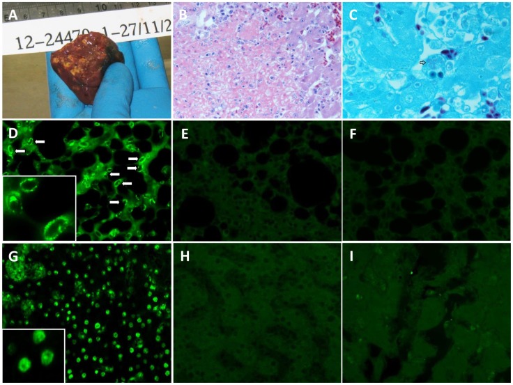
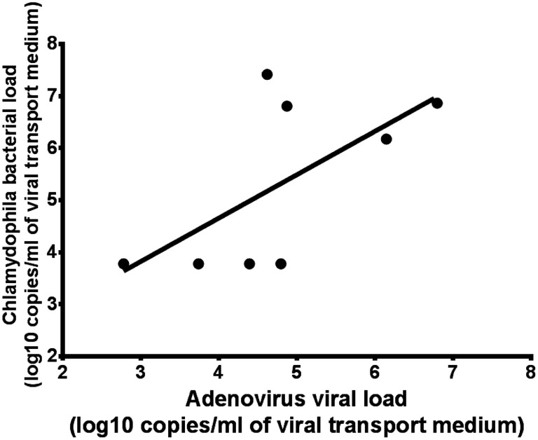
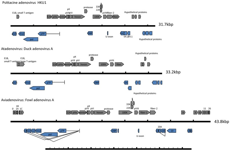
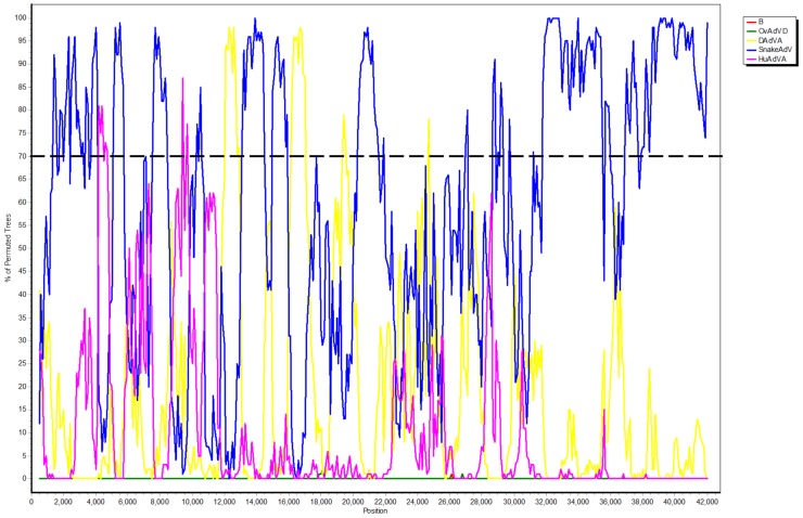
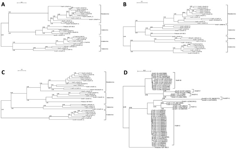
Chlamydophila psittaci is found worldwide, but is particularly common among psittacine birds in tropical and subtropical regions. While investigating a human psittacosis outbreak that was associated with avian chlamydiosis in Hong Kong, we identified a novel adenovirus in epidemiologically linked Mealy Parrots, which was not present in healthy birds unrelated to the outbreak or in other animals. The novel adenovirus (tentatively named Psittacine adenovirus HKU1) was most closely related to Duck adenovirus A in the Atadenovirus genus. Sequencing showed that the Psittacine adenovirus HKU1 genome consists of 31,735 nucleotides. Comparative genome analysis showed that the Psittacine adenovirus HKU1 genome contains 23 open reading frames (ORFs) with sequence similarity to known adenoviral genes, and six additional ORFs at the 3' end of the genome. Similar to Duck adenovirus A, the novel adenovirus lacks LH1, LH2 and LH3, which distinguishes it from other viruses in the Atadenovirus genus. Notably, fiber-2 protein, which is present in Aviadenovirus but not Atadenovirus, is also present in Psittacine adenovirus HKU1. Psittacine adenovirus HKU1 had pairwise amino acid sequence identities of 50.3-54.0% for the DNA polymerase, 64.6-70.7% for the penton protein, and 66.1-74.0% for the hexon protein with other Atadenovirus. The C. psittaci bacterial load was positively correlated with adenovirus viral load in the lung. Immunostaining for fiber protein expression was positive in lung and liver tissue cells of affected parrots, confirming active viral replication. No other viruses were found. This is the first documentation of an adenovirus-C. psittaci co-infection in an avian species that was associated with a human outbreak of psittacosis. Viral-bacterial co-infection often increases disease severity in both humans and animals. The role of viral-bacterial co-infection in animal-to-human transmission of infectious agents has not received sufficient attention and should be emphasized in the investigation of disease outbreaks in human and animals.
Conflict of interest statement
The authors have declared that no competing interests exist.
Figures
Similar articles
-
Disease surveillance in wild Victorian cacatuids reveals co-infection with multiple agents and detection of novel avian viruses.Vet Microbiol. 2019 Aug;235:257-264. doi: 10.1016/j.vetmic.2019.07.012. Epub 2019 Jul 18. Vet Microbiol. 2019. PMID: 31383310
-
A Chlamydia psittaci Outbreak in Psittacine Birds in Sardinia, Italy.Int J Environ Res Public Health. 2022 Oct 30;19(21):14204. doi: 10.3390/ijerph192114204. Int J Environ Res Public Health. 2022. PMID: 36361084 Free PMC article.
-
First Identification of Chlamydia psittaci in the Acute Illness and Death of Endemic and Endangered Psittacine Birds in Mexico.Avian Dis. 2016 Jun;60(2):540-4. doi: 10.1637/11360-122915-Case. Avian Dis. 2016. PMID: 27309302
-
Chlamydia psittaci: A zoonotic pathogen causing avian chlamydiosis and psittacosis.Virulence. 2024 Dec;15(1):2428411. doi: 10.1080/21505594.2024.2428411. Epub 2024 Dec 2. Virulence. 2024. PMID: 39541409 Free PMC article. Review.
-
[Avian chlamydiosis (parrot disease, psittacosis, ornithosis)].Tijdschr Diergeneeskd. 2012 Sep;137(9):588-93. Tijdschr Diergeneeskd. 2012. PMID: 23025204 Review. Dutch. No abstract available.
Cited by
-
Sialic Acid Receptors: The Key to Solving the Enigma of Zoonotic Virus Spillover.Viruses. 2021 Feb 8;13(2):262. doi: 10.3390/v13020262. Viruses. 2021. PMID: 33567791 Free PMC article. Review.
-
Molecular Characterisation of a Novel and Highly Divergent Passerine Adenovirus 1.Viruses. 2020 Sep 17;12(9):1036. doi: 10.3390/v12091036. Viruses. 2020. PMID: 32957674 Free PMC article.
-
The FDA-Approved Drug Nelfinavir Inhibits Lytic Cell-Free but Not Cell-Associated Nonlytic Transmission of Human Adenovirus.Antimicrob Agents Chemother. 2020 Aug 20;64(9):e01002-20. doi: 10.1128/AAC.01002-20. Print 2020 Aug 20. Antimicrob Agents Chemother. 2020. PMID: 32601166 Free PMC article.
-
Identification of two novel adenoviruses in smooth-billed ani and tropical screech owl.PLoS One. 2020 Feb 28;15(2):e0229415. doi: 10.1371/journal.pone.0229415. eCollection 2020. PLoS One. 2020. PMID: 32109945 Free PMC article.
-
The (Re-)Emergence and Spread of Viral Zoonotic Disease: A Perfect Storm of Human Ingenuity and Stupidity.Viruses. 2023 Jul 27;15(8):1638. doi: 10.3390/v15081638. Viruses. 2023. PMID: 37631981 Free PMC article.
References
-
- Centre for Health Protection (2013) Psittacosis outbreak in an animal management centre, 2012. Communicable Diseases Watch 10: 9–10.
Publication types
MeSH terms
LinkOut - more resources
Full Text Sources
Other Literature Sources
Medical
