. 2015 Apr;17(4):262-70.
doi: 10.1038/gim.2014.174. Epub 2014 Dec 4.

Comprehensive analysis of patients with Stargardt macular dystrophy reveals new genotype-phenotype correlations and unexpected diagnostic revisions

Affiliations

Comprehensive analysis of patients with Stargardt macular dystrophy reveals new genotype-phenotype correlations and unexpected diagnostic revisions

Jacques Zaneveld et al. Genet Med. 2015 Apr.

Abstract

Purpose: Stargardt macular dystrophy (STGD) results in early central vision loss. We sought to explain the genetic cause of STGD in a cohort of 88 patients from three different cultural backgrounds.

Methods: Next-generation sequencing using a novel capture panel was used to search for disease-causing mutations. Patients with undetermined causes were clinically reexamined and tested for copy-number variations as well as intronic mutations.

Results: We determined the cause of disease in 67% of our patients. Our analysis identified 35 novel ABCA4 alleles. Eleven patients had mutations in genes not previously reported to cause STGD. Finally, 45% of our patients with unsolved causes had single deleterious mutations in ABCA4, a recessive disease gene. No likely pathogenic copy-number variations were identified.

Conclusion: This study expands our knowledge of STGD by identifying dozens of novel alleles that cause the disease. The frequency of single mutations in ABCA4 among STGD patients is higher than that among controls, indicating that these mutations contribute to disease. Disease in 11 patients was explained by mutations outside ABCA4, underlining the need to genotype all retinal disease genes to maximize genetic diagnostic rates. Few ABCA4 mutations were observed in our French Canadian patients. This population may contain an unidentified founder mutation. Our results indicate that copy-number variations are unlikely to be a major cause of STGD.


Figures

Figure 1
Figure 1
High quality sequencing results were obtained, with consistently high coverage across the targeted region. Almost the entire target was covered sufficiently to call SNPs. Error bars display one standard deviation.
Figure 2
Figure 2
Representative CGH microarray from a patient with one hit in ABCA4. Despite a very high probe density, no significant CNVs were found in any of the 12 patients with one mutation in ABCA4
Figure 3
Figure 3
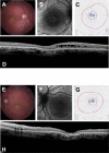
A,E) Fundus photographs of patient 69 show a bilateral atrophic maculopathy with yellow retinal flecks while the retinal vasculature and optic discs are within normal limits. B,F) Fundus auto-fluorescence images show an incomplete rim of hyper-fluorescence in the right macular area and a complete hyper-fluorescent ring in left macula. C,G) GVF shows central scotoma in both eyes. D,H) OCT studies show loss of the IS/OS junctions, a disorganized external limiting membrane and outer nuclear layer, thinning of the outer nuclear layer in the fovea and sub-foveal edema. There were marked cystic spaces in the inner nuclear layer.
Figure 4
Figure 4
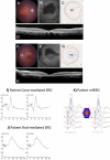
A,E) Fundus photographs of patient 55 show an atrophic maculopathy involving the fovea in the OD with clumps of pigmentation in the macular area. The OS shows an atrophic maculopathy sparing the inferior half of fovea. Retinal vessels and optic discs are within normal limits. B,F) Fundus auto-fluorescence images show macular atrophy appears dark black involving the fovea in the OD and in the OS inferior half of fovea is spared. C,G) Goldmann Visual Fields show an absolute central scotoma OU. D,H) OCT shows bilateral loss of IS/OS junctions and decreased retinal thickness. Accumulation of auto-fluorescent photoreceptor debris in the foveal area is seen in the OD. I) Cone mediated ERG a-waves are within normal amplitude limits (OU) and peak times are delayed (0D) and within normal limits (OS). The b-waves are within the lower limits of normal amplitude (OU) with peak times that are delayed (0D) and within normal limits (OS). J) The rod-mediated ERG b-wave is within normal amplitude and peak time limits (OU). K) mfERGs are attenuated in amplitude at all eccentricities.

References

    1. Burke TR, Tsang SH. Allelic and phenotypic heterogeneity in ABCA4 mutations. Ophthalmic genetics. 2011;32(3):165–174. - PMC - PubMed
    1. Molday RS, Zhang K. Defective lipid transport and biosynthesis in recessive and dominant Stargardt macular degeneration. Progress in lipid research. 2010;49(4):476–492. - PMC - PubMed
    1. Kaplan J, Gerber S, Larget-Piet D, et al. A gene for Stargardt's disease (fundus flavimaculatus) maps to the short arm of chromosome 1. Nature genetics. 1993;5(3):308–311. - PubMed
    1. Yang Z, Chen Y, Lillo C, et al. Mutant prominin 1 found in patients with macular degeneration disrupts photoreceptor disk morphogenesis in mice. The Journal of clinical investigation. 2008;118(8):2908–2916. - PMC - PubMed
    1. Zhang K, Kniazeva M, Han M, et al. A 5-bp deletion in ELOVL4 is associated with two related forms of autosomal dominant macular dystrophy. Nature genetics. 2001;27(1):89–93. - PubMed

Publication types

Substances