. 2015 Feb 18;137(6):2140-54.
doi: 10.1021/ja510147n. Epub 2015 Feb 6.

Stimuli-responsive nanomaterials for biomedical applications

Affiliations

Stimuli-responsive nanomaterials for biomedical applications

Angela P Blum et al. J Am Chem Soc. .

Abstract

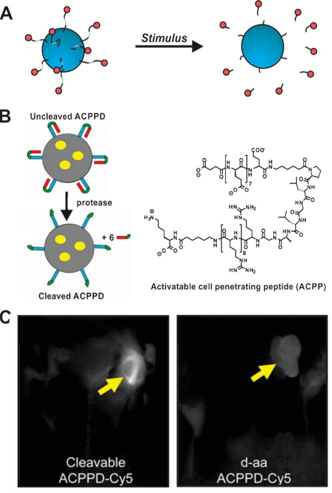
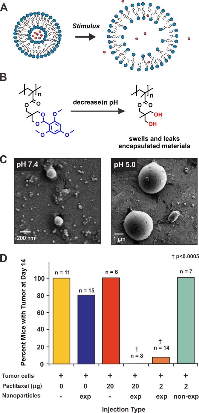
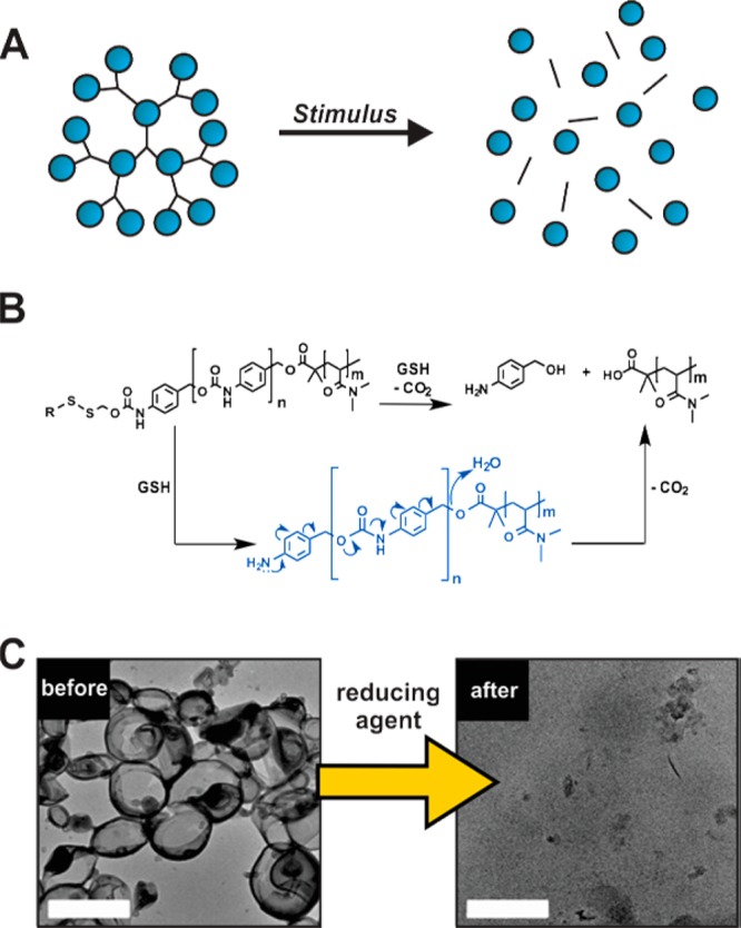
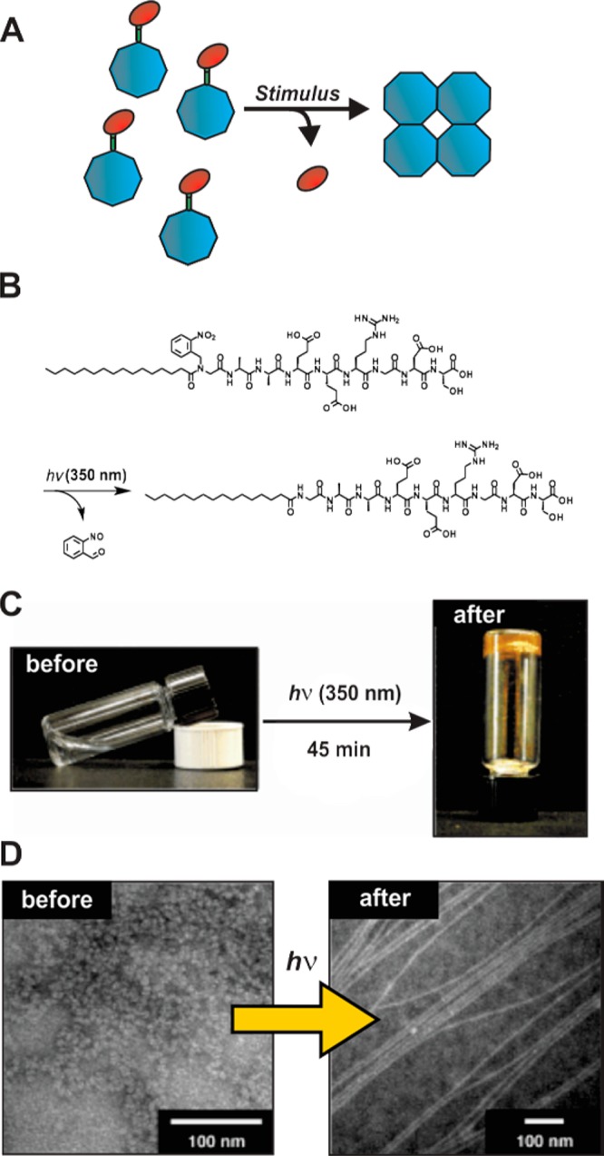
Nature employs a variety of tactics to precisely time and execute the processes and mechanics of life, relying on sequential sense and response cascades to transduce signaling events over multiple length and time scales. Many of these tactics, such as the activation of a zymogen, involve the direct manipulation of a material by a stimulus. Similarly, effective therapeutics and diagnostics require the selective and efficient homing of material to specific tissues and biomolecular targets with appropriate temporal resolution. These systems must also avoid undesirable or toxic side effects and evade unwanted removal by endogenous clearing mechanisms. Nanoscale delivery vehicles have been developed to package materials with the hope of delivering them to select locations with rates of accumulation and clearance governed by an interplay between the carrier and its cargo. Many modern approaches to drug delivery have taken inspiration from natural activatable materials like zymogens, membrane proteins, and metabolites, whereby stimuli initiate transformations that are required for cargo release, prodrug activation, or selective transport. This Perspective describes key advances in the field of stimuli-responsive nanomaterials while highlighting some of the many challenges faced and opportunities for development. Major hurdles include the increasing need for powerful new tools and strategies for characterizing the dynamics, morphology, and behavior of advanced delivery systems in situ and the perennial problem of identifying truly specific and useful physical or molecular biomarkers that allow a material to autonomously distinguish diseased from normal tissue.


Figures

Figure 1
Figure 1
Stimuli-driven direct release or activation strategies. (A) Cartoon scheme depicting the direct release of drugs (red circles) or activation of diagnostic agents following initiation by a stimulus. (B) A literature example of a dendrimer (called an activatable cell-penetrating dendrimer, ACPPD) decorated with activatable cell-penetrating peptides (ACPPs) that also contains encapsulated Cy 5 dye for fluorescence imaging or gadolinium cargo for use in MRI diagnostics (yellow circles). In this design, enzymes upregulated in cancer cells (MMPs) facilitate cleavage of the ACPP hairpin, exposing a polyarginine cell penetrating peptide motif, which facilitates the entry of the cargo-carrying nanoparticle. Prior to cleavage, the ACPP forms a hairpin by non-covalent interactions between segments of polyglutamic acid and polyargine (the cell-penetrating motif) that flank the recognition sequence of the enzyme. Upon cleavage of the peptide hairpin, the polyglutamic acid segment is released, exposing the polyarginine fragment, which can then penetrate cells. (C) Fluorescence images of mice 48 h post injection of either the cleavable ACPPD with encapsulated Cy 5, or a non-cleavable ACPPD (d-amino acid control) variant. In these images, there is a substantial increase in florescence at tumors only when the particles with the cleavable ACPP are administered, illustrating that this method can be used to target cancer cells and internalize while carrying useful diagnostic or therapeutic cargo. Panels B and C are adapted from Olson et al. with permission from the National Academy of Sciences.
Figure 2
Figure 2
Expansile nanoparticle systems. (A) A general cartoon describing systems that release encapsulated drugs (red circles) by expansion into a fenestrated structure upon activation by a stimulus. (B) Chemical structures of expansile nanoscale particles containing 2,4,6-trimethoxylbenzaldehyde-derived acetals that hydrolyze at pH ≤ 5 to yield diol-containing scaffolds, which form micrometer-scale hydrogels. The pH of activation is in line with the pH of cellular lysosomes, and so these materials have been used to release encapsulated drugs inside cellular organelles upon internalization. (C) Scanning electron microscopy images of the acetal described in B at pH 7.4, and the diol hydrolysis product at pH 5.0. Note that the diameter of the material expands 350-fold in response to mildly acidic environments. (D) Experimental data from in vivo studies in which C57BL/6 mice are injected with Lewis lung carcinoma cells alongside paclitaxel-loaded expansile (exp) and non-expansile particles (non-exp, which contain related benzaldehyde-derived acetals) and appropriate controls. Note that only mice that received the drug-loaded exapansile particles were free from tumors. Panels C and D are adapted from Colby et al. with permission from the Royal Society of Chemistry and Griset et al. with permission from the American Chemical Society, respectively.
Figure 3
Figure 3
Stimuli-responsive strategies invoking a gatekeeping mechanism. (A) A cartoon illustration of the gatekeeping strategy in which the pores (green cylinders) of mesoporous silica nanoparticles (MSNPs, depicted as blue spheres) are blocked by a gate (yellow crosses). Stimulus activation results in opening of the gate and release of encapsulated imaging agents or therapeutics (red spheres). Note that other nanomaterials such as core–shell particles also employ similar approaches to release cargo. (B) A literature example of a pH-activated MSNP. In this example, a β-cyclodextrin (β-CD) is used to cap the pores of drug- or fluorophore-carrying MSNPs. At physiological pH, the β-CD encapsulates aromatic amines that are appended to the periphery of the MSNP, blocking the nanopore and entrapping cargo. Protonation of the amines following a decrease in pH results in the release of the cyclodextrin gate, enabling free diffusion of the pore contents. (C) Fluorescent images illustrating doxorubicin release from the β-CD-gated MSNPs described in panel B after internalization of the materials into acidified endosomal compartments of KB-13 cells. Release of doxorubicin was also correlated with a decrease in cell viability. Neutralization of lysosomal pH by the addition of NH4Cl results in inhibition of doxorubicin release and toxcity, providing support for the proposed mechanism of activation. Panels B and C are adapted from Meng et al. with permission from the American Chemical Society.
Figure 4
Figure 4
Stimuli-driven disassembly processes. (A) Cartoon illustrating a generic disassembly event in which a nanoscale material breaks down into smaller fragments, releasing encapsulated or chemically appended drugs (blue spheres). (B) A literature example of a deploymerizaton event that is initiatied under reductive conditions. Here, polymersomes assembled from block copolymers composed of a self-immolative poly(benzyl carbamate) block and a hydrophilic poly(N,N-dimethylacrylamide) (PDMA) block. In this report, the self-immolative block was caged with either perylen-3-yl, o-nitrobenzyl, or disulfide moieties (as depicted), which uncage in response to visible light (420 nm), UV light (365 nm), or reductive agents, respectively. Upon activation, the block copolymer depolymerizes into 4-aminobenzyl alcohol, carbon dioxide, and PDMA. These polymersomes were also loaded with doxorubicin or campothecin, and payload release coincident with depolymerization was observed. (C) Transmission electron microscopy of the block copolymer before and after treatment with dithiothreitol. Scale bars are 1 μm. Panels B and C are adapted from Liu et al. with permission from the American Chemical Society.
Figure 5
Figure 5
Stimuli-induced assembly events. (A) Cartoon illustrating a generic assembly event in which an uncaging event occurs (loss of red blocking unit) to enable the assembly of many unimers (blue octagons). Note that we define an assembly event as a unimer or ill-defined structure assembling into a much larger stucture of higher order. Aggregation is a related process in which unimers or disordered phases assemble into larger, amphorphous aggregates. (B) Chemical structures depicting a literature example of an o-nitrobenzyl “caged” amphiphilic peptide that becomes amphiphillic and self-assembles into hydrogel-forming fibers upon removal of the caging group (i.e., the red oval in scheme A) by 350 nm light. (C) Photograph of the caged amphiphile and the hydrogel formed after removal of the o-nitrobenzyl cage. (D) Transmission electron microscopy images of the system before and after exposure to UV light. Panels B–D are adapted from Muraoka et al. with permission from John Wiley and Sons Ltd.
Figure 6
Figure 6
Materials that respond to various stimuli by changing intra- and inter-molecular packing parameters resulting in transitions between distinct morphologies or phases. (A) A cartoon depicting spherical nanoparticles transitioning into cylindrical structures upon the introduction of a stimulus. (B) Polymers composed of poly(N-isopropylacrylamide) (PNIPAAm) and ethyltryptophan (EtTrp) organized along a polyphosphazene backbone assemble into discrete phases in solution. These phases are formed by non-covalent molecular interactions and can be disrupted by the introduction of other molecules, leading to changes in the overall material morphology. Upon the introduction of a small molecule (indomethacin) that hydrogen-bonds with the polymer, disruption of intra- and inter-polymer interactions results in reorganization of the material and ultimately rearrangement into a different preferred phase. (C) Transmission electron microscopy images depicting a phase transition from network-like bicontinuous rods to vesicular or multilamellar large compound structures. Panels B and C are adapted from Zhang et al. with permission from John Wiley and Sons Ltd.
Figure 7
Figure 7
Nanomaterials capable of autonomous motion. These materials are often prepared by incorporating motors that use catalysis to generate chemical concentration gradients and propel themselves through solution via self-electrophoresis. (A) Cartoon depiction of the motion of a nanomaterial generated via the depletion of chemical fuel placed on one side of the object. (B) Platinum–copper nanorods that catalyze the reduction of iodine to iodide while oxidizing copper at the opposite end of the rod. The reduction that takes place at the platinum end of the nanorod generates a flow of electrons toward the platinum end of the material. This electron flow generates a charge differential, thus inducing fluid movement toward the platinum segment and propelling the nanorod in the opposite direction of the fluid. (C) Optical microscopy snapshots tracking movement of Pt–Cu nanorods over time (images (g) and (h) depict instances of surface-immobilized nanorods). Panels B and C are adapted from Liu et al. with permission from the American Chemical Society.
Figure 8
Figure 8
Strategy for facilitating direct accumulation of nanomaterial-derived scaffolds in tumor tissue. (A) A general scheme depicting the design of the system consisting of a spherical particle composed of polymeric peptide-amphiphiles, where the hydrophilic portion is a substrate for tumor-associated MMPs. As nanoscale spherical assemblies, the nanoparticles are free to circulate into and out of tissue until they encounter locations with a large abundance of MMPs, indicative of cancer or inflammation. Upon cleavage of the peptide, fragmented amphiphiles reassemble into a large, interconnected scaffold that that is too large to re-enter circulation. (B) Fluorescence data of tumor-burdened mice 1 h or 3 days post intra-tumoral injection of the MMP-responsive nanoparticles labeled with Alexafluor-647. Enzyme-responsive particles (prepared from l-amino acids) are assembled as designed and are retained in the tumor tissue for at least a week, at which point the animal is sacrificed. Non-responsive particles (comprised of d-amino acids) are rapidly cleared from the tumor tissue. We envision that responsive systems such as these could be used to trigger accumulation of drug-loaded particles that could be further activated to release drugs in a tissue-specific fashion or passively via long-term degradation. Figure adapted from Chien et al. with permission from the American Chemical Society.

References

    1. Lipinski C. A.; Lombardo F.; Dominy B. W.; Feeney P. J. Adv. Drug Delivery Rev. 2001, 46, 3. - PubMed
    1. Vlieghe P.; Lisowski V.; Martinez J.; Khrestchatisky M. Drug Discovery Today 2010, 15, 40. - PubMed
    1. Widakowich C.; de Castro G. Jr.; de Azambuja E.; Dinh P.; Awada A. Oncologist 2007, 12, 1443. - PubMed
    1. Matsumura Y.; Maeda H. Cancer Res. 1986, 46, 6387. - PubMed
    1. Barenholz Y. J. Controlled Release 2012, 160, 117. - PubMed

Publication types

Substances