Review
. 2014 Dec 4;7(1):a016659.
doi: 10.1101/cshperspect.a016659.

DNA strand exchange and RecA homologs in meiosis

Affiliations
Review

DNA strand exchange and RecA homologs in meiosis

M Scott Brown et al. Cold Spring Harb Perspect Biol. .

Abstract

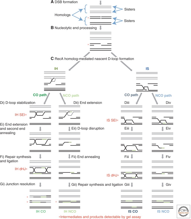
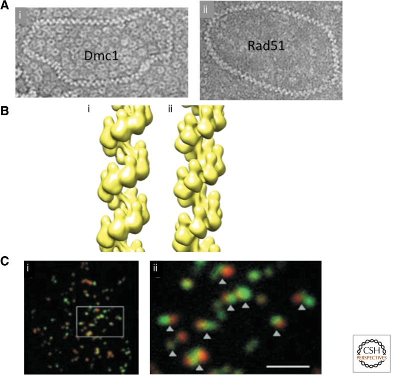
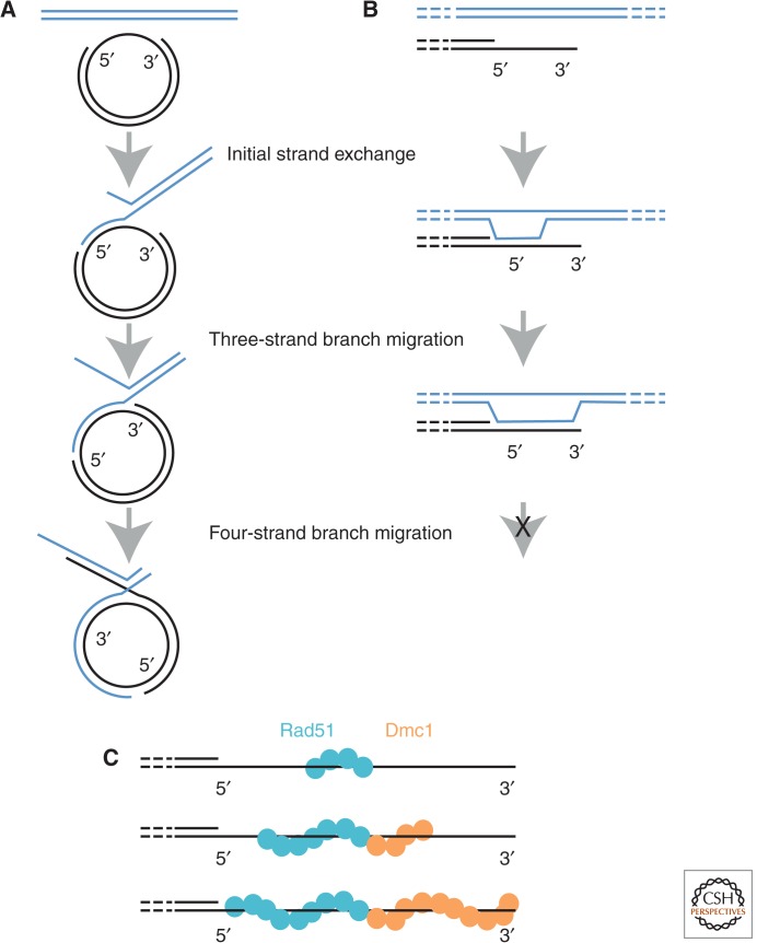
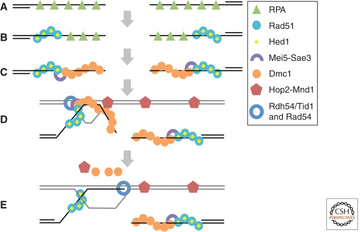
Homology search and DNA strand-exchange reactions are central to homologous recombination in meiosis. During meiosis, these processes are regulated such that the probability of choosing a homolog chromatid as recombination partner is enhanced relative to that of choosing a sister chromatid. This regulatory process occurs as homologous chromosomes pair in preparation for assembly of the synaptonemal complex. Two strand-exchange proteins, Rad51 and Dmc1, cooperate in regulated homology search and strand exchange in most organisms. Here, we summarize studies on the properties of these two proteins and their accessory factors. In addition, we review current models for the assembly of meiotic strand-exchange complexes and the possible mechanisms through which the interhomolog bias of recombination partner choice is achieved.


Figures

Figure 1.
Figure 1.
Meiotic recombination pathways. Four distinct recombination pathways can repair a meiotic DSB. Both strands of all four sister chromatids are shown with black and gray lines representing the two homologous chromosomes. Red asterisks indicate recombination intermediates and products that can be observed by Southern blots. (A) Meiotic recombination is initiated by the programmed introduction of DSBs. (B) 5′ to 3′ nucleolytic resection generates 3′ single-strand DNA (ssDNA) tails on both sides of the DSB. (C) RecA homologs locate an intact repair template on a homologous chromatid or the sister chromatid and catalyze strand exchange, generating a nascent D-loop intermediate. This D-loop structure is not stable enough to be observed in physical assays. (Di) Interhomolog (IH) crossover (CO) intermediates are formed when the nascent strand invasion of a homologous chromatid is stabilized. The resulting joint molecule (JM) is called an IH SEI. (Ei) Restorative DNA synthesis from the invading 3′ end (shown in green) extends the D-loop, allowing annealing of the second end of the DSB. (Fi) Further DNA synthesis and ligation of the remaining nicks generates an IH double Holliday junction (dHJ). (Gi) Resolution of the IH dHJ generates an IH CO in which the flanking DNA sequences are reciprocally exchanged. (Dii–Gii) IH NCOs are formed by a synthesis-dependent strand-annealing (SDSA) mechanism when an IH nascent JM is not stabilized. (Dii) DNA synthesis extends the 3′ end of a nascent D-loop. (Eii) The D-loop is disrupted. (Fii) This extended 3′ end anneals to the ssDNA tract on the opposite end. (Gii) Following further DNA synthesis and ligation, an IH NCO is formed. There is no reciprocal exchange of flanking regions in this recombination product. (Diii–Giii) Beside the choice of repair template, an IS CO is formed through the same biochemical steps as an IH CO. (Div–Giv) Similarly, an IS NCO forms through the same SDSA steps as an IH NCO. DSB, double-strand break; IS, intersister; NCO, noncrossover.
Figure 2.
Figure 2.
Microscopic analysis of strand-exchange proteins. (A) Electron micrographs of human (i) Dmc1, and (ii) Rad51filaments coating a 1312 bp circular dsDNA plasmid. Note the high density of toroids in the background of the Dmc1 image (From Sheridan et al. 2008; reprinted, with permission, from Oxford University Press © 2008.) (B) Helical reconstructions of human (i) Dmc1, and (ii) Rad51 filaments (courtesy of E. Egelman). (C) Surface spread S. cerevisiae meiotic nuclei immunostained for Rad51 (green) and Dmc1 (red). (i) Low magnification view, and (ii) blow up of region indicated. Scale bar, 2 μm. (From Shinohara et al. 2000; reprinted, with permission, from the American Society of Plant Biologists © 1999.)
Figure 3.
Figure 3.
Directionality of Dmc1- versus Rad51-mediated branch migration. (A) Diagramatic representation of the four-strand reaction using substrates that can undergo conversion from three-strand branch-migration reactions to four-strand reactions. The left side of the linear duplex substrate (blue) is homologous to the ssDNA gap on the circular substrate. Strand exchange initiates in the ssDNA gap, branch migration extends the tract of heteroduplex to the ssDNA–dsDNA junction at the 3′ end of the ssDNA region. Then, further 5′-3′ branch migration results in reciprocal stand exchange via the four-strand reaction. (B) Inferred consequence of 5′-3′ branch migration on D-loops formed in vivo. Ends at DSB sites are processed to have 3′ overhanging ssDNA tails. Branch migration is expected to proceed to the 3′ end, but not be able to carry out a four-strand reaction because an end has been reached. (C) Model for strand-exchange filament elongation on 3′ ssDNA tails. Rad51 (blue) nucleates filament formation. Dmc1 filaments (orange) are seeded at the end of a Rad51 filament. The direction of filament elongation is proposed to be the same as the direction of branch migration for the four-strand reaction. Thus, the Rad51 filament is elongated in the 3′–5′ direction, the Dmc1 filament elongated in the 5′–3′ direction. This will tend to completely coat the entire ssDNA region, and perhaps lead to extension of the Rad51 filament into the flanking dsDNA.
Figure 4.
Figure 4.
Working model for assembly and function of Rad51–Dmc1 recombinosomes. (A) RPA binds to ssDNA regions formed by nucleolytic resection of DNA ends. (B) Rad51, with the aid of mediator proteins (not shown), displaces RPA. Rad51 is prevented from forming D-loops by the inhibitory protein Hed1. (C) Mei5–Sae3 promotes initiation of Dmc1 filaments at the end of a Rad51 filament. Once initiated, Dmc1 filaments elongate on DNA by homotypic protomer–protomer interactions. (D) Dmc1 carries out a homology search culminating in formation of a segment of heteroduplex DNA. Efficient formation of D-loops by Dmc1 requires interaction of the searching filament with a complex of Hop2–Mnd1 bound to the target dsDNA. (E) The Rdh54/Tid1 translocase, or the Rad54 translocase, binds the Rad51 filament and translocates along the heteroduplex, simultaneously displacing Dmc1 and extending the heteroduplex tract to the 3′ end. The end is, thus, rendered accessible for initiation of DNA synthesis.
Figure 5.
Figure 5.
HIS4::LEU2 DSB hot spot. (A) Structure of the HIS4::LEU2 DSB hot spot. The paternal and maternal homologs are shown in gray and black, respectively. A single DSB hot spot is located downstream from the LEU2 gene, which was inserted adjacent to the HIS4 locus (Cao et al. 1990). XhoI restriction endonuclease cleavage sites (X) are engineered such that cleavage of the paternal and maternal homologs yields fragments of distinct sizes on Southern blots (probe location in green). (B) A representative 1D gel, showing the structure of chromatids at the HIS4::LEU2 DSB hot spot throughout a meiotic time course experiment. *A meiosis-specific band resulting from gene conversion of the XhoI site closest to the DSB site. (C) Single time point of a representative 2D gel showing branched molecules (SEIs, dHJs, and multichromatid joint molecules [mcJMs]) migrating off of the linear arc. (D) Schematic of the various species observable at the HIS4::LEU2 DSB hot spot. Note that although both products and intermediates of meiotic recombination are observable, crosslinking of the DNA by ultraviolet light before DNA purification is required to stabilize the branched JMs. SEI nomenclature is from Kim et al. 2010. (From data in Figure 2 in Oh et al. 2007; modified, with permission.)
Figure 6.
Figure 6.
IH bias models. (A) Barrier to sister chromatid recombination (BSCR). In response to DSB formation, a local recombination deficient zone (red) is created on the recipient homolog. Because there are no DSBs at the allelic position on the homolog, the donor homolog remains a recombination proficient zone (green). (B) Anchor pad model. The Rad51 filament (blue) end of the DSB invades the sister chromatid. Further Rad51 polymerization beyond the D-loop (light blue) creates an “anchor pad” that blocks invasion of the sister chromatid by the Dmc1 end of the DSB. Left with no other choice, the Dmc1 end invades a homologous chromatid. In WT cells, this IH nascent interaction proceeds to an IH dHJ. In rec8 cells, the nascent IH interaction has an equal probability of forming an IH or an IS dHJ. Note that the version of the model shown has Rad51 and Dmc1 loaded on opposite DSB ends (Hong et al. 2013). However, the model could be modified to account for loading of the two RecA homologs on both ends, (C) steric hindrance model 1. A scaffold structure, dependent on Rad51, projects the Dmc1 filament away from the recipient homolog axis. The scaffold precludes invasion of the sister chromatid, (D) steric hindrance model 2. The DSB ends are held in close proximity to the recipient homolog axis, whereas the sister chromatid loop is distant from the axis. (E) Hop2–Mnd1 creates recombination proficient zones distant from DSBs. Hop2–Mnd1 associates with chromatin nonspecifically, locally clustering DNA. A DSB on the recipient homolog results in local depletion of Hop2–Mnd1. Aided by Hop2–Mnd1-mediated clustering of chromatin, the Dmc1 filament is able to simultaneously sample discontinuous regions of chromatin for homology, but precluded from searching in the vicinity of the DSB owing to the lack of Hop2–Mnd1. Note that, except for the cartoon of the anchor pad model in B, Rad51 and Dmc1 are both shown on both ends of a DSB. However, the originally proposed version of the anchor pad model (Hong et al. 2013) could be modified to account for symmetric RecA homolog loading.

References

    1. Akamatsu Y, Tsutsui Y, Morishita T, Siddique MSP, Kurokawa Y, Ikeguchi M, Yamao F, Arcangioli B, Iwasaki H 2007. Fission yeast Swi5/Sfr1 and Rhp55/Rhp57 differentially regulate Rhp51-dependent recombination outcomes. EMBO J 26: 1352–1362. - PMC - PubMed
    1. Alexeev A, Mazin A, Kowalczykowski SC 2003. Rad54 protein possesses chromatin-remodeling activity stimulated by the Rad51–ssDNA nucleoprotein filament. Nat Struct Biol 10: 182–186. - PubMed
    1. Anderson LK, Reeves A, Webb LM, Ashley T 1999. Distribution of crossing over on mouse synaptonemal complexes using immunofluorescent localization of MLH1 protein. Genetics 151: 1569–1579. - PMC - PubMed
    1. Argunhan B, Farmer S, Leung W-K, Terentyev Y, Humphryes N, Tsubouchi T, Toyoizumi H, Tsubouchi H 2013. Direct and indirect control of the initiation of meiotic recombination by DNA damage checkpoint mechanisms in budding yeast. PLoS ONE 8: e65875. - PMC - PubMed
    1. Baumann P, Benson FE, West SC 1996. Human Rad51 protein promotes ATP-dependent homologous pairing and strand transfer reactions in vitro. Cell 87: 757–766. - PubMed

LinkOut - more resources