Review
. 2014 Dec 4;7(3):a020602.
doi: 10.1101/cshperspect.a020602.

Central nervous system regenerative failure: role of oligodendrocytes, astrocytes, and microglia

Affiliations
Review

Central nervous system regenerative failure: role of oligodendrocytes, astrocytes, and microglia

Jerry Silver et al. Cold Spring Harb Perspect Biol. .

Abstract

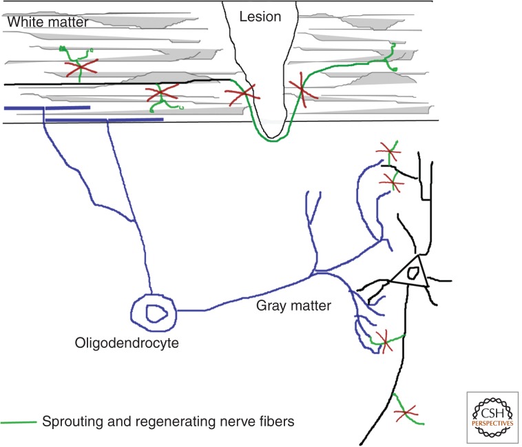
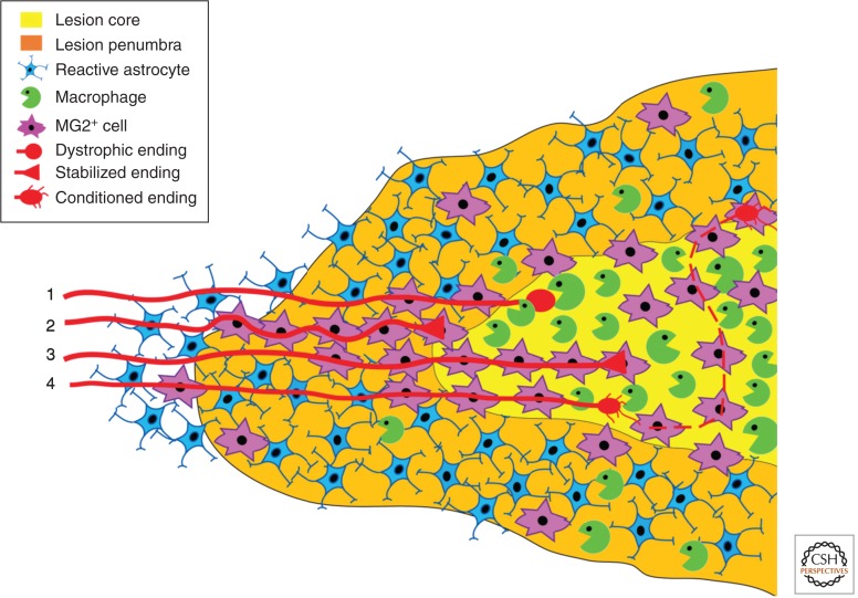
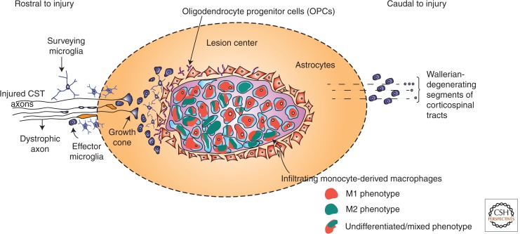
Animal studies are now showing the exciting potential to achieve significant functional recovery following central nervous system (CNS) injury by manipulating both the inefficient intracellular growth machinery in neurons, as well as the extracellular barriers, which further limit their regenerative potential. In this review, we have focused on the three major glial cell types: oligodendrocytes, astrocytes, and microglia/macrophages, in addition to some of their precursors, which form major extrinsic barriers to regrowth in the injured CNS. Although axotomized neurons in the CNS have, at best, a limited capacity to regenerate or sprout, there is accumulating evidence that even in the adult and, especially after boosting their growth motor, neurons possess the capacity for considerable circuit reorganization and even lengthy regeneration when these glial obstacles to neuronal regrowth are modified, eliminated, or overcome.


Figures

Figure 1.
Figure 1.
Oligodendrocytes express neurite growth inhibitory proteins, including the membrane protein Nogo-A, on their cell surface and CNS myelin. These proteins inhibit branch formation along the mature axon in white matter, but they also impair compensatory and regenerative fiber growth following axonal injury. In gray matter, the lower levels of these inhibitory proteins allow some structural remodeling of dendritic and axonal arbors and connections to occur, but these processes can still be potentiated by neutralization or deletion of the neurite growth inhibitors in the mature CNS.
Figure 2.
Figure 2.
Schematic representation of the proximal end of a dorsal column crush lesion 7 d after injury. GFAP+ astrocytes (blue) have pulled away from the lesion core, which is now populated by NG2+ cells (purple) and phagocytic ED1+ macrophages (green). The fibroblastic and ependymal cell types are not displayed, but are also plentiful in the lesion core. Dorsal root ganglion neurons (red) attempt to regenerate into the lesion core. (1) Typical axon with a dystrophic growth cone that has become susceptible to macrophage attack. (2) Typical axon that has undergone macrophage-mediated retraction back to NG2+ cells and stabilized. (3) Atypical axon that has stabilized further distally within the lesion core on a contiguous bridge of NG2+ cells. (4) Growth cone of a neuron that has been stimulated or conditioned and able to overcome macrophage-induced axonal dieback and extend into the lesion core on NG2+ cells. (From Busch et al. 2010; reprinted, with express permission, from the Journal of Neuroscience and the investigators of this review.)
Figure 3.
Figure 3.
Schematic of microglia and MDM reactions elicited by SCI. After injury, the lesion center (also referred to as “epicenter” in contusion lesions) becomes filled with phagocytic macrophages derived from blood monocyte precursors. These cells become enlarged as they phagocytose lipid and cell debris. These and other stimuli in the lesion prime an M1 macrophage phenotype (red). Only a subset of macrophages become “alternatively” activated (i.e., M2 macrophages, green). Some cells remain undifferentiated or adopt a heterogeneous phenotype (orange/green mix). Macrophages in the lesion center are “walled off” by reactive astrocytes, which create a scar. OPCs interdigitate between scar-forming astrocytes and are drawn toward the lesion edge by undefined factors. Complete OPC differentiation into myelinating oligodendrocytes may require factors derived from (M2) microglia subsets, which often lie outside the lesion microenvironment (gradient fill). Microglia exist in intact spinal cord as sentinel cells, which continuously survey the microenvironment. After injury or in response to subtle changes in homeostasis, microglia become activated and transform morphologically and phenotypically into effector microglia. Depending on the composition of factors present in the microenvironment, microglia can become polarized to become M1 or M2 effector cells. Rostral to the site of injury, surveying and effector microglia colocalize with damaged axons, a subset of which are undergoing dieback, but also with a subset that are stabilized or attempting to grow. Caudal to the lesion, descending axons undergo Wallerian degeneration (WD). Various factors released during WD activate microglia (and macrophages). It is common to see effector microglia (and, presumably, a subset of MDMs) colocalized with WD axon segments. CST, corticospinal tract.

References

    1. Abel R, Baron HC, Casha S, Harms J, Hurlbert J, Kucher K, Maier D, Thietje R, Weidner N, Curt A 2011. Therapeutic anti-Nogo-A antibodies in acute spinal cord injury: Safety and pharmacokinetic data from an ongoing first-in-human trial. ISCOS Meeting 2011, Washington, DC.
    1. Aguayo AJ, Rasminsky M, Bray GM, Carbonetto S, McKerracher L, Villegas-Prez M, Vidal-Sanz M, Carter DA 1991. Degenerative and regenerative responses of injured neurons in the central nervous system of adult mammals. Phil Trans R Soc B 331: 337–343. - PubMed
    1. Ahmed Z, Dent RG, Leadbeater WE, Smith C, Berry M, Logan A 2005. Matrix metalloproteases: Degradation of the inhibitory environment of the transected optic nerve and the scar by regenerating axons. Mol Cell Neurosci 28: 64–78. - PubMed
    1. Akbik, Feras V, Sarah M, Bhagat, Pujan R, Patel, William BJ, Cafferty, Stephen M, Strittmatter 2013. Anatomical plasticity of adult brain is titrated by Nogo receptor 1. Neuron 77: 859–866. - PMC - PubMed
    1. Alilain WA, Horn KP, Hu H, Dick TE, Silver J 2011. Functional regeneration of respiratory pathways after spinal cord injury. Nature 475: 196–200. - PMC - PubMed