Review
. 2015 Mar;64(3):504-17.
doi: 10.1136/gutjnl-2014-307891. Epub 2014 Dec 4.

MicroRNAs: new players in IBD

Affiliations
Review

MicroRNAs: new players in IBD

R Kalla et al. Gut. 2015 Mar.

Erratum in

Abstract

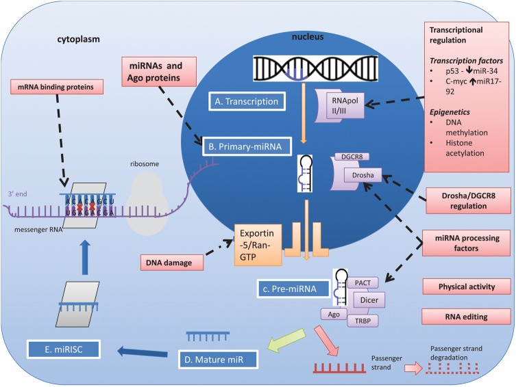
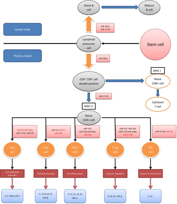
MicroRNAs (miRNAs) are small non-coding RNAs, 18-23 nucleotides long, which act as post-transcriptional regulators of gene expression. miRNAs are strongly implicated in the pathogenesis of many common diseases, including IBDs. This review aims to outline the history, biogenesis and regulation of miRNAs. The role of miRNAs in the development and regulation of the innate and adaptive immune system is discussed, with a particular focus on mechanisms pertinent to IBD and the potential translational applications.

Keywords: CELLULAR IMMUNOLOGY; CROHN'S DISEASE; INTESTINAL TRACT; T LYMPHOCYTES; ULCERATIVE COLITIS.


Figures

Figure 1
Figure 1
Pubmed microRNA (miRNA) citations in Gastroenterology and Inflammatory Bowel Diseases (IBD). Search terms used were as follows: Gastroenterology: (miRNA OR MicroRNA) AND (Gastroenterology OR IBD OR Inflammatory Bowel Disease OR Crohn’s Disease OR Ulcerative Colitis OR Colon OR Stomach OR Intestine OR Oesophagus OR Oesophagus OR Rectum) NOT mirna[author]; IBD: (miRNA OR MicroRNA) AND (IBD OR Inflammatory Bowel Disease OR Crohn’s Disease OR Ulcerative Colitis) NOT mirna[author]; miRNA: (miRNA OR MicroRNA) NOT mirna[author]; Each search was run for print publication dates for each year from 2001 to 2014. Citations were normalised to the total number of Pubmed indexed articles during the same time period (nb, the term microRNA was introduced in 2001).
Figure 2
Figure 2
miRNA biogenesis and regulation. (A) Processing begins in the nucleus where primary miRNA transcripts (pri-miR) are transcribed by RNA polymerase II or RNA polymerase III. (B) Nuclear cleavage of pri-miRNA is performed by a protein complex consisting of the RNAse-III-type enzyme Drosha and DGCR8 (DiGeorge critical region 8), which generates a 60–70 nucleotide sequence called pre-miRNA. Drosha cleavage generates a 2 nucleotide 3' overhang which appears to be a key biogenesis step. DCGR8 acts as an anchor on the stem loops of the target miRNA, allowing Drosha to correctly position on the pri-miRNA. Mirtrons are similar in structure but do not undergo Drosha/DGCR8 processing. (C) pre-miRNA is transported from the nucleus to the cytoplasm by the Exportin-5 (Exp5) — RanGTP complex. Correct binding of the double stranded stem and 3' regions to the RanGTP structure stabilises the miRNA, preventing degradation and facilitating the correct transport of pre-miRNA. (D) Final cleavage of the hairpin loop is performed by Dicer (RNAse III like enzyme) with co-factors: Tar RNA binding protein (TRBP); and protein activator of double-stranded RNA-dependent protein kinase (PACT). (E) The remaining 22 nucleotide RNA duplex is incorporated with Ago proteins, forming a pre-RNA induced silencing complex (pre-RISC). The duplex is separated within Ago proteins into a single stranded mature miRNA and its passenger strand. The mature miRNA strand is retained to form RISC which is eventually destined for mRNA repression/cleavage while its passenger strand undergoes degradation. miRNA recognises its target via 6-8 nucleotide sequence at the 5' end of the miRNA however the binding site can vary. Examples of regulatory elements in miRNA biogenesis. Transcriptional regulation Transcription factors can influence miRNA expression by binding directly to promoter elements. Examples include c-Myc binding and upregulating miR-17–92 cluster and p53interaction with miR-34. miRNAs and argonaute (Ago) proteins as regulators mature miRNAs can act as regulators of miRNA processing either as an auto-regulatory loop or for other miRNAs (e.g. the biogenesis of let-7). RNA editing Once transcribed, miRNAs can undergo editing, which can influence miRNA target specificity. RNA editing occurs in ∼6% of human miRNAs with some studies reporting higher levels of RNA editing (50%). RNA editing is miRNA gene- and tissue-specific (e.g A to I edited members of the miR-376 family specifically within the human cortex). Drosha/DGCR8 The Drosha-DGCR8 complex can undergo post-transcription self-regulation, which allows circulatory negative feedback once sufficient microprocessor activity is available. Cross-regulation between Drosha and DGCR8 may assist in homeostatic control of miRNA biogenesis. miRNA processing factors Specific proteins can either directly or indirectly up-regulate or downregulate the maturation of select miRNAs. A nucleo-cytoplasmic protein with dual functionality is heterogeneous nuclear ribonucleoprotein A1 (hnRNP-A1) which facilitates nuclear pri-miR-18a processing. Physical activity - Physiological changes such as exercise can induce modifications in the miRNA biogenesis machinery. Following 3 hours of endurance exercise in an untrained male, there is upregulation of Drosha, Dicer and Exp5 mRNA levels. DNA damage - DNA damage can promote post transcriptional processing of primary and precursor miRNAs which play a role in the initiation, activation and maintenance of the DNA damage response. DNA damage accelerates nuclear export of pre-miRNAs via Exp5- nucleopore-Nup153 interaction. mRNA binding proteins - mRNA binding proteins bind to the 3-UTR elements of the target mRNA and can either enhance or reverse translational repression by influencing mRNA-miRNA complex interaction.
Figure 3
Figure 3
Examples of miRNA circuits. Tsang and Milo describe two distinct circuits, Type I and Type II that incorporate miRNAs in their regulatory machinery. (A) In Type I circuits, upstream transcription factors will positively coregulate miRNA and their target mRNA. One such example is the repression of E2F1 by miR-17-5p, both of which are activated by the transcription factor c-Myc. It has been suggested that the function of such circuits is to define and maintain target-protein homoeostasis, especially in cells that are ultrasensitive to target mRNA abundance. (B) Type II circuits allow transcriptional activation or repression (positive or negative feedback loop) of a target gene by an upstream factor with associated synergistic miRNA expression. If an mRNA is to be repressed, transcription factors will downregulate the mRNA directly and also upregulate the relevant miRNA. If however a mRNA is to be upregulated, this would occur directly by the transcription factor with synergistic miRNA repression.
Figure 4
Figure 4
MicroRNAs (miRNAs) and the adaptive immune system. This diagram displays a developmental flow chart of the adaptive immune system, mainly T cells. The miRNAs highlighted in black promote the differentiation and/or function of their respective T cell populations while those highlighted in red are inhibitors of these processes. Cytokines released by each T cell subtype are also summarised. MHC, major histocompatibility complex; Th, T helper cell; Tfh, T follicular helper cell; Treg, regulatory T cell; IL, interleukin; TNF, tumour necrosis factor; IFN, interferon; TGF, transforming growth factor.
Figure 5
Figure 5
MicroRNAs (miRNAs) and autophagy (adapted with permission from Ventham et al, Gastroenterology). This diagram summarises the influence of miRNAs within different components of autophagy. Altered sequence in the immunity related GTPase family M protein (IRGM) gene results in an impaired binding site for miR-196. The consequent reduction in miR-196 activity results in IRGM upregulation and causes ineffective bacterial clearance of adherent invasive Escherichia coli (AIEC) in the intestinal cells of patients with Crohn's disease. ATG16L1 has also been shown to be a target of a host of miRNAs. miR-106B and miR-93 repress ATG16L1 mRNA translation, thereby disrupting the autophagy pathway and bacterial clearance of AIEC. miR-30C and miR-130A have also been shown to directly target ATG16L1 and ATG5. Similarly, miR-142-3p has also been shown to negatively regulate ATG16L1 and autophagy. Finally, NOD2 has been shown to induce the expression of miR-29 to limit IL-23 release, indirectly influencing the Th17 pathway in human dendritic cell lines. Polymorphisms in NOD2 impair the ability to express miR-29 resulting in exaggerated IL-23 induced inflammation. Recently, a set of miRNAs that directly target NOD2 expression, miR-192, miR-495, miR-512 and miR-671 have also been described albeit in a different cell line (colonic epithelial HCT116 cells).

Similar articles

Cited by

References

    1. Burisch J, Jess T, Martinato M, et al. . The burden of inflammatory bowel disease in Europe. J Crohns Colitis 2013;7:322–37. - PubMed
    1. Jostins L, Ripke S, Weersma RK, et al. . Host-microbe interactions have shaped the genetic architecture of inflammatory bowel disease. Nature 2012;491:119–24. - PMC - PubMed
    1. Esteller M. Non-coding RNAs in human disease. Nat Rev Genet 2011;12:861–74. - PubMed
    1. Xu W-D, Pan H-F, Li J-H, et al. . MicroRNA-21 with therapeutic potential in autoimmune diseases. Expert Opin Ther Targets 2013;17:659–65. - PubMed
    1. Shi C, Liang Y, Yang J, et al. . MicroRNA-21 knockout improve the survival rate in DSS induced fatal colitis through protecting against inflammation and tissue injury. PLoS ONE 2013;8:e66814. - PMC - PubMed