

Targeting the neurovascular unit in brain trauma
- PMID: 25475543
- PMCID: PMC6495706
- DOI: 10.1111/cns.12359
Targeting the neurovascular unit in brain trauma
Abstract
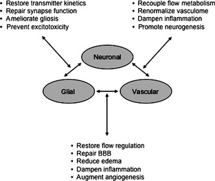
Although the neurovascular unit was originally developed as a conceptual framework for stroke, it is now recognized that these cell-cell interactions play critical roles in many other CNS disorders as well. In brain trauma, perturbations within the neurovascular unit may be especially important. Changes in neurovascular coupling may disrupt blood flow and metabolic regulation. Disruption of transmitter release-reuptake kinetics in neurons and astrocytes may augment excitotoxicity. Alterations in gliovascular signaling may underlie blood-brain barrier disruptions and traumatic edema. Perturbations in cell-cell signaling between all neuronal, glial, and vascular compartments may increase susceptibility to cell death. Finally, repairing the brain after trauma requires the integrated restoration of all neural, glial, and vascular connectivity for effective functional recovery. Just as in stroke, saving neurons alone may also be insufficient for treating brain trauma. In this minireview, we attempt to briefly highlight some of these pathways to underscore the importance of rescuing the entire neurovascular unit in brain trauma.
Keywords: Endothelial; Glia; Neuron; Neuroprotection; Traumatic brain injury.
© 2014 John Wiley & Sons Ltd.
Conflict of interest statement
The authors declare no conflict of interest.
Figures
References
-
- Martin NA, Patwardhan RV, Alexander MJ, et al. Characterization of cerebral hemodynamic phases following severe head trauma: Hypoperfusion, hyperemia, and vasospasm. J Neurosurg 1997;87:9–19. - PubMed
-
- Golding EM, Robertson CS, Bryan RM Jr. The consequences of traumatic brain injury on cerebral blood flow and autoregulation: A review. Clin Exp Hypertens 1999;21:299–332. - PubMed
Publication types
MeSH terms
Grants and funding
LinkOut - more resources
Full Text Sources
Other Literature Sources
