

Diagnostic accuracy of cone-beam CT compared with panoramic images in predicting retromolar canal during extraction of impacted mandibular third molars
- PMID: 25475767
- PMCID: PMC4320425
- DOI: 10.4317/medoral.19930
Diagnostic accuracy of cone-beam CT compared with panoramic images in predicting retromolar canal during extraction of impacted mandibular third molars
Abstract
Objectives: The clinical significance of the existence of a retromolar canal and of its neurovascular content is not yet clear.The aim of the present study was to assess the visibility, diameter and course of the mandibular retromolar canal (MRC) using cone beam computed tomography (CBCT) scan--had been taken for pre-operative radiographic evaluation of impacted mandibular third molars--compared to panoramic radiographs.
Study design: Subjects eligible for study enrollment were those who underwent preoperative CBCT scan for the extraction of impacted mandibular third molars were determined to be extremely close to the mandibular canal on panoramic radiographs. Radiographs were screened for the presence and course of retromolar canals, and linear measurements.
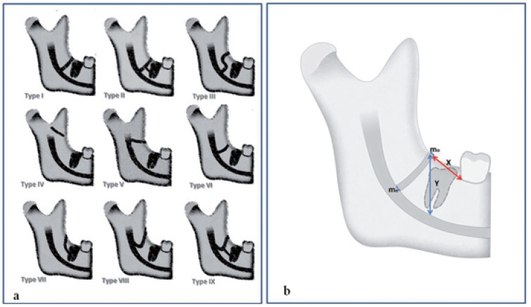
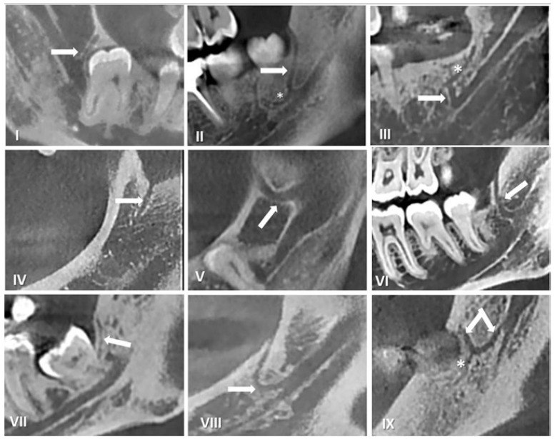
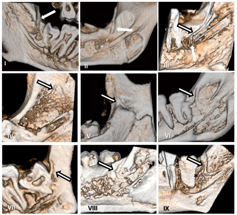
Results: 947 hemimandibles in 632 patients were examined.A total of 253 MRCs (144 left, 109 right) were detected with CBCT images (26.7%). Only 29 of these canals were also seen on the corresponding panoramic radiographs. Most MRCs had a vertical course (type VI, 28.46%), followed by slightly curved (type I, 26.09%). The visibility of the MRC on the OPGs, according to the increase in the diameter, was not statistically significant for both sides (p >.05).Statistically difference were found for the width at the point of origin from the mandibular canal (p: .037), the mean distance from the MRC to the second molar (p: .042) and height of MRC when compared the gender.
Conclusions: The findings suggest that the MRC isn't a rare anatomical structure. This study therefore clearly establishes the incidence and importance of the MRC. The detection of the presence of the MRC using CBCT may be crucial for extraction of mandibular third molars.
Conflict of interest statement
Figures
Similar articles
-
Radiographic study of the mandibular retromolar canal: an anatomic structure with clinical importance.J Endod. 2011 Dec;37(12):1630-5. doi: 10.1016/j.joen.2011.09.007. Epub 2011 Oct 26. J Endod. 2011. PMID: 22099895
-
Evaluation by dental cone-beam computed tomography of the incidence and sites of branches of the inferior dental canal that supply mandibular third molars.Br J Oral Maxillofac Surg. 2016 Dec;54(10):1116-1120. doi: 10.1016/j.bjoms.2016.08.007. Epub 2016 Aug 26. Br J Oral Maxillofac Surg. 2016. PMID: 27576162
-
A comparative study of cone-beam computed tomography and conventional panoramic radiography in assessing the topographic relationship between the mandibular canal and impacted third molars.Oral Surg Oral Med Oral Pathol Oral Radiol Endod. 2007 Feb;103(2):253-9. doi: 10.1016/j.tripleo.2006.06.060. Epub 2006 Sep 1. Oral Surg Oral Med Oral Pathol Oral Radiol Endod. 2007. PMID: 17234544
-
[The retromolar canal (foramen retromolare). Overview and case report].Schweiz Monatsschr Zahnmed. 2011;121(9):821-34. Schweiz Monatsschr Zahnmed. 2011. PMID: 21987358 Review. French, German.
-
Efficacy of CBCT for assessment of impacted mandibular third molars: a review - based on a hierarchical model of evidence.Dentomaxillofac Radiol. 2015;44(1):20140189. doi: 10.1259/dmfr.20140189. Dentomaxillofac Radiol. 2015. PMID: 25135317 Free PMC article. Review.
Cited by
-
Observation of retromolar canals on cone beam computed tomography.Oral Radiol. 2020 Oct;36(4):365-370. doi: 10.1007/s11282-019-00414-0. Epub 2019 Nov 15. Oral Radiol. 2020. PMID: 31732909
-
Incidence and Anatomical Properties of Retromolar Canal in an Iranian Population: A Cone-Beam Computed Tomography Study.Int J Dent. 2020 Mar 9;2020:9178973. doi: 10.1155/2020/9178973. eCollection 2020. Int J Dent. 2020. PMID: 32211048 Free PMC article.
-
Factors Influencing the Onset of Intra- and Post- Operative Complications Following Tooth Exodontia: Retrospective Survey on 1701 Patients.Antibiotics (Basel). 2019 Dec 13;8(4):264. doi: 10.3390/antibiotics8040264. Antibiotics (Basel). 2019. PMID: 31847095 Free PMC article.
-
Correlating the clinical assessment of impacted mandibular third molars with panoramic radiograph and intraoral periapical radiograph.J Int Soc Prev Community Dent. 2016 Dec;6(Suppl 3):S219-S225. doi: 10.4103/2231-0762.197198. J Int Soc Prev Community Dent. 2016. PMID: 28217540 Free PMC article.
-
Evaluation of retromolar canals on cone beam computerized tomography scans and digital panoramic radiographs.Oral Maxillofac Surg. 2017 Sep;21(3):307-312. doi: 10.1007/s10006-017-0632-3. Epub 2017 Jun 1. Oral Maxillofac Surg. 2017. PMID: 28573345
References
-
- Gadbail AR, MankarGadbail MP, Hande A, Chaudhary MS, Gondivkar SM, Korde S. Tumor angiogenesis: role in locally aggressive biological behavior of ameloblastoma and keratocysticodontogenic tumor. Head Neck. 2013;35:329–34. - PubMed
-
- Naitoh M, Hiraiwa Y, Aimiya H, Ariji E. Observation of bifid mandibular canal using cone-beam computerized tomography. Int J Oral Maxillofac Implants. 2009;24:155–9. - PubMed
-
- Bilecenoglu B, Tuncer N. Clinical and anatomical study of retromolar foramen and canal. J Oral Maxillofac Surg. 2006;64:1493–7. - PubMed
-
- von Arx T, Hanni A, Sendi P, Buser D, Bornstein MM. Radiographic study of the mandibular retromolar canal: an anatomic structure with clinical importance. J Endod. 2011;37:1630–5. - PubMed
-
- Pires CA, Bissada NF, Becker JJ, Kanawati A, Landers MA. Mandibular incisive canal: cone beam computed tomography. Clin Implant Dent Relat Res. 2012;14:67–73. - PubMed
Publication types
MeSH terms
LinkOut - more resources
Full Text Sources
Other Literature Sources
