. 2014 Dec 5;16(6):489.
doi: 10.1186/s13058-014-0489-4.

Luminal breast cancer metastases and tumor arousal from dormancy are promoted by direct actions of estradiol and progesterone on the malignant cells

Affiliations

Luminal breast cancer metastases and tumor arousal from dormancy are promoted by direct actions of estradiol and progesterone on the malignant cells

Ndiya Ogba et al. Breast Cancer Res. .

Abstract

Introduction: Luminal, estrogen receptor-positive (ER(+)) breast cancers can metastasize but lie dormant for years before recurrences prove lethal. Understanding the roles of estrogen (E) or progestin (P) in development of luminal metastases or in arousal from dormancy is hindered by few preclinical models. We have developed such models.

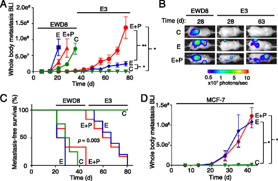
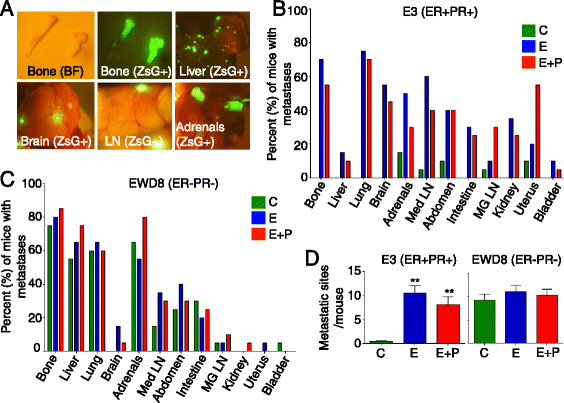
Methods: Immunocompromised, ovariectomized (ovx'd) mice were intracardiac-injected with luminal or basal human breast cancer cells. Four lines were tested: luminal ER(+)PR(+) cytokeratin 5-negative (CK5(-)) E3 and MCF-7 cells, basal ER(-)PR(-)CK5(+) estrogen withdrawn-line 8 (EWD8) cells, and basal ER(-)PR(-)CK5(-) MDA-MB-231 cells. Development of micrometastases or macrometastases was quantified in ovx'd mice and in mice supplemented with E or P or both. Metastatic deposits were analyzed by immunohistochemistry for luminal, basal, and proliferation markers.

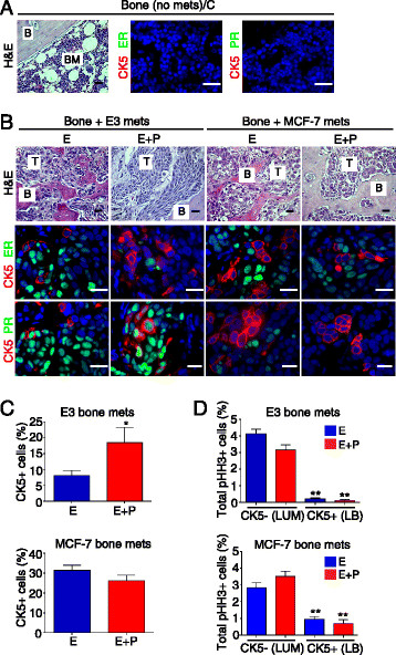
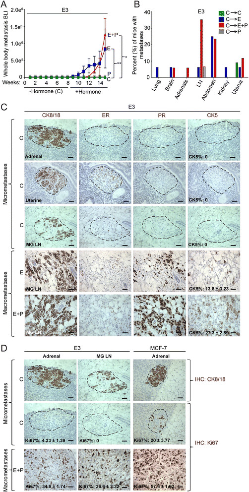
Results: ER(-)PR(-) cells generated macrometastases in multiple organs in the absence or presence of hormones. By contrast, ovx'd mice injected with ER(+)PR(+) cells appeared to be metastases-free until they were supplemented with E or E+P. Furthermore, unlike parental ER(+)PR(+)CK5(-) cells, luminal metastases were heterogeneous, containing a significant (6% to 30%) proportion of non-proliferative ER(-)PR(-)CK5(+) cells that would be chemotherapy-resistant. Additionally, because these cells lack receptors, they would also be endocrine therapy-resistant. With regard to ovx'd control mice injected with ER(+)PR(+) cells that appeared to be metastases-free, systematic pathologic analysis of organs showed that some harbor a reservoir of dormant micrometastases that are ER(+) but PR(-). Such cells may also be endocrine therapy- and chemotherapy-resistant. Their emergence as macrometastases can be triggered by E or E+P restoration.

Conclusions: We conclude that hormones promote development of multi-organ macrometastases in luminal disease. The metastases display a disturbing heterogeneity, containing newly emergent ER(-)PR(-) subpopulations that would be resistant to endocrine therapy and chemotherapy. Similar cells are found in luminal metastases of patients. Furthermore, lack of hormones is not protective. While no overt metastases form in ovx'd mice, luminal tumor cells can seed distant organs, where they remain dormant as micrometastases and sheltered from therapies but arousable by hormone repletion. This has implications for breast cancer survivors or women with occult disease who are prescribed hormones for contraception or replacement purposes.


Figures

Figure 1
Figure 1
Hormones are necessary for luminal tumor metastases. (A) Ovariectomized (Ovx’d) non-obese diabetic/severe combined immunodeficient gamma (NSG) mice were intracardiac (IC)-injected with 105 luciferase and ZsGreen-tagged basal-like estrogen withdrawn-line 8 (EWD8) or luminal E3 cells, and implanted with cellulose control (C) or with estrogen (E) or estrogen + progestin (E+P)-releasing pellets. Weekly quantitation used in vivo bioluminescent imaging (BLI). Data are presented as mean ± standard error of the mean (SEM) of BLI signal (n = 20 per treatment group). *P <0.05, **P <0.005, Student’s t test. (B) Representative luciferase BLI whole-body images. (C) Kaplan-Meier survival curves (n = 20 per treatment group); P = 0.003, log-rank test. (D) Ovx’d NSG mice were IC injected with 5 × 105 luciferase and ZsGreen-tagged luminal MCF-7 cells and implanted with C, E, or E+P pellets. Weekly in vivo luciferase BLI quantitation. Data are presented as mean ± SEM of BLI signal (n = 20 per treatment group). *P <0.05, Student’s t test.
Figure 2
Figure 2
Hormones control metastases of luminal but not basal-like tumor cells. (A) Ovariectomized (Ovx’d) non-obese diabetic/severe combined immunodeficient gamma (NSG) mice were intracardiac-injected with 105 luciferase and ZsGreen-tagged basal-like EWD8 or luminal E3 cells and implanted with cellulose (C) or with estrogen (E) or estrogen + progestin (E+P)-releasing pellets. Representative images at necropsy of ZsG fluorescent metastases in different organs are shown. BF, bright field image (n = 20 per treatment group). (B) Bar graph shows the percentage of mice in each treatment group—C (green), E (blue), and E+P (red)—with E3 metastases to distant organs. Data are presented as mean percentages per group (n = 20 per treatment group). (C) Bar graph shows the percentage of mice in each treatment group—C (green), E (blue), and E+P (red)—with EWD8 metastases to distant organs. Data are presented as mean percentages per group (n = 20 per treatment group). (D) Number of ZsG+ metastatic sites per mouse at necropsy for E3 and EWD8 cells under C (green), E (blue), or E+P (red) conditions. Data are presented as mean ± standard error of the mean (SEM) (n = 20 per treatment group). **P <0.005, Student’s t test.
Figure 3
Figure 3
Homogeneous ER +PR +CK5 luminal E3 and MCF-7 cells generate heterogeneous bone metastases containing ER PR CK5 +luminobasal cells. (A) Representative hematoxylin and eosin (H&E) and immunohistochemistry (IHC) of control (C) bones lacking metastases despite intracardiac (IC) injection of E3 or MCF-7 cells. H&E: (B) bone, (BM) marrow. Scale bars: 50 μm. IHC: estrogen receptor (ER) or progesterone receptor (PR) (green), cytokeratin 5 (CK5) (red), and 4′,6-diamino-2-phenylindole (DAPI) (blue) by immunofluorescent (IF) staining (n = 5 per group). Scale bars: 20 μm. (B) H&E and IHC of E- or E+P-treated mice with bones containing metastases after IC injection of E3 or MCF-7 cells. H&E: bone (B) and tumor (T) cells (n = 5 per group). Scale bars: 50 μm. IHC: ER or PR (green), CK5 (red) and DAPI (blue) by IF staining (n = 5 per group). Scale bars: 20 μm. (C) Percentage of CK5+ cells in E- or E+P-treated E3 and MCF-7 bone metastases. Mean ± standard error of the mean (SEM) values are shown (n = 5 per group). *P <0.05, Student’s t test. (D) Proliferation rates of CK5+ and CK5 cells measured by phosphor-histone H3 (pHH3) in E- and E+P-treated E3 and MCF-7 bone metastases. Mean ± SEM values are shown (n = 5 per group). **P <0.005, Student’s t test.
Figure 4
Figure 4
Hormones reactivate dormant luminal micrometastases. (A) Ovariectomized (Ovx’d) non-obese diabetic/severe combined immunodeficient gamma (NSG) mice were intracardiac (IC)-injected with 105 luciferase- and ZsG-tagged E3 cells without hormone supplementation (cellulose, or C) and monitored for 8 weeks. At week 8, the C pellet was removed and replaced with C (n = 11), estrogen (E) (n = 16), progestin (P) (n = 15), or estrogen + progestin (E+P) (n = 17)-releasing pellets for another 7 to 8 weeks. Weekly bioluminescent luciferase imaging (BLI) imaging in live mice by in vivo imaging systems (IVIS). Data are presented as mean ± standard error of the mean (SEM) of BLI signal. *P <0.05, **P <0.005, Student’s t test. (B) Bar graph shows the percentage of mice in each treatment group—C (green), E (blue), E+P (red), and P (gray)—with E3 metastases in distant organs without (C) or with E, P, or E+P restoration. Data are presented as mean percentages per group. (C) Immunohistochemistry (IHC) for CK8/18, ER, PR, and CK5 in organs with E3 micrometastases (C) and macrometastases (E or E+P). For percentage of CK5+ cells, mean ± SEM values are shown (n = 3 per group). Scale bars: 50 μm. (D) IHC for CK8/18 and nuclear proliferation marker (Ki67) in organs with E3 or MCF-7 micrometastases (C) and macrometastases (E+P). For percentage of Ki67+ cells, mean ± SEM values are shown (n = 3 per group). Scale bars: 50 μm.

Similar articles

Cited by

References

    1. Dunnwald LK, Rossing MA, Li CI. Hormone receptor status, tumor characteristics, and prognosis: a prospective cohort of breast cancer patients. Breast Cancer Res. 2007;9:R6. doi: 10.1186/bcr1639. - DOI - PMC - PubMed
    1. Hammond ME, Hayes DF, Dowsett M, Allred DC, Hagerty KL, Badve S, Fitzgibbons PL, Francis G, Goldstein NS, Hayes M, Hicks DG, Lester S, Love R, Mangu PB, McShane L, Miller K, Osborne CK, Paik S, Perlmutter J, Rhodes A, Sasano H, Schwartz JN, Sweep FC, Taube S, Torlakovic EE, Valenstein P, Viale G, Visscher D, Wheeler T, Williams RB, et al. American Society of Clinical Oncology/College of American Pathologists guideline recommendations for immunohistochemical testing of estrogen and progesterone receptors in breast cancer. J Clin Oncol. 2010;28:2784–2795. doi: 10.1200/JCO.2009.25.6529. - DOI - PMC - PubMed
    1. Demicheli R, Biganzoli E, Ardoino I, Boracchi P, Coradini D, Greco M, Moliterni A, Zambetti M, Valagussa P, Gukas ID, Bonadonna G. Recurrence and mortality dynamics for breast cancer patients undergoing mastectomy according to estrogen receptor status: different mortality but similar recurrence. Cancer Sci. 2010;101:826–830. doi: 10.1111/j.1349-7006.2009.01472.x. - DOI - PMC - PubMed
    1. Zhang XH, Giuliano M, Trivedi MV, Schiff R, Osborne CK. Metastasis dormancy in estrogen receptor-positive breast cancer. J Clin Oncol. 2013;19:6389–6397. - PMC - PubMed
    1. Kennecke H, Yerushalmi R, Woods R, Cheang MC, Voduc D, Speers CH, Nielsen TO, Gelmon K. Metastatic behavior of breast cancer subtypes. J Clin Oncol. 2010;28:3271–3277. doi: 10.1200/JCO.2009.25.9820. - DOI - PubMed

Publication types

MeSH terms