. 2014;15(12):551.
doi: 10.1186/s13059-014-0551-7.

Transcriptome-wide modulation of splicing by the exon junction complex

Transcriptome-wide modulation of splicing by the exon junction complex

Zhen Wang et al. Genome Biol. 2014.

Abstract

Background: The exon junction complex (EJC) is a dynamic multi-protein complex deposited onto nuclear spliced mRNAs upstream of exon-exon junctions. The four core proteins, eIF4A3, Magoh, Y14 and MLN51, are stably bound to mRNAs during their lifecycle, serving as a binding platform for other nuclear and cytoplasmic proteins. Recent evidence has shown that the EJC is involved in the splicing regulation of some specific events in both Drosophila and mammalian cells.

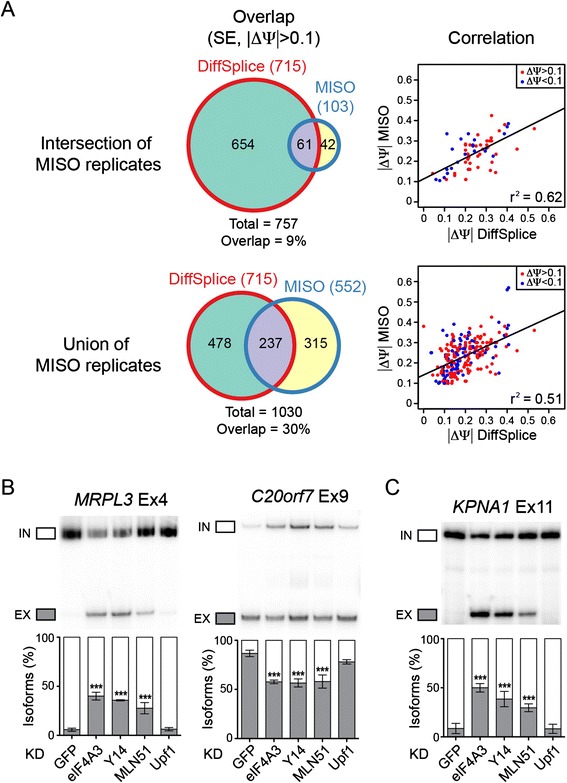
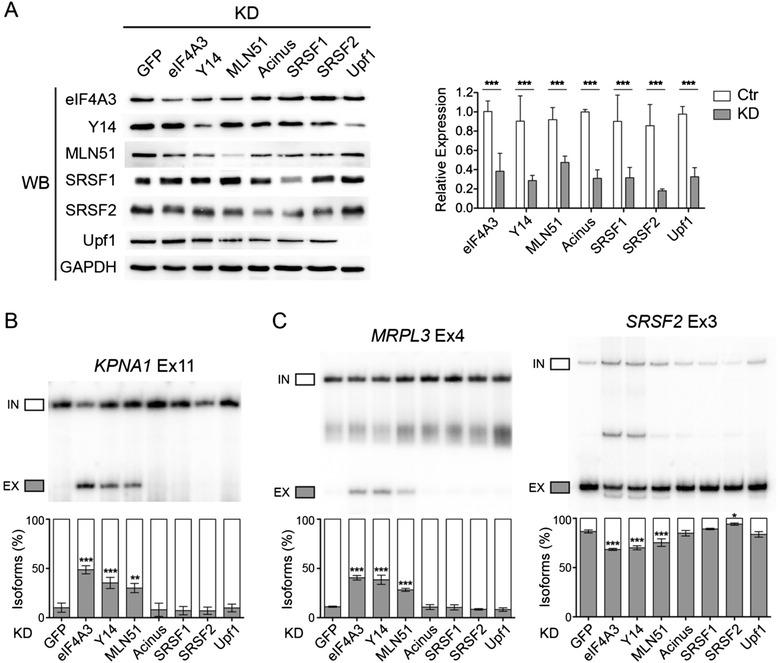
Results: Here, we show that knockdown of EJC core proteins causes widespread alternative splicing changes in mammalian cells. These splicing changes are specific to EJC core proteins, as knockdown of eIF4A3, Y14 and MLN51 shows similar splicing changes, and are different from knockdown of other splicing factors. The splicing changes can be rescued by a siRNA-resistant form of eIF4A3, indicating an involvement of EJC core proteins in regulating alternative splicing. Finally, we find that the splicing changes are linked with RNA polymerase II elongation rates.

Conclusion: Taken together, this study reveals that the coupling between EJC proteins and splicing is broader than previously suspected, and that a possible link exists between mRNP assembly and splice site recognition.


Figures

Figure 1
Figure 1
Identification and validation of splicing changes in EJC KD cells. (A) Venn diagrams showing the overlap between significant splicing events (cassette exons) identified with MISO and DiffSplice (left panels), using a threshold of 0.1 for ΔΨ. Correlation between the ΔΨ values predicted by MISO and DiffSplice are shown on the right, and Pearson’s correlation coefficient (r2) is indicated. (B,C) RT-PCR validation of two alternative exons (B) and one constitutive exon (C) in EJC and Upf1 KD cells. MRPL3 is predicted to increase in exon skipping (ΔΨ >0.1) in EJC KD whereas C20orf7 is predicted to increase in exon inclusion (ΔΨ <0.1) in EJC KD. KPNA1 is predicted to increase in exon skipping (ΔΨ >0.1) in EJC KD. The quantification of triplicate experiments are shown below as mean ± SD. ***P <0.001, one-way ANOVA.
Figure 2
Figure 2
The splicing changes in EJC KD can be rescued by overexpression of siRNA-resistant eIF4A3. (A) Western blot showing the expression level of siRNA-resistant FLAG-eIF4A3 (WT) and a mutant that does not form EJC (D401KE402R, Mut) in the background of control and endogenous eIF4A3 KD condition. GAPDH is shown as a loading control. (B) Immunoprecipitation of FLAG-eIF4A3WT and FLAG-eIF4A3Mut in control and eIF4A3 KD cells. One/15th of input was loaded, and the immunoprecipitated samples were blotted for EJC components. GAPDH is used as a negative control. (C) RT-PCR of alternative splicing patterns in control and eIF4A3 KD cells with eIF4A3WT or Mut. Three representative genes were selected, with one constitutive exon (KPNA1) and two alternative exons (MRPL3, ΔΨ >0; C20orf7, ΔΨ <0). The quantification of triplicate experiments are shown below as mean ± SD. *P < 0.05; **P < 0.01; ***P < 0.001, one-way ANOVA.
Figure 3
Figure 3
EJC-dependent splicing changes are specific to EJC core protein. (A) Western blot (left) and qPCR (right) showing the KD efficiency of EJC core proteins as well as EJC associated protein Acinus, and SR proteins SRSF1 and SRSF2. GAPDH is used as a control. (B,C) RT-PCR of splicing patterns of representative constitutive exons (B) and alternative exons (C) in different KD conditions. The quantification of triplicate experiments are shown below as mean ± SD. *P < 0.05; **P < 0.01; ***P < 0.001, one-way ANOVA.
Figure 4
Figure 4
Characteristics of EJC-dependent exons. (A) Box plot of intron lengths for flanking introns, comparing EJC-dependent exon inclusion (+), exon skipping (−) and exons without splicing changes (Ctr). The P value is calculated with Mann–Whitney-Wilcoxon Test, and is indicated. (B) Box plot of intron lengths for flanking introns, comparing EJC-dependent constitutive exons (Con) and EJC-independent constitutive exons (Ctr Con). (C) RNA splicing map showing the normalised eIF4A3 CLIP reads in the upstream exon, cassette exon and downstream exon, 100 nt from the exon-intron border. EJC-dependent exon inclusion (+) are shown as red, exon skipping (−) are shown as blue, and control exons with has no splicing changes are shown as grey. The CLIP reads were normalised to mRNA expression level and exon length, and the height of CLIP reads at a position is the sum of all normalised CLIP reads at that position for each category of exons.
Figure 5
Figure 5
EJC-dependent alternative splicing is linked with transcription. (A) Western blot of control and eIF4A3 KD cells treated with different concentration of transcription elongation inhibitors (DRB, FP and CPT) for 16 h. GAPDH is used as a control. (B) RT-PCR of splicing patterns of representative exons in cells treated with different inhibitors. The quantification of triplicate experiments are shown below as mean ± SD. *P <0.05; **P <0.01; ***P <0.001, one-way ANOVA.

References

    1. Muller-McNicoll M, Neugebauer KM. How cells get the message: dynamic assembly and function of mRNA-protein complexes. Nat Rev Genet. 2013;14:275–287. doi: 10.1038/nrg3434. - DOI - PubMed
    1. Tange TO, Nott A, Moore MJ. The ever-increasing complexities of the exon junction complex. Curr Opin Cell Biol. 2004;16:279–284. doi: 10.1016/j.ceb.2004.03.012. - DOI - PubMed
    1. Moore MJ, Proudfoot NJ. Pre-mRNA processing reaches back to transcription and ahead to translation. Cell. 2009;136:688–700. doi: 10.1016/j.cell.2009.02.001. - DOI - PubMed
    1. Le Hir H, Andersen GR. Structural insights into the exon junction complex. Curr Opin Struct Biol. 2008;18:112–119. doi: 10.1016/j.sbi.2007.11.002. - DOI - PubMed
    1. Le Hir H, Moore MJ, Maquat LE. Pre-mRNA splicing alters mRNP composition: evidence for stable association of proteins at exon-exon junctions. Genes Dev. 2000;14:1098–1108. - PMC - PubMed

Publication types

MeSH terms

Associated data