. 2014 Dec 5:12:54.
doi: 10.1186/s12951-014-0054-4.

Investigation of size-dependent cell adhesion on nanostructured interfaces

Affiliations

Investigation of size-dependent cell adhesion on nanostructured interfaces

Chiung Wen Kuo et al. J Nanobiotechnology. .

Abstract

Background: Cells explore the surfaces of materials through membrane-bound receptors, such as the integrins, and use them to interact with extracellular matrix molecules adsorbed on the substrate surfaces, resulting in the formation of focal adhesions. With recent advances in nanotechnology, biosensors and bioelectronics are being fabricated with ever decreasing feature sizes. The performances of these devices depend on how cells interact with nanostructures on the device surfaces. However, the behavior of cells on nanostructures is not yet fully understood. Here we present a systematic study of cell-nanostructure interaction using polymeric nanopillars with various diameters.

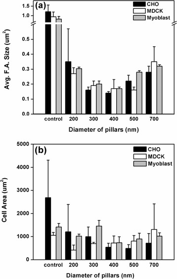
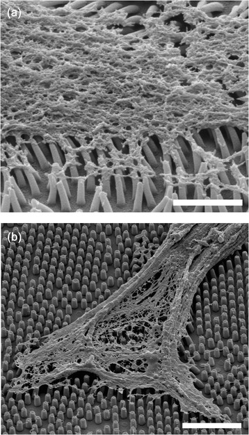
Results: We first checked the viability of cells grown on nanopillars with diameters ranging from 200 nm to 700 nm. It was observed that when cells were cultured on the nanopillars, the apoptosis rate slightly increased as the size of the nanopillar decreased. We then calculated the average size of the focal adhesions and the cell-spreading area for focal adhesions using confocal microscopy. The size of focal adhesions formed on the nanopillars was found to decrease as the size of the nanopillars decreased, resembling the formations of nascent focal complexes. However, when the size of nanopillars decreased to 200 nm, the size of the focal adhesions increased. Further study revealed that cells interacted very strongly with the nanopillars with a diameter of 200 nm and exerted sufficient forces to bend the nanopillars together, resulting in the formation of larger focal adhesions.

Conclusions: We have developed a simple approach to systematically study cell-substrate interactions on physically well-defined substrates using size-tunable polymeric nanopillars. From this study, we conclude that cells can survive on nanostructures with a slight increase in apoptosis rate and that cells interact very strongly with smaller nanostructures. In contrast to previous observations on flat substrates that cells interacted weakly with softer substrates, we observed strong cell-substrate interactions on the softer nanopillars with smaller diameters. Our results indicate that in addition to substrate rigidity, nanostructure dimensions are additional important physical parameters that can be used to regulate behaviour of cells.


Figures

Figure 1
Figure 1
A schematic representation of the fabrication of the polymeric nanopillar arrays. (a) Nanosphere lithography is utilized to produce a single-layer closely packed structure. (b) The diameters of the polystyrene beads are reduced to a specific size by the oxygen-plasma process. (c) A layer of nickel is evaporated on the top of the polystyrene beads. (d) The polystyrene beads are dissolved with dichloromethane. (e) The nickel film serves as the etching mask for the silicon-hole array in ICP etching. (f) The nickel film is removed by the etchant solution. (g) The silicon-hole arrays are used as molds for replication by spin-coating the photo-curable polymers. The UV-curable adhesive is cured under UV radiation for 10 minutes. (h) After being peeled away from the silicon mold, well-ordered periodic polymeric nanopillar arrays are obtained. (i) The polymeric nanopillar arrays are then used to culture cells.
Figure 2
Figure 2
Scanning electron micrographs of polymeric nanopillar arrays made of UV-curable adhesive. The diameters of the nanopillars are (a) 214 ± 13 nm, (b) 322 ± 16 nm, (c) 425 ± 17 nm, (d) 500 ± 19 nm, and (e) 684 ± 17 nm. Scale bars are 1 μm.
Figure 3
Figure 3
Apoptotic cells (percentage) after 24 hours of incubation on nanopillar arrays of varied diameters and controls.
Figure 4
Figure 4
Confocal images of focal adhesions formed on (a), (d), (g) the flat NOA 61 substrates, (b), (e), (h) 200 nm nanopillars and (c), (f), (i) 400 nm nanopillars for myoblast, CHO and MDCK cells, respectively. Green coloration indicates the location of vinculin proteins. Arrows indicate clustering of vinculin in the focal adhesions. Scale bar is 5 μm.
Figure 5
Figure 5
Measured focal adhesion sizes and cell spreading areas on nanopillars. (a) Size of focal adhesions; (b) area of cell spreading measured on various nanopillars. Data are presented as the means ± SD.
Figure 6
Figure 6
SEM images of cells cultured on nanopillars. (a) CHO cells cultured on 300-nm nanopillars. Scale bars are 100 μm. (b) MDCK cells on 200-nm nanopillars. Scale bars are 10 μm. (c) CHO cells cultured on 400-nm nanopillars, Scale bars are 20 μm. (d) CHO cells cultured on 700-nm nanopillars. Scale bars are 10 μm.
Figure 7
Figure 7
SEM images of the internal cell structures after dissolution of the cell membranes. (a) CHO cells cultured on 200-nm nanopillars Scale: 2 μm (b) CHO cells cultured on 400-nm nanopillars Scale bar: 5 μm.
Figure 8
Figure 8
Confocal image of a CHO cell cultured on a 200-nm nanopillar array immunofluorescently stained with (a) tensin (green) and (b) FAK (red) after 24 hours of incubation. (c) Combined DIC and fluorescence image. Scale bar is 20 μm.

References

    1. Geiger B, Bershadsky A, Pankov R, Yamada KM. Transmembrane crosstalk between the extracellular matrix–cytoskeleton crosstalk. Nat Rev Mol Cell Biol. 2001;2:793–805. doi: 10.1038/35099066. - DOI - PubMed
    1. Zamir E, Geiger B. Molecular complexity and dynamics of cell-matrix adhesions. J Cell Sci. 2001;114:3583–3590. - PubMed
    1. Zamir E, Katz M, Posen Y, Erez N, Yamada KM, Katz BZ, Lin S, Lin DC, Bershadsky A, Kam Z, Geiger B. Dynamics and segregation of cell-matrix adhesions in cultured fibroblasts. Nat Cell Biol. 2000;2:191–196. doi: 10.1038/35008607. - DOI - PubMed
    1. Morgan MR, Humphries MJ, Bass MD. Synergistic control of cell adhesion by integrins and syndecans. Nat Rev Mol Cell Biol. 2007;8:957–969. doi: 10.1038/nrm2289. - DOI - PMC - PubMed
    1. Parsons JT, Horwitz AR, Schwartz MA. Cell adhesion: integrating cytoskeletal dynamics and cellular tension. Nat Rev Mol Cell Biol. 2010;11:633–643. doi: 10.1038/nrm2957. - DOI - PMC - PubMed

Publication types