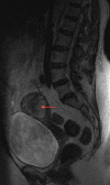
Uterine lipoleiomyoma
- PMID: 25477364
- PMCID: PMC4256602
- DOI: 10.1136/bcr-2014-207763
Uterine lipoleiomyoma
Abstract
We report a case of a patient who was found to have a uterine lipoleiomyoma on ultrasound and MRI, which was later confirmed with histological evidence. Uterine lipoleiomyomas are rare benign tumours that are often misdiagnosed on imaging, leading to unnecessary invasive procedures. Increased awareness of the tumour and its characteristics on imaging can aid future preoperative diagnosis.
2014 BMJ Publishing Group Ltd.
Figures


References
-
- Avritscher R, Iyer RB, Ro J et al. . Lipoleiomyoma of the uterus. AJR Am J Roentgenol 2001;177:856. - PubMed
-
- Chan H, Chau M, Lam C et al. . Uterine lipoleiomyoma: ultrasound and computed tomography findings. J HK Coll Radiol 2003;6:30–2.
-
- Chawla A, Krantikumar R, Raut A et al. . Radiological case of the month. Appl Radiol 2004;33:38–40.
-
- Lee S, Chae H, Jeong B et al. . A clinical review of uterine lipoleiomyoma: a study for value and limitations of radiologic evaluation in preoperative diagnosis of lipoleiomyoma. Korean J Obstet Gynecol 2012;55:953–7.
Publication types
MeSH terms
LinkOut - more resources
Full Text Sources
Other Literature Sources
Medical
