. 2015 Jan;43(1):544-52.
doi: 10.1093/nar/gku1289. Epub 2014 Dec 4.

A novel RNA molecular signature for activation of 2'-5' oligoadenylate synthetase-1

Affiliations

A novel RNA molecular signature for activation of 2'-5' oligoadenylate synthetase-1

Virginia K Vachon et al. Nucleic Acids Res. 2015 Jan.

Abstract

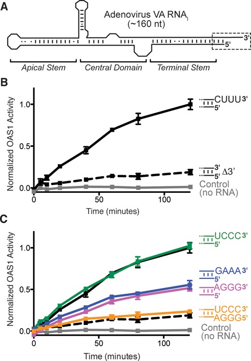
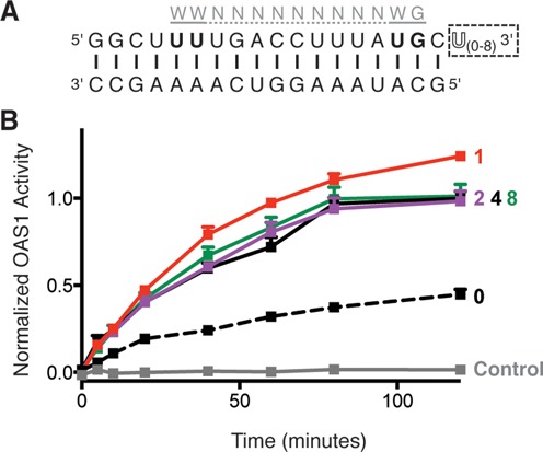
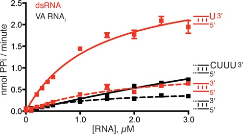
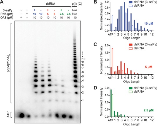
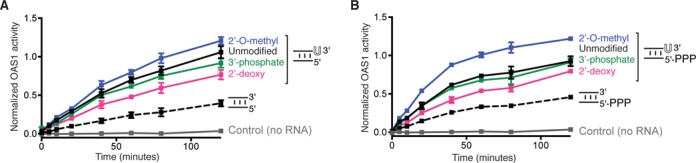
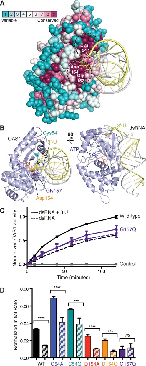
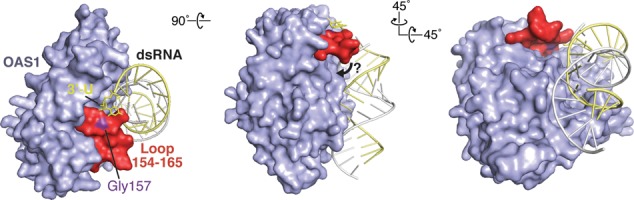
Human 2'-5' oligoadenylate synthetase-1 (OAS1) is central in innate immune system detection of cytoplasmic double-stranded RNA (dsRNA) and promotion of host antiviral responses. However, the molecular signatures that promote OAS1 activation are currently poorly defined. We show that the 3'-end polyuridine sequence of viral and cellular RNA polymerase III non-coding transcripts is critical for their optimal activation of OAS1. Potentiation of OAS1 activity was also observed with a model dsRNA duplex containing an OAS1 activation consensus sequence. We determined that the effect is attributable to a single appended 3'-end residue, is dependent upon its single-stranded nature with strong preference for pyrimidine residues and is mediated by a highly conserved OAS1 residue adjacent to the dsRNA binding surface. These findings represent discovery of a novel signature for OAS1 activation, the 3'-single-stranded pyrimidine (3'-ssPy) motif, with potential functional implications for OAS1 activity in its antiviral and other anti-proliferative roles.


Figures

Figure 1.
Figure 1.
Activation of OAS1 by adenovirus VA RNAI requires its single-stranded pyrimidine-rich 3′-end. (A) VA RNAI secondary structure and domain organization. The Terminal Stem 3′-end contains a single-stranded sequence (CUUU-3′; dashed box and shown as sequence in subsequent panels). (B) Chromogenic assay of OAS1 activity shows that deletion of the wild-type 3′-end single-stranded sequence (Δ3′; dashed line) dramatically reduces OAS activation by full-length VA RNAI (CUUU-3′; solid line). (C) Alternate pyrimidine-rich (UCCC-3′) or purine-rich (AGGG-3′ or GAAA-3′) single-stranded 3′-ends promote full and partial activation of OAS1, respectively. Addition of a complementary 5′-end extension to fully base pair the UCCC-3′ sequence (5′-AGGG/UCCC-3′) attenuates OAS1 activation to the same extent as when the 3′-end single-stranded sequence is absent. The data in panels (B) and (C) are normalized to wild-type VA RNAI (CUUU-3′).
Figure 2.
Figure 2.
The 3′-ssPy motif potentiates OAS1 activation by a simple dsRNA duplex containing an OAS1 consensus sequence. (A) Sequence of the 18-bp dsRNA duplex highlighting the location of the known OAS1 consensus sequence (gray) and additional 3′-end single-stranded uridine residues (3′-ssPy motif; dashed box). (B) Analysis of OAS1 activation by 18-bp dsRNA duplexes with zero, one, two, four or eight single-stranded 3′-end uridine residues using the chromogenic assay. In both panels data are normalized to the 18-bp dsRNA with four 3′-end single-stranded U residues.
Figure 3.
Figure 3.
Kinetic analysis of OAS1 activation by RNAs with and without a 3′-ssPy motif. OAS1 activity over a range of VA RNAI and 18-bp dsRNA duplex concentrations both with (solid lines) and without (dashed lines) the 3′-ssPy motif (3′-CUUU and 3′-U, respectively). Data were fit using non-linear regression to obtain the kinetic parameters (Vmax and Kapp) shown in Table 1.
Figure 4.
Figure 4.
The 3′-ssPy motif causes an increase in OAS1 activity but not an altered accumulation of specific product lengths. (A) Phosphorimager analysis of denaturing PAGE showing OAS1 synthesis of 2′-5′ oligoadenylates in the presence of 18-bp dsRNA activator with and without the 3′-ssPy motif at three different concentrations (10, 5 and 2.5 μM). OAS1 activation at a single concentration of the known activator poly(I).poly(C) RNA is shown for comparison. (B–D) Quantification of 2′-5′ oligoadenylate product band intensities in the presence of (B) 10-μM, (C) 5-μM and (D) 2.5-μM 18-bp dsRNA activator with (solid bars) or without (open bars) the 3′-ssPy motif. All bands were normalized to the most intense product band produced (i.e. n = 4 oligoadenylate in the lane third from left, corresponding to 10-μM dsRNA with a 3′-ssPy). Remaining α-32P-ATP is shown on the far-left of the x-axis.
Figure 5.
Figure 5.
3′-end modifications have minor effects on 3′-ssPy activity and are relatively unaltered by 5′-ppp on the reverse strand of 18-bp RNA. (A) Assays of OAS1 activation by dsRNA duplexes with chemical modifications to the 3′-ssPy motif ribose group: 2′-O-methyl, 2′-deoxyribose and 3′-phosphate. dsRNA with unmodified 3′-ssPy (one single-stranded 3′-end uridine) and without a 3′-ssPy motif are shown for comparison. None of the modifications reduced activity to the level of the dsRNA lacking a 3′-ssPy motif but subtle changes in activation suggest an influence of the ribose sugar pucker on 3′-ssPy motif activity. (B) As panel (A) but for dsRNA duplexes with a 5′-triphosphate group on the complementary strand. The 5′-triphosphate group did not alter relative effects of the modifications on 3′-ssPy motif potency but does appear to modestly enhance activation by the 3′-ssPy motif with 2′-O-methyl ribose modification. In all panels data are normalized to the 18-bp dsRNA with the unmodified 3′-ssPy motif.
Figure 6.
Figure 6.
OAS1 G157 is a critical mediator of 3′-ssPy motif action. (A) Consurf analysis of OAS1 run using the X-ray crystal structure of the human protein (PDB ID: 4IG8) determined in complex with the 18-bp dsRNA duplex. Two highly conserved patches (dashed circles) are located adjacent to the approximate position of the modeled 3′-ssPy motif. (B) Two orthogonal views of the OAS1-dsRNA structure with mutated protein residues and modeled 3′-ssPy motif highlighted. (C) Chromogenic assay showing the effect of the 3′-ssPy on wild-type and G157Q mutant OAS1 activity. (D) Comparison of initial rate of reaction for wild-type and mutant OAS1 proteins activated by the 18-bp dsRNA duplex with (solid bar) or without (striped bar) an additional 3′-end single-stranded uridine residue (3′-ssPy motif). One-way ANOVA: P ≤ 0.0001 (****), P between 0.0001 and 0.001 (***) and not significant (ns; P ≥ 0.05). In panels (C–D), data are normalized to wild-type OAS1 activation by 18-bp dsRNA with the 3′-ssPy motif.
Figure 7.
Figure 7.
Model for 3′-ssPy motif action. Surface representations of the OAS1–dsRNA complex with modeled 3′-ssPy motif (3′-U). The OAS1 loop comprising residues 154 to 165 and the mutated residue that ablates dependence on the 3′-ssPy motif (Gly157) are highlighted. The three views are related by the rotations shown and the ‘left’ image corresponds to the orientation shown in Figure 6. A potential ‘closure’ of the 154–165 loop mediated by 3′-ssPy interaction at the surface comprising G157 is denoted with an arrow (center image).

References

    1. Takeuchi O., Akira S. Pattern recognition receptors and inflammation. Cell. 2010;140:805–820. - PubMed
    1. Zust R., Cervantes-Barragan L., Habjan M., Maier R., Neuman B.W., Ziebuhr J., Szretter K.J., Baker S.C., Barchet W., Diamond M.S., et al. Ribose 2′-O-methylation provides a molecular signature for the distinction of self and non-self mRNA dependent on the RNA sensor Mda5. Nat. Immunol. 2011;12:137–143. - PMC - PubMed
    1. Kowalinski E., Lunardi T., McCarthy A.A., Louber J., Brunel J., Grigorov B., Gerlier D., Cusack S. Structural basis for the activation of innate immune pattern-recognition receptor RIG-I by viral RNA. Cell. 2011;147:423–435. - PubMed
    1. Cole J.L. Activation of PKR: an open and shut case. Trends Biochem. Sci. 2007;32:57–62. - PMC - PubMed
    1. Yoneyama M., Kikuchi M., Natsukawa T., Shinobu N., Imaizumi T., Miyagishi M., Taira K., Akira S., Fujita T. The RNA helicase RIG-I has an essential function in double-stranded RNA-induced innate antiviral responses. Nat. Immunol. 2004;5:730–737. - PubMed

Publication types

MeSH terms

LinkOut - more resources