. 2014 Dec 5;346(6214):1238-42.
doi: 10.1126/science.1259587.

Chromatin decondensation is sufficient to alter nuclear organization in embryonic stem cells

Affiliations

Chromatin decondensation is sufficient to alter nuclear organization in embryonic stem cells

Pierre Therizols et al. Science. .

Abstract

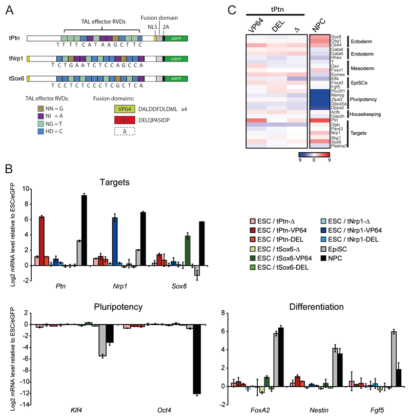
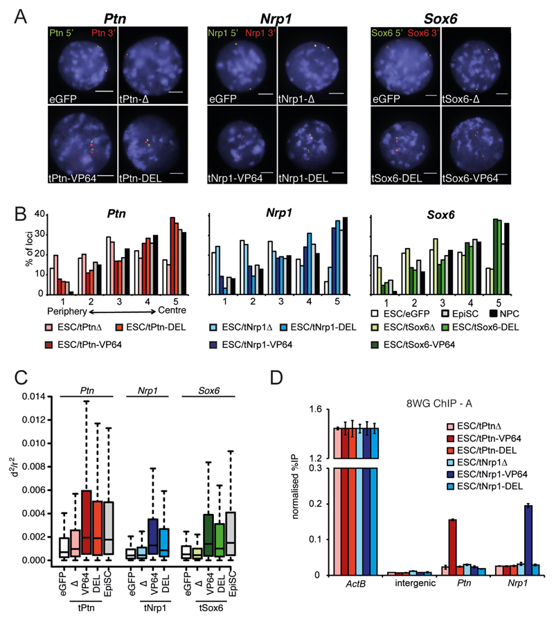
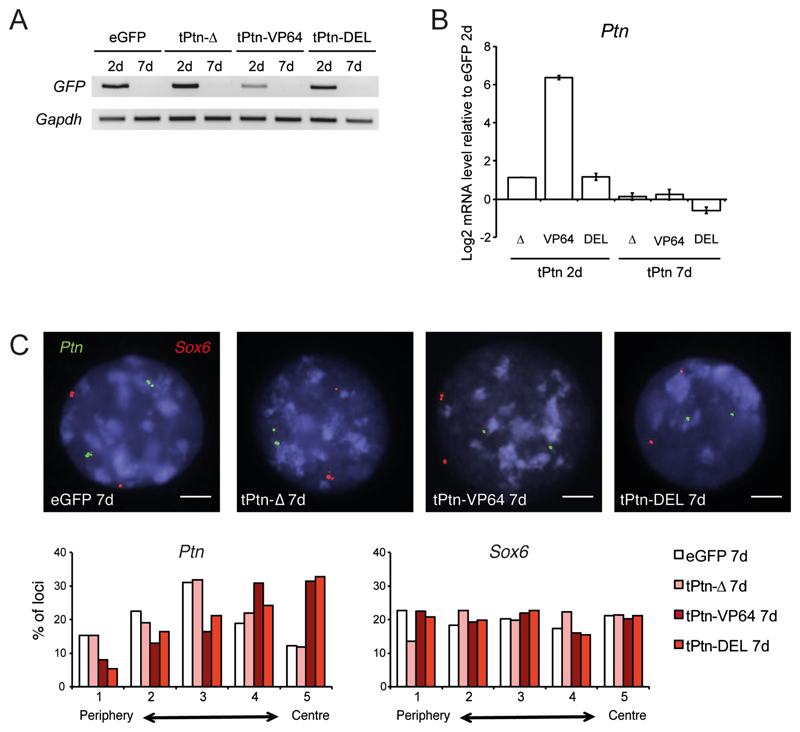
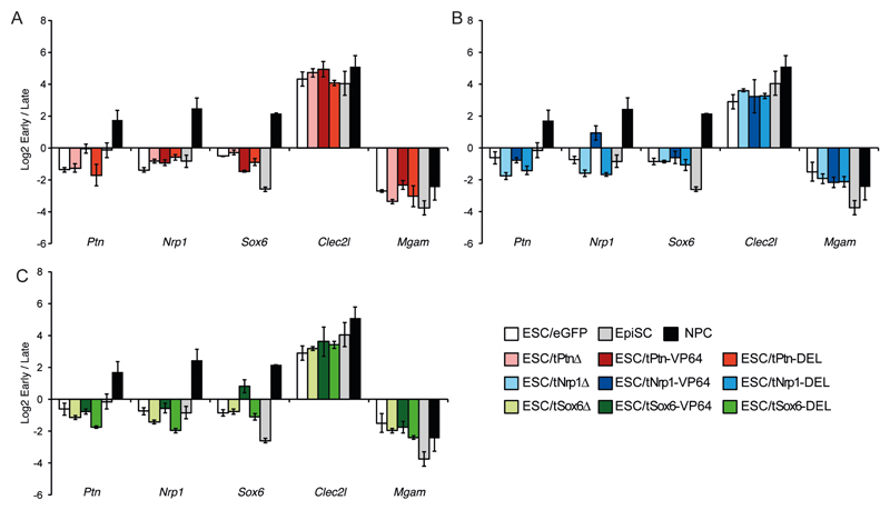
During differentiation, thousands of genes are repositioned toward or away from the nuclear envelope. These movements correlate with changes in transcription and replication timing. Using synthetic (TALE) transcription factors, we found that transcriptional activation of endogenous genes by a viral trans-activator is sufficient to induce gene repositioning toward the nuclear interior in embryonic stem cells. However, gene relocation was also induced by recruitment of an acidic peptide that decondenses chromatin without affecting transcription, indicating that nuclear reorganization is driven by chromatin remodeling rather than transcription. We identified an epigenetic inheritance of chromatin decondensation that maintained central nuclear positioning through mitosis even after the TALE transcription factor was lost. Our results also demonstrate that transcriptional activation, but not chromatin decondensation, is sufficient to change replication timing.


Figures

Figure. 1
Figure. 1. TALE-TF induces transcriptional activation in mouse ESC.
(A) Schematics of TAL-effector constructs specific for Ptn, Nrp1 and Sox6. DNA binding domains fused to either 4 repeats of VP16 (VP64) or DELQPASIDP peptide (13). Constructs (Δ) with the VP64 domain removed serve as a negative control. Expression of eGFP from the same construct via a self-cleaving (2A) peptide allows for isolation of cells expressing the construct by FACS. (B) Mean (+/- s.e.m.) log2 mRNA level, established by RT-qPCR, for TALE target genes (Ptn, Nrp1, Sox6), genes involved in pluripotency (Oct4, Klf4) or differentiation (FoxA2, Nestin, Fgf5) in ESCs transfected with the different TALE vectors. Expression is shown relative to eGFP transfection. Expression changes are also shown for the differentiation of ESCs to EpiSCs or NPCs. n = 3 biological replicates. (C) Heatmap showing 27 genes from the expression microarray selected to represent different differentiation states, as well as genes surrounding the regions targeted by the TAL-effectors. Log2 transformed mRNA ratios for ESCs transfected by the different tPtn constructs relative to eGFP transfection, or for cells differentiated into NPCs compared to levels in ESCs, are displayed using the color code shown.
Figure. 2
Figure. 2. Synthetic chromatin decondensation causes gene relocalization.
(A) FISH with probes flanking Ptn (left), Nrp1 (center) and Sox6 (right), in nuclei of ESCs transfected by eGFP or the -Δ, -VP64 and -DEL TALEs targeting Ptn (left); Nrp1 (center); and Sox6 (right). Nuclei were counter- stained with DAPI (blue). Scale bar = 5 μm. (B) Distribution of Ptn (left), Nrp1 (center) and Sox6 (right) hybridization signals across 5 concentric shells eroded from the periphery (shell 1) through to the center (shell 5) of the nucleus (Fig. S1A) in ESCs transfected by eGFP or the different TALEs constructs, or for ESCs differentiated to EpiSCs and NPCs. n= 100-150 nuclei for at least 2 biological replicates. (C) Box plots showing the distribution of normalized squared interprobe distances (d2/r2) at Ptn (left), Nrp1 (center) and Sox6 (right) in ESCs transfected by eGFP (open bars) or the corresponding TALEs. Un-transfected EpiSCs differentiated from ESCs are shown for comparison (dark gray). Shaded boxes show the median and interquartile range of the data. n = 100-150 nuclei. Statistical analysis is in Table S1. (D) ChIP for RNAPII (8WG antibody) at Actb, Ptn, and Nrp1 promoters and at an intergenic negative contro, in ESCs transfected by tPtn-Δ (light red), tPtn-VP64 (dark red), tPtn-DEL (red), tNrp1-Δ (light blue), tNrp1-VP64 (dark blue), tNrp1-DEL (blue). Enrichment is shown as mean % input bound ± SD over three technical replicates of biological replicate A (replicate B is shown in Fig. S5).
Figure. 3
Figure. 3. Central nuclear positioning is maintained after the loss of gene induction.
(A) RT-PCR of Gfp mRNA in ESC, 2 (2d) or 7 days (7d) after transfection with tPtn-Δ, tPtn-VP64 and tPtn-DEL. Gapdh serves as a constitutively expressed control. (B) Mean (+/- s.e.m.) log2 mRNA level for Ptn in ESCs 2 (left) or 7 days (right) after transfection with tPtn-Δ, tPtn-VP64 and tPtn-DEL. Expression is shown relative to eGFP transfection (n = 3; 2 biological and 1 technical replicates). (C) FISH at Ptn (green) and Sox6 (red), in ESCs 7 days after transfection by eGFP, tPtn-Δ, tPtn-VP64 or tPtn-DEL. Scale bar = 5 μm. Histograms below quantify the nuclear distribution of Ptn (left) and Sox6 (right) signals - as in Fig. 2B - for ESCs 7 days after transfection by eGFP (open bars), tPtn-Δ (light red), tPtn-VP64 (dark red) and and tPtn-DEL (red). n = 100-150 nuclei for each condition for each of at least 2 biological replicates.
Figure. 4
Figure. 4. Synthetic activation of transcription but not nuclear re-positioning shifts replication timing.
Mean (+/- s.e.m.) log2 ratio of early: late S phase fraction for Ptn, Nrp1, Sox6, Clec2l and Mgam, in ESCs after transfection with eGFP (white), tPtn-Δ (light gray), tPtn-VP64 (dark red), tPtn-DEL (light red), tNrp1-Δ (light blue), tNrp1-VP64 (dark blue), tNrp1-DEL (blue), tSox6-Δ (light green), tSox6-VP64 (dark green), tSox6-DEL (green). For comparison the changes in replication timing during the differentiation of ESCs to EpiSCs or NPCs are included.

Comment in

References

    1. Meister P, Taddei A. Building silent compartments at the nuclear periphery: a recurrent theme. Current opinion in genetics & development. 2013;23:96–103. - PubMed
    1. Akhtar A, Gasser SM. The nuclear envelope and transcriptional control. Nature reviews. Genetics. 2007;8:507–517. - PubMed
    1. Guelen L, Pagie L, Brasset E, Meuleman W, Faza MB, Talhout W, et al. Domain organization of human chromosomes revealed by mapping of nuclear lamina interactions. Nature. 2008;453:948–951. - PubMed
    1. Finlan LE, Sproul D, Thomson I, Boyle S, Kerr E, Perry P, et al. Recruitment to the nuclear periphery can alter expression of genes in human cells. PLoS genetics. 2008;4:e1000039. - PMC - PubMed
    1. Reddy KL, Zullo JM, Bertolino E, Singh H. Transcriptional repression mediated by repositioning of genes to the nuclear lamina. Nature. 2008;452:243–247. - PubMed

Publication types