. 2014 Dec 5;346(6214):1258236.
doi: 10.1126/science.1258236.

An ancient defense system eliminates unfit cells from developing tissues during cell competition

Affiliations

An ancient defense system eliminates unfit cells from developing tissues during cell competition

S N Meyer et al. Science. .

Abstract

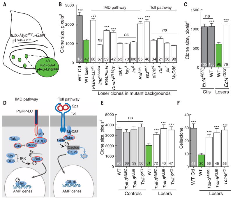
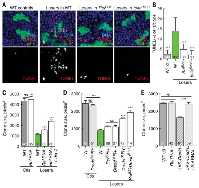
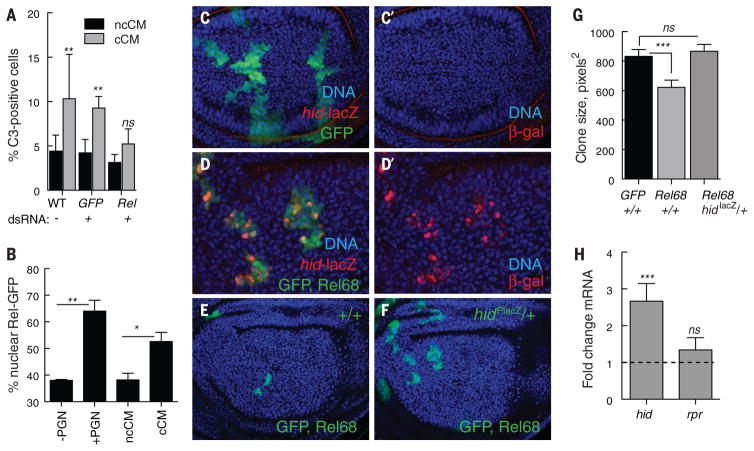
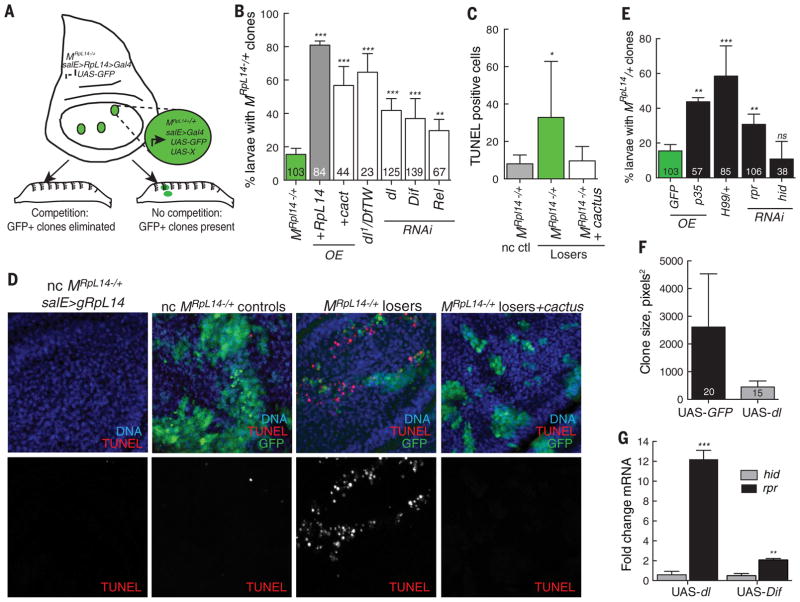
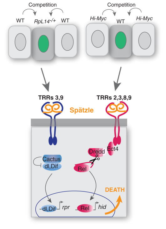
Developing tissues that contain mutant or compromised cells present risks to animal health. Accordingly, the appearance of a population of suboptimal cells in a tissue elicits cellular interactions that prevent their contribution to the adult. Here we report that this quality control process, cell competition, uses specific components of the evolutionarily ancient and conserved innate immune system to eliminate Drosophila cells perceived as unfit. We find that Toll-related receptors (TRRs) and the cytokine Spätzle (Spz) lead to NFκB-dependent apoptosis. Diverse "loser" cells require different TRRs and NFκB factors and activate distinct pro-death genes, implying that the particular response is stipulated by the competitive context. Our findings demonstrate a functional repurposing of components of TRRs and NFκB signaling modules in the surveillance of cell fitness during development.


Figures

Fig. 1
Fig. 1. Select components of the IMD and Toll pathways are required to eliminate WT loser cells in Myc-induced cell competition
(A) Schematic of the assay for Myc-induced cell competition. See the materials and methods section for details. (B) Mean clone size in wing discs of the indicated genotype. In all figures, gray bars show the mean size of control clones (generated in the absence of cell competition), green bars show the mean size of loser clones in WTwing discs, and white bars show the mean size of loser clones in wing discs of the indicated genotype. The number of clones scored for each genotype is indicated in each graph. Mutations in PGRP-LC, BG4/FADD, Dredd, and Rel of the IMD pathway and mutations in the Toll pathway gene spz suppress elimination of the loser cells, resulting in a significant increase in clone growth. Mutations in most genes in the Toll pathway (including dl, Dif, and MyD88) encoding a TIR-domain protein do not alter the outcome of WT loser cells. (C) The TIR-domain protein Ect4/dSarm is required to eliminate WT loser cells in wing discs. The Ect44273 mutant does not affect control wing disc clones but completely suppresses elimination of loser cells. (D) Simplified schematic of the Drosophila innate immune pathways. In the IMD pathway, signaling from the transmembrane protein PGRP-LC, IMD/RIP, FADD, and DREDD/Caspase-8 causes endo-proteolytic cleavage of Relish (Rel), removing an autoinhibitory domain and allowing the Rel-homology domain (RHD/Rel-68) to translocate to the nucleus and activate AMP genes. In the Toll pathway, binding of Spz to the Toll receptor recruits a complex consisting of Myd88, Tube/IRAK-4, and Pelle/ IRAK-1, which phosphorylates Cactus/IκB and targets it for degradation. This releases Dorsal or Dif for activation of distinct AMP genes in the nucleus. Components outlined in red denote those also required for Myc-induced cell competition. P, phosphorylation. (E and F). Null mutations in Toll-3, Toll-8, or Toll-9 suppress elimination of loser cells, increasing loser clone size (E) and cell number per clone (F), but do not alter control clones. Error bars in this figure are SEM. All P values are relative to WT loser clones except in (E) at left—they are relative to WTcontrol clones. ***P < 0.001. ns, not significant (Mann-Whitney test).
Fig. 2
Fig. 2. Dredd-mediated Rel activation eliminates loser cells
(A) Rel and Tollo are required for death of loser cells. TUNEL assays showing cell death in control clones, WT loser clones, and loser clones in RelE20 and tolloR23B mutant backgrounds 24 hours after clone induction. Many WT loser cells are TUNEL-positive, but their death is suppressed in the mutant backgrounds. (B) Quantification of data from (A). (C) Expression of Rel-RNAi (or Rel-RNAi and Dicer to enhance RNAi efficiency) within loser cells suppresses their loss but does not affect control clones. (D) Dredd and Rel are critical for elimination of WT loser cells. Loss of Dredd suppresses the loss of loser clones but does not affect the growth of control clones. Whereas DreddB118−/+ has little effect on loser death, in combination with RelE20−/+ it strongly suppresses death. (E) Expression of Dredd in the absence of competition reduces clone size, but this is blocked by coexpression of Rel RNAi. The number of clones scored for each genotype is indicated. All P values are relative to WT loser clone size except in (C): ***P < 0.001. ns, not significant (Mann-Whitney test).
Fig. 3
Fig. 3. Rel triggers death of WT loser cells by inducing the proapoptotic factor Hid
(A) Rel is required to kill naïve S2 cells. cCM from control co-cultures or those treated with dsRNA against GFP increased death of naïve cells, as measured by activated caspase-3 staining. Cell death was prevented by dsRNA against Rel. Both cocultures and naïve cells in these assays were treated with the indicated dsRNA. Error bars denote SD. (B) Cell competition increases nuclear translocation of Rel. Nuclear localization of Rel-GFP in trans-fected WT-S2 cells treated ± PGN, noncompetitive (nc) CM, or competitive (c) CM for 4 hours is shown. Error bars indicate SD. (C and D) Wing disc with control, GFP-expressing cell clones (C) and cell clones expressing UAS-Rel68 (activated Rel) and UAS-GFP (D) from animals carrying a lacZ reporter insertion in the hid locus (hid-lacZ, red). DNA is stained with Hoechst (blue). hid-lacZ is not induced in control clones (C and C′). Rel68-expressing clones activate expression of hid-lacZ (D and D′), grow poorly, and are small compared with controls (E and G) but are rescued by the mutation caused by the hidPlacZ insertion (F and G). Clones in (D) are shown at higher magnification to facilitate resolution of hid-lacZ expression. (G) Average clone size of control clones (GFP), Rel68-expressing clones in a +/+ background, and Rel68-expressing clones in the hidPlacZ/+ mutant background. Error bars denote SEM. (H) Expression of UAS-Rel68 in WTwing discs induces hid mRNA but not rpr mRNA in quantitative RT-PCR experiments on RNA isolated from wing discs. Error bars indicate SD. ***P < 0.001, **P < 0.01, *P < 0.05. ns, not significant (Mann-Whitney test).
Fig. 4
Fig. 4. Competitive context influences selection of the TRR-NFκB signaling module and proapoptotic inducer. (A)
Scheme for larval screening of innate immune pathway components in competition between WT and MRpL14/+ cells. GFP-positive wing disc clones (MRpL14/+ loser cells) are generated in a background of essentially WTcells (MRpL14/+ rescued with gRpL14); see materials and methods for details.The GFP-MRpL14/+ loser clones are competitively eliminated and thus not present in larvae. If competition is suppressed by overexpression or RNAi, the GFP-positive wing disc clones persist in the larvae. (B) Competitive elimination of MRpL14/+ loser cells is predominantly triggered by Dorsal and Dif. Overexpression of Cactus, RNAi against dl or Dif, or dl1/Df(2L)TW119, a deficiency that removes both dl and Dif, each suppress loss of MRpL14/+ loser cells. Over-expression of UAS-RpL14 was used as a control for clone survival. See fig. S6 for the full data set. (C and D) Cell death mediates MRpL14/+ cell competition. (C) Apoptosis, marked by TUNEL staining, is high in MRpL14/+ loser cells but is reduced when Dorsal and Dif activity are blocked by UAS-cactus expression. (D). TUNEL staining of wing discs with UAS-GFP–expressing clones, quantified in (C). Left to right: control noncompetitive (nc) MRpL14/+ wing disc rescued by gRpL14 expression; control nc act>Gal4, UAS-GFP clones generated in unrescued MRpL14/+wing disc; MRpL14/+ loser clone generated in MRpL14/+ salE>gRpL14 disc; and MRpL14/+ loser clone expressing UAS-cactus generated in MRpL14/+ salE>gRpL14 disc. (E) Competitive elimination of MRpL14/+ loser cells requires cell death induced by Reaper (Rpr). Elimination of MRpL14/+ loser clones is prevented by expression of the apoptosis inhibitor p35; by one copy of the H99 deficiency that removes hid, rpr, and other proapoptotic genes; and by RNAi against rpr, but not RNAi against hid. Number of larvae scored per genotype is indicated. (F) dorsal-expressing clones are smaller than WT sibling clones. Mitotic recombination generated GFP-positive clones that express UAS-dorsal and WTGFP-negative sibling clones.The graph shows the mean clone size of the indicated number of clones. (G) Expression of UAS-dorsal and UAS-Dif induces expression of rpr, but not hid, in quantitative RT-PCR experiments on RNA isolated from wing discs. Error bars in (B) and (E) to (G) denote SD; error bars in (C) indicate SEM. ***P < 0.001, **P < 0.01, *P < 0.05 (Student’s t test).
Fig. 5
Fig. 5. A model of the role of TRR-NFκB modules in cell competition
A model for TRR-NFκB function in cell competition, incorporating data from both competitive contexts. See text for full description.We propose that in both Minute- and Myc-induced competitive contexts, loser cells are eliminated through a signaling pathway mediated by Spz,TRRs, and NFκB function. Context-and/or genetic-dependent interactions that cause Rel activation trigger expression of Hid, whereas those activating Dif and Dorsal trigger expression of Rpr. Apoptosis is the ultimate fate of both loser populations. Red denotes genes required for Myc-induced competition, blue indicates those required for competition in M/+ mosaics, and orange denotes shared requirements.

Comment in

References

    1. Buss LW. The Evolution of Individuality. Princeton Univ. Press; Princeton, NJ: 1987.
    1. King N. The unicellular ancestry of animal development. Dev Cell. 2004;7:313–325. doi: 10.1016/j.devcel.2004.08.010. - DOI - PubMed
    1. Niklas KJ, Newman SA. The origins of multicellular organisms. Evol Dev. 2013;15:41–52. doi: 10.1111/ede.12013. - DOI - PubMed
    1. Dejosez M, Ura H, Brandt VL, Zwaka TP. Safeguards for cell cooperation in mouse embryogenesis shown by genome-wide cheater screen. Science. 2013;341:1511–1514. doi: 10.1126/science.1241628. - DOI - PubMed
    1. Johnston LA. Competitive interactions between cells: Death, growth, and geography. Science. 2009;324:1679–1682. doi: 10.1126/science.1163862. - DOI - PMC - PubMed

Publication types

MeSH terms