Review
. 2015 Feb;68(2):100-10.
doi: 10.1136/jclinpath-2014-202685. Epub 2014 Dec 4.

Diffuse lung disease of infancy: a pattern-based, algorithmic approach to histological diagnosis

Affiliations
Free PMC article
Review

Diffuse lung disease of infancy: a pattern-based, algorithmic approach to histological diagnosis

Jane E Armes et al. J Clin Pathol. 2015 Feb.
Free PMC article

Abstract

Diffuse lung disease (DLD) of infancy has multiple aetiologies and the spectrum of disease is substantially different from that seen in older children and adults. In many cases, a specific diagnosis renders a dire prognosis for the infant, with profound management implications. Two recently published series of DLD of infancy, collated from the archives of specialist centres, indicate that the majority of their cases were referred, implying that the majority of biopsies taken for DLD of infancy are first received by less experienced pathologists. The current literature describing DLD of infancy takes a predominantly aetiological approach to classification. We present an algorithmic, histological, pattern-based approach to diagnosis of DLD of infancy, which, with the aid of appropriate multidisciplinary input, including clinical and radiological expertise and ancillary diagnostic studies, may lead to an accurate and useful interim report, with timely exclusion of inappropriate diagnoses. Subsequent referral to a specialist centre for confirmatory diagnosis will be dependent on the individual case and the decision of the multidisciplinary team.

Keywords: HISTOPATHOLOGY; LUNG; PAEDIATRIC PATHOLOGY.


Figures

Figure 1
Figure 1
The functional unit of the lung: terminal bronchiole (bottom right), alveolar ducts and alveoli within a lung lobule bounded by an inter-lobular septum (top left to centre). Note the artery accompanying the bronchiole: the vein lies separately in the inter-lobular septum. Note also how thin the alveolar septa are: they contain a single capillary. Connective tissue is largely confined to the sheath around the bronchiole and artery and to the interlobular septum.
Figure 2
Figure 2
A muscular pulmonary artery with well-formed internal and external elastic lamina, demonstrable by elastic stains (Elastic van Gieson). The artery has a thick collagenous adventitial coat that merges with the collagen around the accompanying bronchiole seen in part at top right (the pulmonary veins have a much thinner collagenous adventitia). Note that there is a layer of elastic tissue present in the mucosa of the bronchiole. The bronchiole and artery should generally be about equal in diameter.
Figure 3
Figure 3
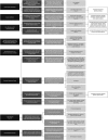
An algorithmic, histological, pattern-based approach to diagnosis of diffuse lung disease of infancy. IHC, immunohistochemistry; PAS, periodic acid Schiff; SpB, surfactant protein B; SpC, surfactant protein C.
Figure 4
Figure 4
Acinar dysplasia. (A) Thoracic contents from a 24-week fetal death in utero showing bilateral, extremely small, granular lungs. The lungs are scarcely bigger than the atrial appendages. (B) Microscopic appearance of acinar dysplasia with airway development limited to bronchial and bronchiolar structures only, with no distal airways development. This imparts a distinctly lobular appearance, reminiscent of the pseudoglandular stage of fetal lung development. These lobules are surrounded by abundant loose mesenchyme.
Figure 5
Figure 5
Congenital alveolar dysplasia in a 2-month-old infant, born at 37 weeks gestation. The lung abnormality is diffuse and shows development reminiscent of the late canalicular/early saccular stage of fetal lung maturation. There are angulated distal airspaces, and mesenchymal crests are present. Many of the epithelial cells lining the airspaces are columnar. The neonate was ventilated and subsequently died. The abnormality must be diffusely present throughout the lungs to make the diagnosis.
Figure 6
Figure 6
Alveolar growth abnormality with pulmonary interstitial glycogenosis. (A) There is diffuse enlargement and simplification of alveolar spaces. The interstitium is thickened. In addition, there is oedema of the interlobular septum, with distension of the veins and lymphatics and focal deposition of haemosiderin. (B) Thick alveolar septae due to oval and spindle cells with cytoplasmic glycogen demonstrated by periodic acid Schiff. There is no significant interstitial inflammatory cell infiltrate.
Figure 7
Figure 7
Alveolar capillary dysplasia with misalignment of the pulmonary veins. (A) There is characteristic interstitial thickening with broadened alveolar septa. Capillaries, where they can be identified, are not situated immediately subjacent to alveolar epithelium, but are in the middle of the septa at a distance from the airspaces. There is muscular medial hypertrophy of a small muscular pulmonary artery, indicating pulmonary arterial hypertension. Within the same fibrous sheath as the artery and bronchiole, there are vascular structures (4 o'clock and 11 o'clock) that have thin, muscular walls and contain blood in their lumina. These are misaligned pulmonary veins (elsewhere in the biopsy specimen no veins could be identified in the interlobular septa). Several small dilated lymphatic vessels are also present. (B) CD31 immunohistochemistry highlights the abnormally placed capillaries that lie centrally within the alveolar septa.
Figure 8
Figure 8
Chronic pneumonitis of infancy with known surfactant protein C deficiency. (A) There is diffuse interstitial thickening and type II pneumocyte hyperplasia without significant alveolar proteinosis or desquamative pneumonia. (B) At high power, type II pneumocyte hyperplasia is obvious and the thickened interstitium contains scattered chronic inflammatory cells.
Figure 9
Figure 9
ABAC3 deficiency. Interstitial thickening with florid type II pneumocyte hyperplasia and pulmonary alveolar proteinosis due to genetically proven ABCA3 deficiency. The eosinophilic debris within the alveolar spaces has a characteristic ‘glassy’ appearance. There are abundant intra-alveolar macrophages. There is also hyperplasia of smooth muscle in the interstitium.
Figure 10
Figure 10
Surfactant protein B deficiency. (A) Interstitial thickening, and a striking quantity of granular, proteinaceous debris within alveolar spaces. Macrophages are not especially prominent, and there is little type II pneumocyte hyperplasia evident in this field. (B) Immunohistochemical staining with antibody to surfactant protein B is negative. Type 2 pneumocyte hyperplasia is more evident than in the H&E section. (C) Immunohistochemical staining with antibody to surfactant protein B in control lung tissue. Staining of surfactant protein B is present in type II pneumocytes lining the alveolar walls, and to a lesser extent, in the alveolar spaces.
Figure 11
Figure 11
Pulmonary arterial hypertension. (A) Cellular intimal thickening of small muscular pulmonary arteries. (B) Plexiform lesion. The parent artery is present to the left of the field and shows only mild intimal thickening. Several branches are present in its adventitial coat, and these show severe concentric, fibrous and myxoid intimal thickening with deposition of fibrinoid. Within the lumen of one vessel (3 o'clock), there is cellular proliferation with the formation of slit-like vascular lumina, creating a plexiform lesion. Surrounding this there are thin-walled dilated vessels, the whole forming a complex vascular lesion.
Figure 12
Figure 12
Pulmonary veno-occlusive disease. A large septal vein is shown in semi-longitudinal section. The original muscular media is evident as a thin cuff around the vessel, and the lumen is occluded by myxoid and fibrous and cellular intimal proliferation. There is a residual narrow and tortuous lumen. There is surrounding severe alveolar capillary congestion. There is also lymphatic dilatation.
Figure 13
Figure 13
Persistent pulmonary hypertension of the newborn. The lung is immature (thickened interstitium with double capillary loops). There are multiple profiles of an artery accompanying a respiratory bronchiole. The artery is thick-walled and muscular and is contracted—the endothelial cells are crowded together and pushed into the lumen in a hobnail pattern (Elastic Van Gieson). After birth, under the influence of increasing oxygen tension these arteries should normally dilate. In persistent pulmonary hypertension of the newborn, the majority of intra-acinar arteries in the lung have this constricted appearance.
Figure 14
Figure 14
Lymphangiectasia. A bronchovascular bundle that shows large numbers of greatly dilated lymphatic vessels ramifying in the collagen surrounding the bronchiole and muscular pulmonary artery (Elastic Van Gieson).
Figure 15
Figure 15
Neuroendocrine cell hyperplasia of infancy, showing increased numbers of neuroendocrine cells in a small airway, identified by antibombesin immunohistochemistry.

References

    1. Kurland G, Deterding RR, Hagood JS, et al. . An official American Thoracic Society clinical practice guideline: classification, evaluation, and management of childhood interstitial lung disease in infancy. Am J Respir Crit Care Med 2013;188:376–94. - PMC - PubMed
    1. Langston C, Dishop MK. Diffuse lung disease in infancy: a proposed classification applied to 259 diagnostic biopsies. Pediatr Dev Pathol 2009;12:421–37. - PubMed
    1. Rice A, Tran-Dang MA, Bush A, et al. . Diffuse lung disease in infancy and childhood: expanding the chILD classification. Histopathology 2013;63:743–55. - PubMed
    1. Deutsch GH, Young LR, Deterding RR, et al. . Diffuse lung disease in young children: application of a novel classification scheme. Am J Respir Crit Care Med 2007;176:1120–8. - PMC - PubMed
    1. Sebire NJ, Malone M, Ashworth M, et al. . Diagnostic pediatric surgical pathology. New York: Churchill Livingstone Elsevier, 2010; Chapter 12, Respiratory Pathology.

MeSH terms