

A procedure to evaluate the efficiency of surface sterilization methods in culture-independent fungal endophyte studies
- PMID: 25477934
- PMCID: PMC4204985
- DOI: 10.1590/s1517-83822014000300030
A procedure to evaluate the efficiency of surface sterilization methods in culture-independent fungal endophyte studies
Abstract
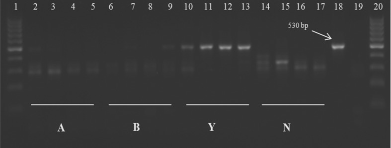
Extraneous DNA interferes with PCR studies of endophytic fungi. A procedure was developed with which to evaluate the removal of extraneous DNA. Wheat (Triticum aestivum) leaves were sprayed with Saccharomyces cerevisiae and then subjected to physical and chemical surface treatments. The fungal ITS1 products were amplified from whole tissue DNA extractions. ANOVA was performed on the DNA bands representing S. cerevisiae on the agarose gel. Band profile comparisons using permutational multivariate ANOVA (PERMANOVA) and non-metric multidimensional scaling (NMDS) were performed on DGGE gel data, and band numbers were compared between treatments. Leaf surfaces were viewed under variable pressure scanning electron microscopy (VPSEM). Yeast band analysis of the agarose gel showed that there was no significant difference in the mean band DNA quantity after physical and chemical treatments, but they both differed significantly (p < 0.05) from the untreated control. PERMANOVA revealed a significant difference between all treatments (p < 0.05). The mean similarity matrix showed that the physical treatment results were more reproducible than those from the chemical treatment results. The NMDS showed that the physical treatment was the most consistent. VPSEM indicated that the physical treatment was the most effective treatment to remove surface microbes and debris. The use of molecular and microscopy methods for the post-treatment detection of yeast inoculated onto wheat leaf surfaces demonstrated the effectiveness of the surface treatment employed, and this can assist researchers in optimizing their surface sterilization techniques in DNA-based fungal endophyte studies.
Keywords: DNA; endophyte; fungi; surface sterilization.
Figures


References
-
- Anand R, Paul L, Chanway C. Research on endophytic bacteria: recent advances with forest trees. In: Schulz B, Boyle C, Sieber T, editors. Microbial Root Endophytes. Vol. 9. Springer-Verlag; Berlin: 2006. pp. 89–103.
-
- Anderson MJ. A new method for non-parametric multivariate analysis of variance. Austral Ecol. 2001;26:32–46.
-
- Arnold AE, Henk DA, Eells RL, Lutzoni F, Vilgalys R. Diversity and phylogenetic affinities of foliar fungal endophytes in loblolly pine inferred by culturing and environmental PCR. Mycologia. 2007;99:185–206. - PubMed
-
- Backman PA, Sikora RA. Endophytes: An emerging tool for biological control. Biol Cont. 2008;46:1–3.
-
- Baek JM, Kenerley CM. Detection and enumeration of a genetically modified fungus in soil environments by quantitative competitive polymerase chain reaction. FEMS Microbiol Ecol. 1998;25:419–428.
Publication types
MeSH terms
Substances
LinkOut - more resources
Full Text Sources
Other Literature Sources
Molecular Biology Databases