. 2014:2014:520651.
doi: 10.1155/2014/520651. Epub 2014 Nov 16.

Traumatic fracture in a patient with osteopoikilosis

Affiliations

Traumatic fracture in a patient with osteopoikilosis

Adeline Du Mortier et al. Case Rep Orthop. 2014.

Abstract

We report a case of traumatic humeral neck fracture occurring in a patient with osteopoikilosis after a motorcycle accident. The radiograph revealed the fracture but also multiple bone lesions. A few years before, the patient had been operated for a maldiagnosed chondrosarcoma of the humeral head. Osteopoikilosis is a rare benign hereditary bone disease, whose mode of inheritance is autosomal dominant. It is usually asymptomatic and discovered incidentally on radiograph that shows the presence of multiple osteoblastic lesions. It can mimic other bone pathologies, in particular osteoblastic metastases. Osteopoikilosis is a diagnosis that should be kept in mind to avoid misdiagnosis, particularly with regard to cancer metastasis. This disorder does not require any treatment and complications are rare. However, there may be associated anomalies that require follow-up.


Figures
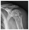
Figure 1
Figure 1
Radiograph of the left shoulder showing a metaphyseal proximal slightly displaced fracture of the humerus and a heterogeneous bone structure with epiphyseal and metaphyseal predominance.
Figure 2
Figure 2
Radiograph of the pelvis showing multiple sclerosis lesions scattered in the pelvis and the upper end of the femur.
Figure 3
Figure 3
Radiograph of the left ankle and of the left foot showing multiple sclerotic spread lesions, observed at the lower part of the tibia and fibula, of the tarsal bones, metatarsal heads and predominant at the epiphyses of the phalanges.
Figure 4
Figure 4
99mTc bone scan revealing uptake only at the site of the fracture of the left humeral head.

References

    1. Ruiz J. P., Monte T. C. S., Blanch-Rubiò J., Ibáñez M. Á. C., Abellò J. C. Osteopoikilosis with involvement of the spine, an atypical presentation. Reumatología Clínica. 2014;10(2):127–129. doi: 10.1016/j.reumae.2013.12.003. - DOI - PubMed
    1. Aghdashi M. A., Aghdashi M. M., Rabiepoor M. Osteopoikilosis: pain as a presenting symptom in three family members. Clinical Medicine Insights: Arthritis and Musculoskeletal Disorders. 2011;4:29–32. doi: 10.4137/CMAMD.S7035. - DOI - PMC - PubMed
    1. Di Primio G. Primer: Benign spotted bones: a diagnostic dilemma. CMAJ. 2011;183(4):456–459. doi: 10.1503/cmaj.091740. - DOI - PMC - PubMed
    1. Ozdemirel A. E., Cakit B. D., Erdem H. R., Koc B. A rare benign disorder mimicking metastasis on radiographic examination: a case report of osteopoikilosis. Rheumatology International. 2011;31(8):1113–1116. doi: 10.1007/s00296-010-1664-2. - DOI - PMC - PubMed
    1. Baasanjav S., Jamsheer A., Kolanczyk M., et al. Osteopoikilosis and multiple exostoses caused by novel mutations in LEMD3 and EXT1 genes respectively—coincidence within one family. BMC Medical Genetics. 2010;11, article 110 doi: 10.1186/1471-2350-11-110. - DOI - PMC - PubMed

LinkOut - more resources