

Measuring Left Ventricular Volumes in Two-Dimensional Echocardiography Image Sequence Using Level-set Method for Automatic Detection of End-Diastole and End-systole Frames
- PMID: 25478488
- PMCID: PMC4253755
- DOI: 10.5812/cardiovascmed.6397
Measuring Left Ventricular Volumes in Two-Dimensional Echocardiography Image Sequence Using Level-set Method for Automatic Detection of End-Diastole and End-systole Frames
Abstract
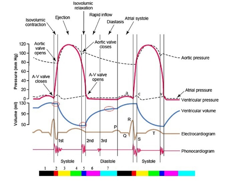
Background: Identifying End-Diastole (ED) and End-Systole (ES) frames is highly important in the process of evaluating cardiac function and measuring global parameters accurately, such as Ejection Fraction (EF), Cardiac Output (CO) and Stroke Volume.
Objectives: The current study aimed to develop a new method based on measuring volume changes in Left Ventricle (LV) during cardiac cycle.
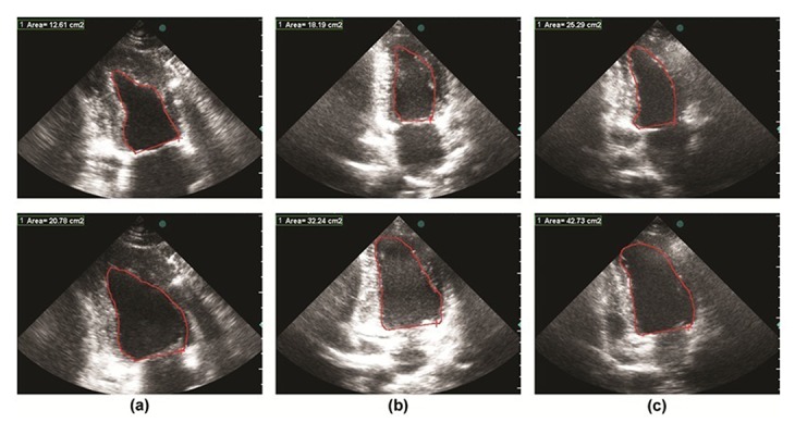
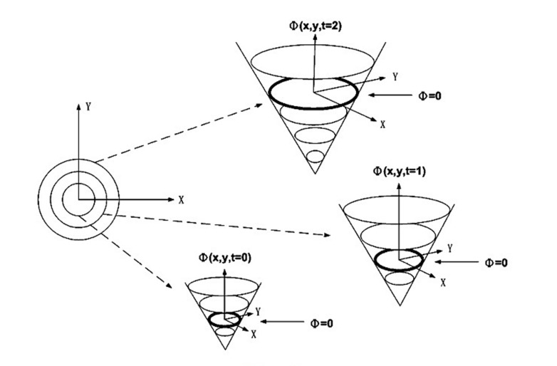
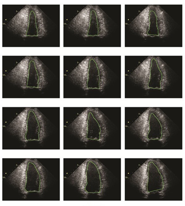
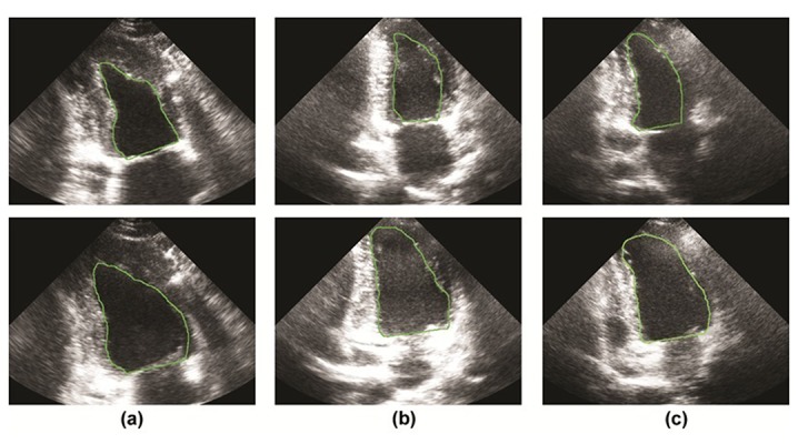
Material and methods: For this purpose, the Level Set method was used both in detecting endocardium border and quantifying cardiac function of all frames.
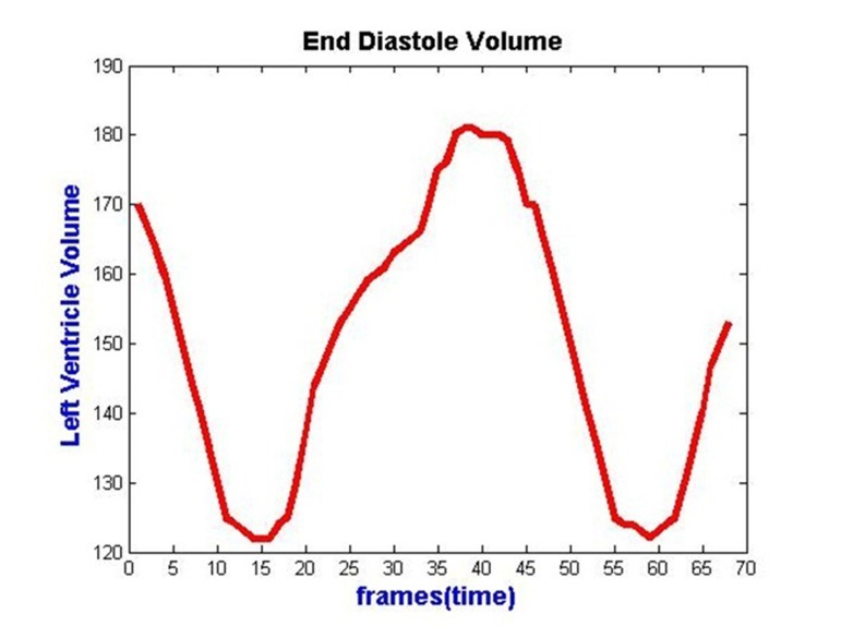
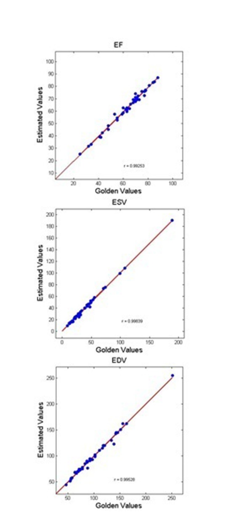
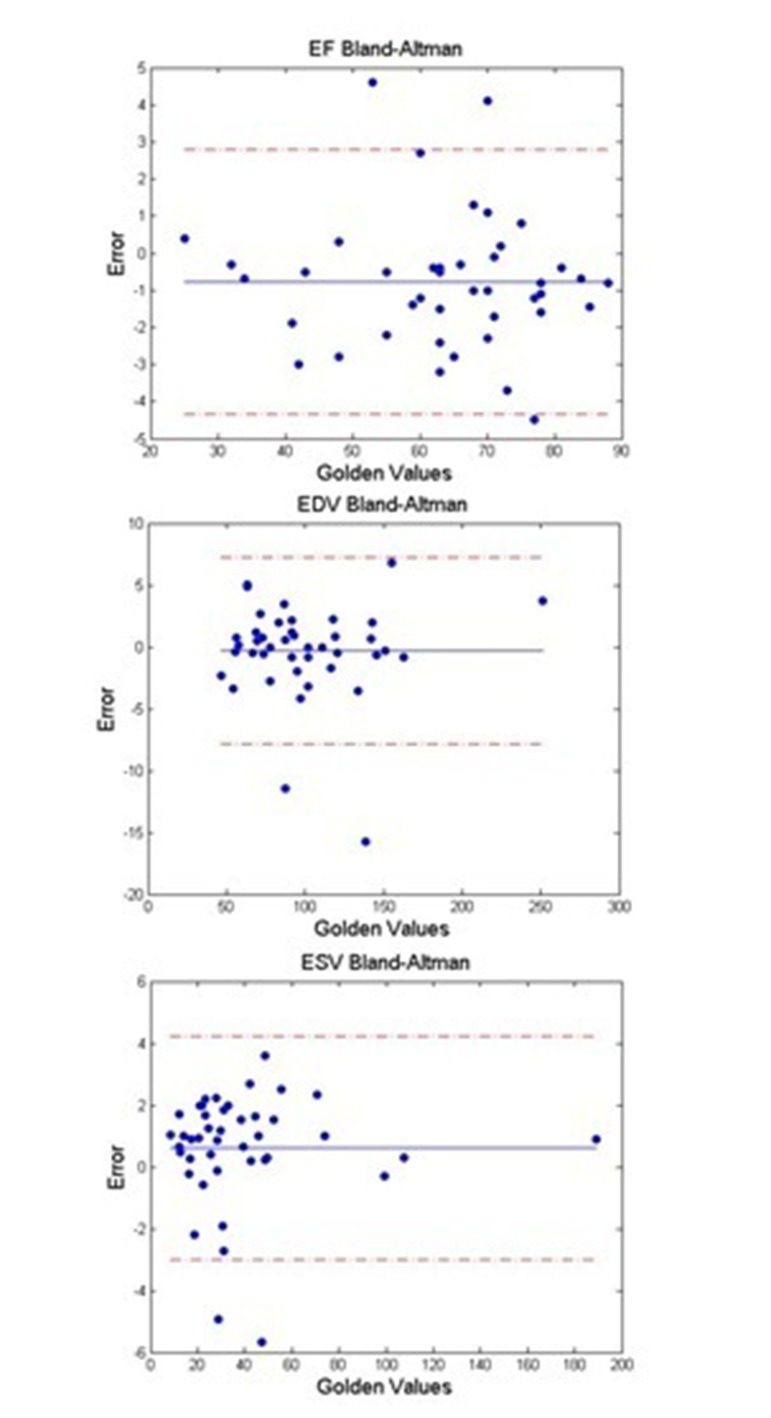
Results: Demonstrating LV volumes displays ED and ES frames and the volumes used in calculating the required parameters.
Conclusions: Since ES and ED frames exist in iso-volumic phases of the cardiac cycle with minimum and maximum values of LV volume signals, such peaks can be utilized in finding related frames.
Keywords: Cardiac Output; Diastole; Echocardiography; Ejection Fraction; Segmentation; Systole.
Figures
Similar articles
-
Automatic detection of end-diastole and end-systole from echocardiography images using manifold learning.Physiol Meas. 2010 Sep;31(9):1091-103. doi: 10.1088/0967-3334/31/9/002. Epub 2010 Jul 23. Physiol Meas. 2010. PMID: 20651421
-
Realization of fully automated quantification of left ventricular volumes and systolic function using transthoracic 3D echocardiography.Cardiovasc Ultrasound. 2018 Jan 23;16(1):2. doi: 10.1186/s12947-017-0121-8. Cardiovasc Ultrasound. 2018. PMID: 29357888 Free PMC article.
-
Accuracy and reproducibility of quantitation of left ventricular function by real-time three-dimensional echocardiography versus cardiac magnetic resonance.Am J Cardiol. 2008 Sep 15;102(6):778-83. doi: 10.1016/j.amjcard.2008.04.062. Epub 2008 Jul 9. Am J Cardiol. 2008. PMID: 18774006
-
Intracardiac ultrasound measurement of volumes and ejection fraction in normal, infarcted, and aneurysmal left ventricles using a 10-MHz ultrasound catheter.Circulation. 1994 Sep;90(3):1481-91. doi: 10.1161/01.cir.90.3.1481. Circulation. 1994. PMID: 8087955
-
Feedback-assisted three-dimensional reconstruction of the left ventricle with MRI.J Magn Reson Imaging. 2003 May;17(5):528-37. doi: 10.1002/jmri.10290. J Magn Reson Imaging. 2003. PMID: 12720262
Cited by
-
CARDIAN: a novel computational approach for real-time end-diastolic frame detection in intravascular ultrasound using bidirectional attention networks.Front Cardiovasc Med. 2023 Oct 6;10:1250800. doi: 10.3389/fcvm.2023.1250800. eCollection 2023. Front Cardiovasc Med. 2023. PMID: 37868778 Free PMC article.
-
Automatic morphological classification of mitral valve diseases in echocardiographic images based on explainable deep learning methods.Int J Comput Assist Radiol Surg. 2022 Feb;17(2):413-425. doi: 10.1007/s11548-021-02542-7. Epub 2021 Dec 12. Int J Comput Assist Radiol Surg. 2022. PMID: 34897594
-
Performance evaluation of computer-aided automated master frame selection techniques for fetal echocardiography.Med Biol Eng Comput. 2023 Jul;61(7):1723-1744. doi: 10.1007/s11517-023-02814-1. Epub 2023 Mar 8. Med Biol Eng Comput. 2023. PMID: 36884143
-
Cardiac phase detection in echocardiography using convolutional neural networks.Sci Rep. 2023 Jun 1;13(1):8908. doi: 10.1038/s41598-023-36047-x. Sci Rep. 2023. PMID: 37264094 Free PMC article.
References
-
- regoratos G, Abrams J, Epstein AE, Freedman RA, Hayes DL, Hlatky MA, et al. ACC/AHA/NASPE 2002 Guideline Update for Implantation of Cardiac Pacemakers and Antiarrhythmia Devices--summary article: a report of the American College of Cardiology/American Heart Association Task Force on Practice Guidelines (ACC/AHA/NASPE Committee to Update the 1998 Pacemaker Guidelines). J Am Coll Cardiol. 2002;40(9):1703–19. doi: 10.1016/S0735-1097(02)02528-7. - DOI - PubMed
-
- Bonow RO, Carabello B, de Leon AC, Jr., Edmunds LH, Jr., Fedderly BJ, Freed MD, et al. Guidelines for the management of patients with valvular heart disease: executive summary. A report of the American College of Cardiology/American Heart Association Task Force on Practice Guidelines (Committee on Management of Patients with Valvular Heart Disease). Circulation. 1998;98(18):1949–84. doi: 10.1161/01.CIR.98.18.1949. - DOI - PubMed
LinkOut - more resources
Full Text Sources