

Eosinophilic Endomyocardial Fibrosis and Strongyloides stercoralis: A Case Report
- PMID: 25478503
- PMCID: PMC4253766
- DOI: 10.5812/cardiovascmed.9370
Eosinophilic Endomyocardial Fibrosis and Strongyloides stercoralis: A Case Report
Abstract
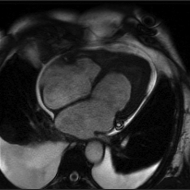
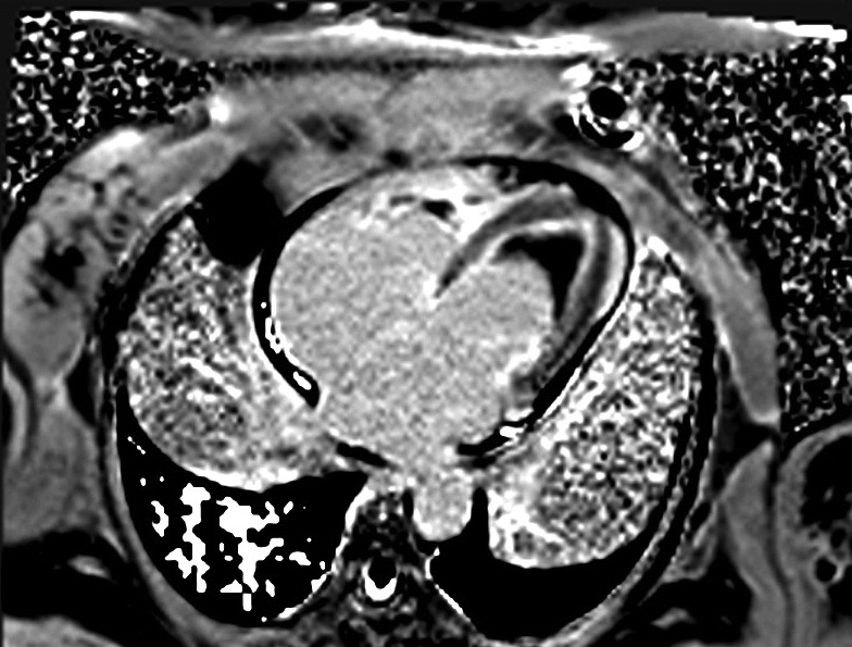
A 64-year-old female with history of previous aortoiliac occlusion and aortoiliac bypass operation four months ago presented with dyspnea, ascites and leg edema. She has been suffering from bloody diarrhea since two weeks earlier. Laboratory data showed important eosinophilia and stool examination was positive for Strongyloides stercoralis. Patient had clinical signs of heart failure. A cardiac MRI revealed hypersignal subendocardium in favor of endomyocardial fibrosis. Hypereosinophilic syndrome is defined by persistent hypereosinophilia for more than 6 months. The association with different etiologies is known but the report of cardiac involvement due to S. stercoralis infection is not very common. Cardiac manifestation is characterized by a restrictive cardiomyopathy due to toxic damage produced by activated eosinophils.
Keywords: Endomyocardial Fibrosis; Magnetic Resonance Imaging; Strongyloides stercoralis.
Figures
References
-
- Acquatella H, Schiller NB, Puigbo JJ, Gomez-Mancebo JR, Suarez C, Acquatella G. Value of two-dimensional echocardiography in endomyocardial disease with and without eosinophilia. A clinical and pathologic study. Circulation. 1983;67(6):1219–26. - PubMed
-
- Ommen SR, Seward JB, Tajik AJ. Clinical and echocardiographic features of hypereosinophilic syndromes. Am J Cardiol. 2000;86(1):110–3. - PubMed
Publication types
LinkOut - more resources
Full Text Sources
Other Literature Sources