. 2014 Dec 5;9(12):e113829.
doi: 10.1371/journal.pone.0113829. eCollection 2014.

A mutation in cnot8, component of the Ccr4-not complex regulating transcript stability, affects expression levels of developmental regulators and reveals a role of Fgf3 in development of caudal hypothalamic dopaminergic neurons

Affiliations

A mutation in cnot8, component of the Ccr4-not complex regulating transcript stability, affects expression levels of developmental regulators and reveals a role of Fgf3 in development of caudal hypothalamic dopaminergic neurons

Peter Koch et al. PLoS One. .

Abstract

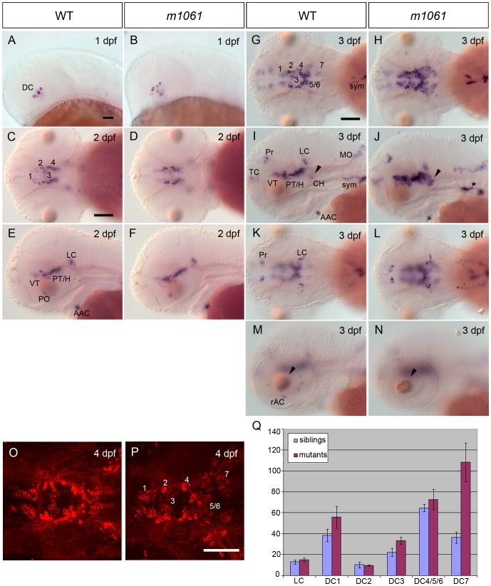
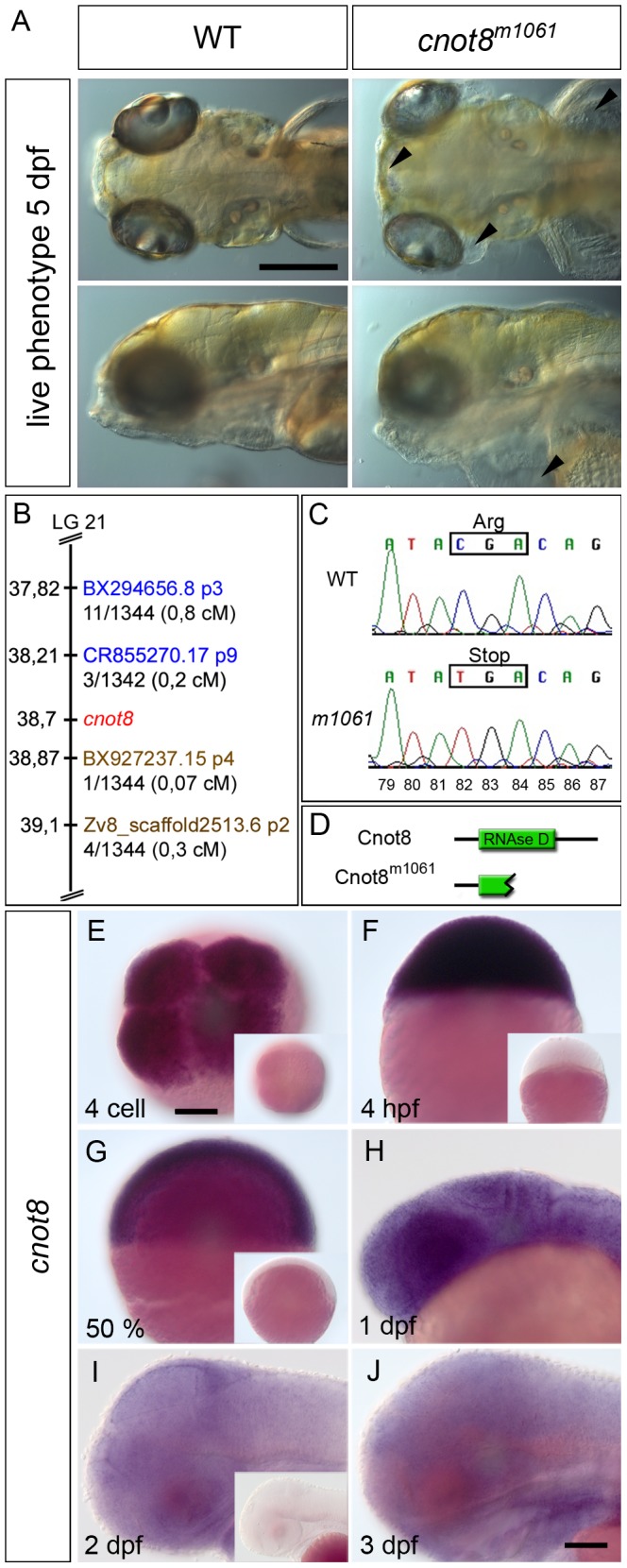
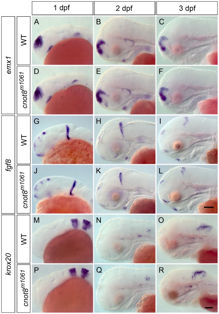
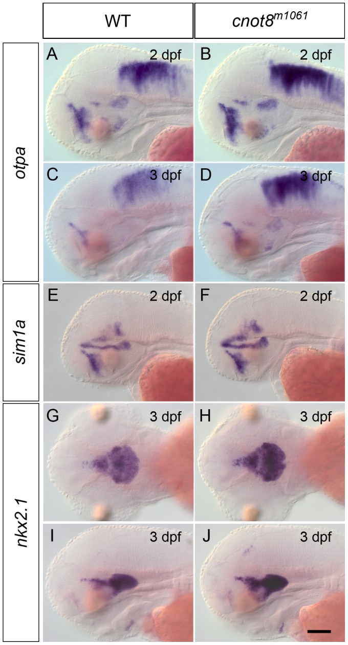
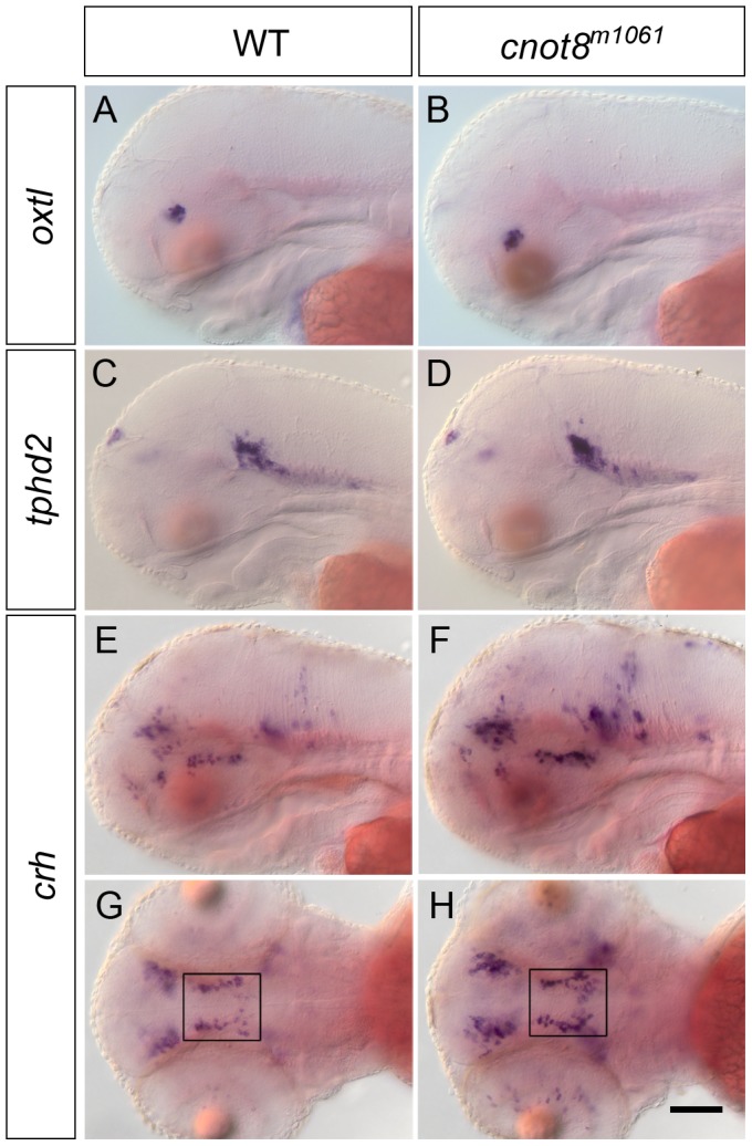
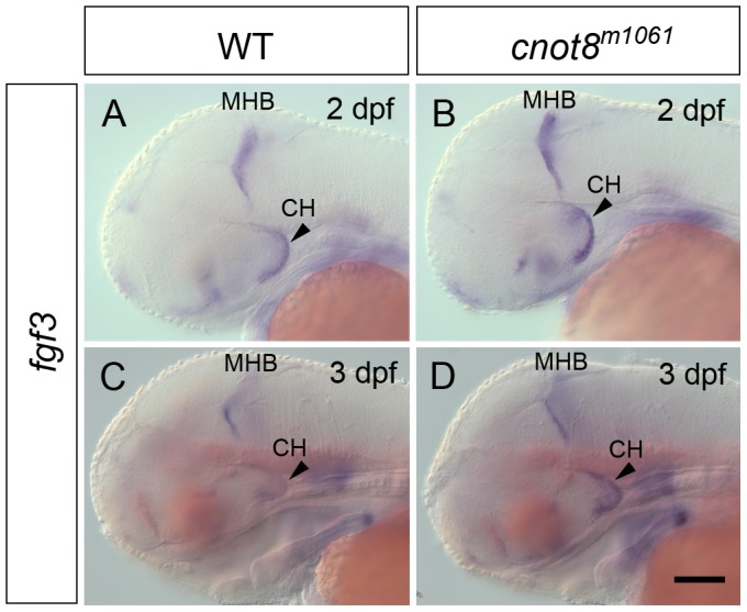
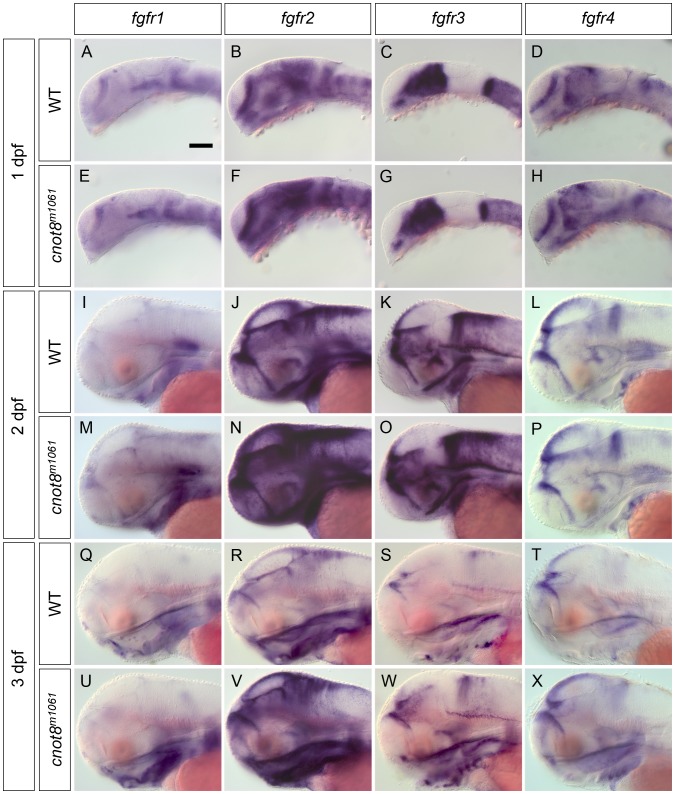
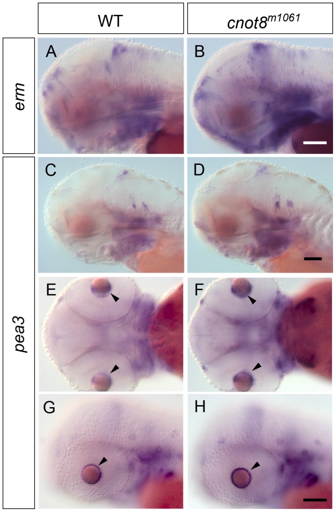
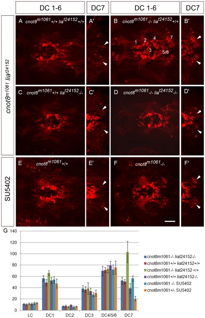
While regulation of the activity of developmental control genes at the transcriptional level as well as by specific miRNA-based degradation are intensively studied, little is known whether general cellular mechanisms controlling mRNA decay may contribute to differential stability of mRNAs of developmental control genes. Here, we investigate whether a mutation in the deadenylation dependent mRNA decay pathway may reveal differential effects on developmental mechanisms, using dopaminergic differentiation in the zebrafish brain as model system. In a zebrafish genetic screen aimed at identifying genes controlling dopaminergic neuron development we isolated the m1061 mutation that selectively caused increased dopaminergic differentiation in the caudal hypothalamus, while other dopaminergic groups were not affected. Positional cloning revealed that m1061 causes a premature stop codon in the cnot8 open reading frame. Cnot8 is a component of the Ccr4-Not complex and displays deadenylase activity, which is required for removal of the poly (A) tail in bulk mRNA turnover. Analyses of expression of developmental regulators indicate that loss of Cnot8 activity results in increased mRNA in situ hybridization signal levels for a subset of developmental control genes. We show that in the area of caudal hypothalamic dopaminergic differentiation, mRNA levels for several components of the FGF signaling pathway, including Fgf3, FGF receptors, and FGF target genes, are increased. Pharmacological inhibition of FGF signaling or a mutation in the fgf3 gene can compensate the gain of caudal hypothalamic dopaminergic neurons in cnot8m1061 mutants, indicating a role for Fgf3 in control of development of this dopaminergic population. The cnot8m1061 mutant phenotype provides an in vivo system to study roles of the Cnot8 deadenylase component of the mRNA decay pathway in vertebrate development. Our data indicate that attenuation of Cnot8 activity differentially affects mRNA levels of developmental control genes.


Conflict of interest statement

Competing Interests: The authors have declared that no competing interests exist.

Figures

Figure 1
Figure 1. m1061 mutants show increased th expression and increase in number of hypothalamic DA neurons from 3 dpf onwards.
Analysis of th gene expression in cnot8m1061 mutants and WT siblings as indicated in header. (A–F) th transcript levels are not altered in m1061/m1061 mutants at 1 and 2 dpf. Embryos were genotyped by PCR. (G–N) m1061 mutants at 3 dpf display stronger WISH signal indicating increased th mRNA levels in DA groups 1 to 7 (arrowhead) and NA sympathetic cells (asterisk). (K, L) NA locus coeruleus (LC) and DA pretectal cells (Pre) do not display altered th expression levels in m1061 mutant embryos. (M, N) The number of DA retinal amacrine cells (rAC) is reduced in m1061 mutant embryos. Additionally m1061 mutants display a lens defect (arrowheads). Genotypes were inferred by th WISH analysis. (O, P) anti-TH immunohistochemistry of m1061 mutant embryo and WT sibling at 4 dpf document DA neurons in the ventral diencephalon. Data confirm a higher cell number of DC7 DA neurons in the caudal hypothalamus of m1061 mutants. Dorsal view z-projections of partial confocal stacks representing the ventral diencephalon are shown. (A, B, E, F, G, H, M, N) lateral views, anterior at left; (C, D, G, H, K.L, O, P) dorsal views, anterior at left. Scale bars 100 µm in A for A, B; in C for C–F; in G for G–N; in P for O, P. Abbreviations: DC - early diencephalic DA group (DC2, DC4), 1 - ventral thalamic DA group, 2,4,5,6 posterior tubercular and hypothalamic Orthopedia-dependent DA groups, 3 - medial hypothalamic DA group, 7 - caudal hypothalamic DA group, VT - ventral thalamus, PT/H - posterior tuberculum/hypothalamus, PO - preoptic area, AAC - arch associated catecholaminergic neurons/carotid body, sym - sympathetic NA neurons, Pr - pretectum, TC - telencephalon, CH - caudal hypothalamus, LC locus coeruleus, MO - medulla oblongata, rAC - retinal amacrine cells. (Q) Quantification of CA cell numbers in m1061 embryos at 4 dpf. Cell counts of DC1-7 and LC TH-expressing cells. Bars show the average number of CA neurons in five m1061 mutants and five WT sibling embryos. Error bars indicate standard deviation. Significance was evaluated using Mann-Whitney test (see text and Table S1).
Figure 2
Figure 2. The live phenotype and positional cloning of the cnot8m1061 mutation.
(A) Comparison of cnot8m1061 mutant to wild-type sibling embryos at 5 dpf; cnot8m1061 mutants display edema formation in the eyes and brain (top row, arrowheads) as well as cardiac and yolk sac (bottom row, arrowhead). Top row dorsal view, bottom row lateral view, anterior to the left. Scale bar 100 µm. (B) Scheme of the m1061 genetic interval. Gene and marker positions were obtained from Ensembl Zv8. CR855270.17 p9 and BX927237 p4 define the interval which comprises the genes zgc:77151, MRP L22, gemin5, cnot8, kibra/WWC1, ARL10 and Nop16 (data not shown). Proximal SSLP markers are highlighted in blue. Distal SSLP markers are highlighted in brown. Numbers provided with SSLP markers reflect recombination events identified per number of meioses analyzed. (C) Sequencing of genomic DNA amplified by PCR from individual m1061 homozygous WT and mutant embryos. The C-to-T mutation at bp 82 of the ORF results in the formation of a premature stop codon in m1061 mutant cnot8 ORF. Numbers (79–87) indicate ORF bp position. (D) Zebrafish Cnot8 comprises 286 amino acids and contains an RNAse D domain which is truncated in cnot8m1061 mutants. (E–J) Expression analysis by WISH using cnot8 antisense probes. Sense controls processed exactly in parallel with the antisense reactions are shown as insets in E, F, G and H to evaluate background stain intensities. (E) Maternal mRNA is detected at 4 cell stage (dorsal view). (F) Ubiquitous expression of cnot8 mRNA at 4 hpf (lateral view). (G, H, I) cnot8 continues to be expressed ubiquitously at 50% epiboly, 1, 2 and 3 dpf (lateral views). Scale bars 100 µm.
Figure 3
Figure 3. cnot8m1061 mutant embryos are not generally delayed in development.
Gene expression analysis of emx1, fgf8 and krox20/egr2b in cnot8m1061 mutants and wild-type siblings at 1, 2 and 3 dpf. (A–F) emx1, (G–L) fgf8 and (M–R) krox20/egr2b. Embryos were genotyped by PCR. All pictures show lateral views. Scale bars 100 µm.
Figure 4
Figure 4. sim1a, otpa and nkx2.1a expression in cnot8m1061 embryos and WT siblings.
Analysis of otpa (A–D), sim1a (E, F) and nkx2.1a (G–J) gene expression in cnot8m1061 mutant embryos and wild-type siblings, stages as indicated. (A–F, I, J) lateral views. (G, H) dorsal views. Embryos were genotyped by PCR. Scale bar 100 µm.
Figure 5
Figure 5. oxtl, tphd2 and crh gene expression in cnot8m1061 embryos at 3 dpf.
(A, B) oxtl, (C, D) tphd2 and (E–H) crh WISH expression analysis in wild-type siblings and cnot8m1061 mutants. (A–F) lateral views. (G, H) dorsal views. Boxes indicate areas of cell counts. Sibling WT embryos developed on average 34.6 crh neurons and cnot8m1061 mutants developed on average 59.4 crh neurons. Significance was evaluated by Mann-Whitney test (p = 0.008). Embryos were genotyped by PCR. Scale bar 100 µm.
Figure 6
Figure 6. fgf3 gene expression is increased in cnot8m1061 mutants.
Analysis of fgf3 gene expression in cnot8m1061 mutants and WT siblings 2 dpf (A, B), and 3 dpf (C, D). All pictures show lateral views. Embryos were genotyped by PCR. Scale bar 100 µm.
Figure 7
Figure 7. The cnot8m1061 mutation affects FGF receptor expression levels.
The expression of the four zebrafish FGF receptor genes fgfr1, 2, 3 and 4 was analyzed by WISH in cnot8m1061 mutants and WT siblings at 1, 2 and 3 dpf. All pictures show lateral views. Embryos were genotyped by PCR. Scale bar 100 µm in A for A–X.
Figure 8
Figure 8. erm and pea3 expression in cnot8m1061 mutants and WT siblings.
(A, B) erm expression in WT siblings and cnot8m1061 mutants at 3 dpf. (C–H) pea3 expression in WT siblings and cnot8m1061 mutants at (C, D) 3 dpf and (E–H) 2 dpf. (E–G) cnot8m1061 mutants show higher pea3 mRNA levels in cells surrounding the lens in comparison to WT siblings (arrowheads). (A–D, G, H) lateral views. (E, F) dorsal views, anterior at left. Embryos were genotyped by PCR. Scale bars 100 µm in B for (A, B), in D for (C, D) and in H for (E–H).
Figure 9
Figure 9. fgf3/lia FGF signaling mediates the increase in caudal hypothalamic DC7 DA neurons in cnot8m1061 mutant embryos.
(A–F) Analysis of DA neurons in cnot8m1061 mutant embryos combined with a mutation in the fgf3 locus (liat24152) or pharmacological suppression of FGF signaling by SU5402. Embryos were fixed at 4 dpf, stained by anti TH immunofluorescence and DA neurons documented by confocal microscopy. Shown are Z-projections of confocal stacks representing the ventral diencephalic DA groups 1 to 7 (dorsal views). Scale bar 50 µm. (A–D) DA neurons in WT, cnot8m1061 or liat24152 single mutant, and cnot8m1061, liat24152 double mutant embryos. Double mutant embryos show loss of cnot8m1061-mediated increase of DC7 caudal hypothalamic DA neurons. (E, F) DA neurons in WT and cnot8m1061 mutant embryos treated with SU5402 from 42 to 48 hpf. Inhibition of FGF signaling by SU5402 reduces the number of DC7 caudal hypothalamic DA neurons in cnot8m1061 mutants below WT levels. (G) Quantification of effects on CA neurons by cell counting of forebrain DA neuronal clusters and locus coeruleus NA neurons in genetic and experimental conditions as indicated in the index at right. Each bar shows the average number of CA neurons in five independent embryos for each experimental condition. Error bars indicate standard deviation. Average DC7 cell numbers: cnot8+/+ lia +/+ 50.8 (WT control); cnot8-/- lia +/+ 103.2; cnot8-/- lia -/- 52.6; cnot8+/+ lia -/- (38.8); cnot8+/+ SU5402 20.6; cnot8-/-SU5402 56.2. Significance was evaluated by Mann-Whitney test. The cell count in single mutant cnot8m1061 (-/-)liat24152 (+/+) is significantly different from single mutant cnot8m1061 (+/+) liat24152 (-/-) embryos (p = 0.008). Comparison of cnot8m1061, liat24152 double mutant and WT embryos reveal no significant difference (p = 0.45). For SU5402 treatments, the number of DC7 DA neurons differs significantly between WT controls and SU5402 treated WT (p = 0.008) and between cnot8m1061 and SU5402-treated cnot8m1061 embryos (p = 0.008). For all other catecholaminergic groups, no significant differences were observed when WT was compared to single mutants, double mutants, or SU5402-treated embryos.

Similar articles

Cited by

References

    1. Schier AF (2007) The Maternal-Zygotic Transition: Death and Birth of RNAs. 316:406–407. - PubMed
    1. Giraldez AJ (2010) microRNAs, the cell's Nepenthe: clearing the past during the maternal-to-zygotic transition and cellular reprogramming. Curr Opin Genet Dev 20:369–375. - PMC - PubMed
    1. Bashirullah A, Cooperstock RL, Lipshitz HD (2001) Spatial and temporal control of RNA stability. 98:7025–7028. - PMC - PubMed
    1. Tadros W, Lipshitz HD (2005) Setting the stage for development: mRNA translation and stability during oocyte maturation and egg activation inDrosophila. 232:593–608. - PubMed
    1. Garneau NL, Wilusz J, Wilusz CJ (2007) The highways and byways of mRNA decay. 8:113–126. - PubMed

Publication types

MeSH terms