Review
. 2014 Dec 5;9(12):e114498.
doi: 10.1371/journal.pone.0114498. eCollection 2014.

Vagal nerve stimulation therapy: what is being stimulated?

Affiliations
Review

Vagal nerve stimulation therapy: what is being stimulated?

Guy Kember et al. PLoS One. .

Abstract

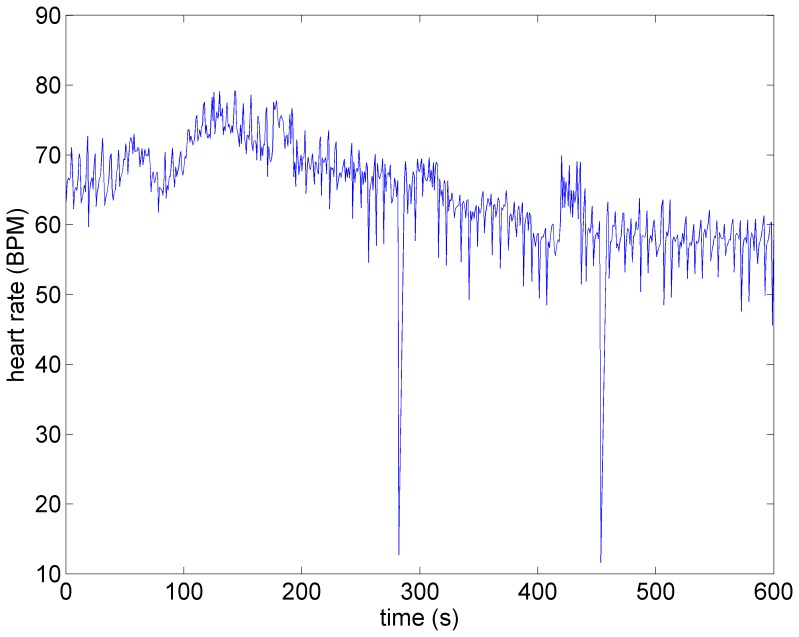
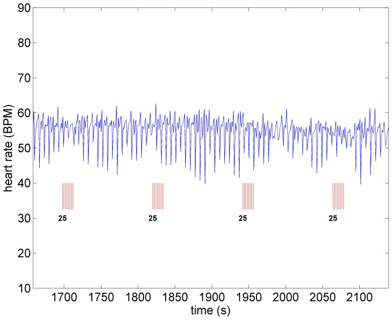
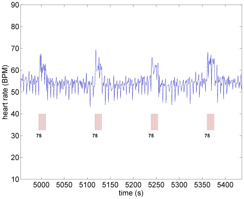
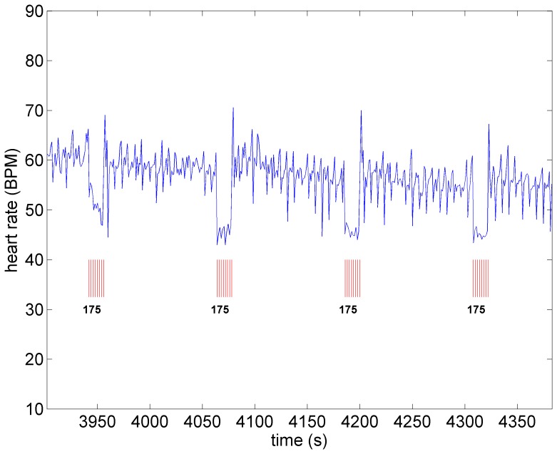
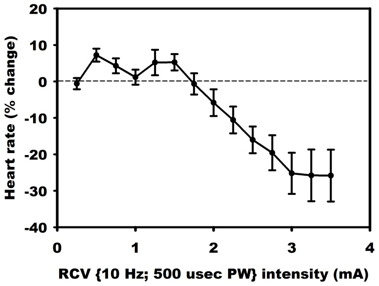
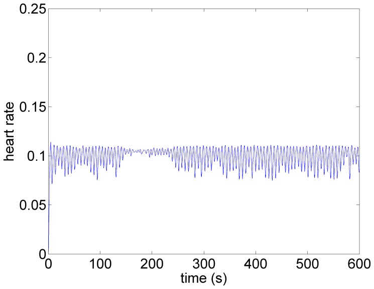
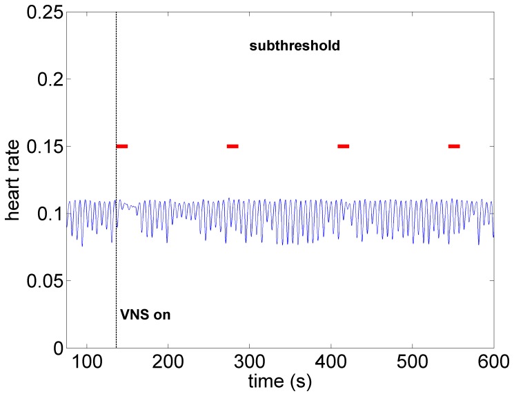
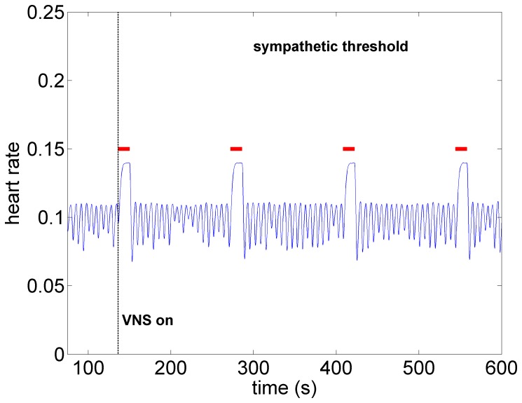
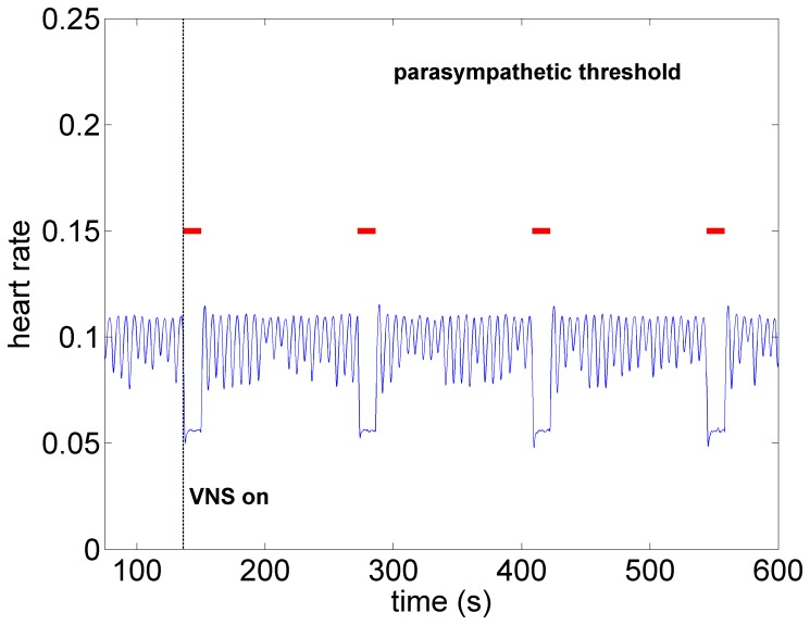
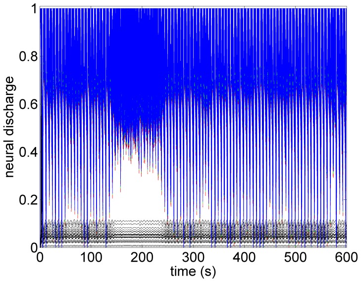
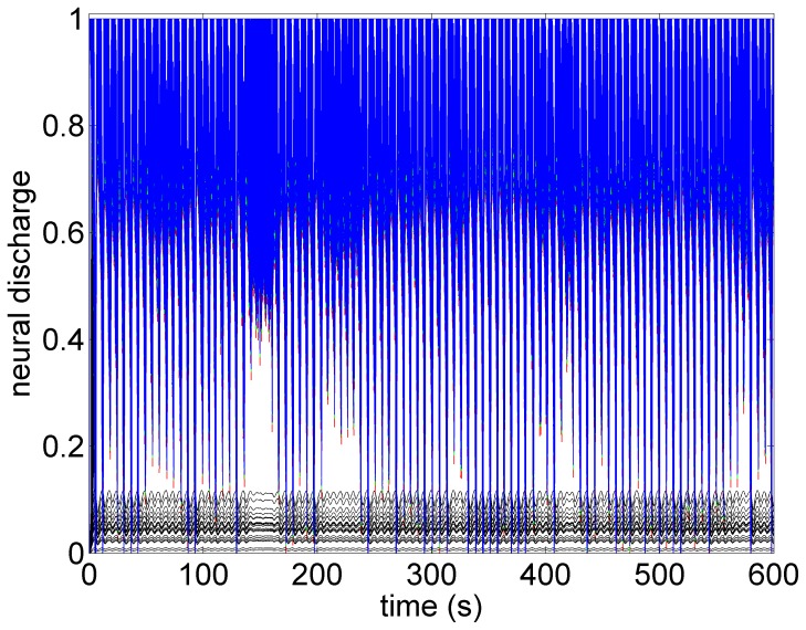
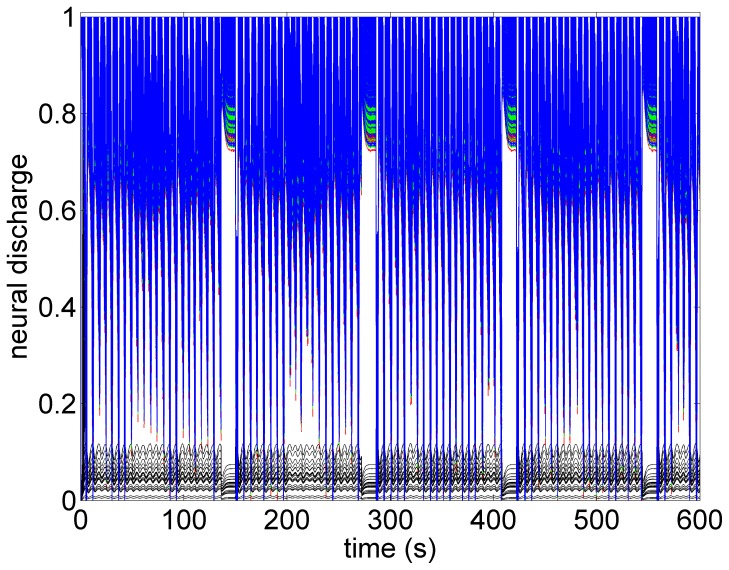
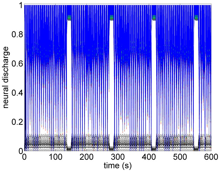
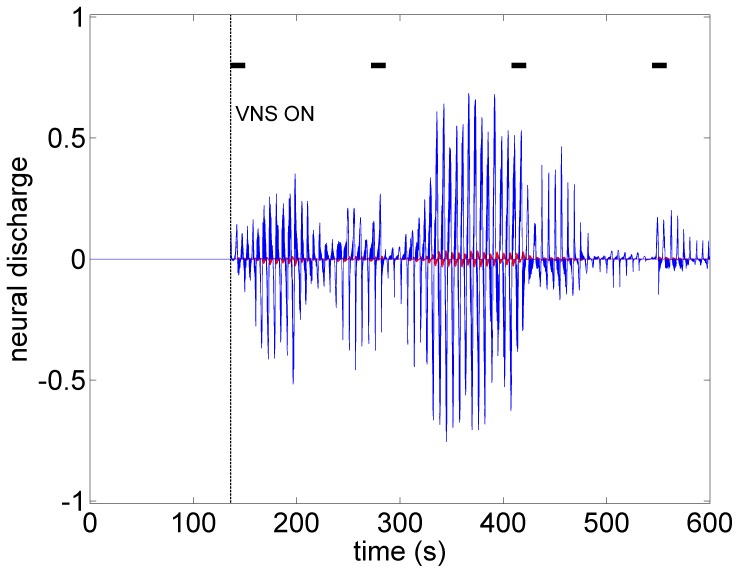
Vagal nerve stimulation in cardiac therapy involves delivering electrical current to the vagal sympathetic complex in patients experiencing heart failure. The therapy has shown promise but the mechanisms by which any benefit accrues is not understood. In this paper we model the response to increased levels of stimulation of individual components of the vagal sympathetic complex as a differential activation of each component in the control of heart rate. The model provides insight beyond what is available in the animal experiment in as much as allowing the simultaneous assessment of neuronal activity throughout the cardiac neural axis. The results indicate that there is sensitivity of the neural network to low level subthreshold stimulation. This leads us to propose that the chronic effects of vagal nerve stimulation therapy lie within the indirect pathways that target intrinsic cardiac local circuit neurons because they have the capacity for plasticity.


Conflict of interest statement

Competing Interests: The experimental part of this study was funded in part by Cyberonics, Inc. The theoretical part was funded fully by The Natural Sciences and Engineering Research Council of Canada. Additionally, JLA and JAA are paid consultants of Cyberonics, Inc. There are no patents, products in development or marketed products to declare. This does not alter the authors' adherence to all the PLOS ONE policies on sharing data and materials. In particular: (a) there are no other competing interests relating to employment, consultancy, patents, products in development, marketed products, etc., and (b) the above does not alter the authors' adherence to PLOS ONE policies on sharing data and materials, to the effect that it places no restrictions on sharing of data and/or materials. (c) GK and MZ declare that they have no competing interests.

Figures

Figure 1
Figure 1. Heart rate in an anesthetized canine under baseline conditions, in the absence of VNS.
Note the variations in heart rate that occur in the normal state.
Figure 2
Figure 2. Heart rate under low level stimulation (0.25 mA) in the same animal as in Figure 1.
No discernible heart rate changes are observed.
Figure 3
Figure 3. Heart rate under moderate level stimulation (0.75 mA) in the same animal as in Figure 1.
Pronounced tachycardia is observed.
Figure 4
Figure 4. Heart rate as the level of stimulation is increased from that in Figure 3 (from 0.75 mA to 1.75 mA).
Pronounced bradycardia is observed.
Figure 5
Figure 5. Average response to VNS from 7 animals that were in the conscious state.
Figure 6
Figure 6. Model simulation under baseline conditions (zero stimulation).
The oscillatory pattern and the variability in that pattern is similar to that observed in the experiment (Figure 1) and is typical at low blood demand and in the presence of low level noise within the system . Brief intervals of resonance, whereby the oscillations are subdued, can be observed in both cases.
Figure 7
Figure 7. Model simulation under subthreshold conditions whereby the direct component of the VSC is not being activated but the indirect component and therefore the local circuit elements of the neural network are being activated at low intensity.
Here, as in the experiment (Figure 2), there are no discernible changes in heart rate. Red bars indicate time intervals when VNS is on.
Figure 8
Figure 8. Model simulation under sympathetic threshold conditions whereby the direct component of the VSC is not being activated but the indirect component is being activated at higher intensity than that in Figure 7.
Pronounced tachycardia is observed, similar to that seen in the experiment under moderate intensity stimulation (Figure 3). Red bars indicate time intervals when VNS is on.
Figure 9
Figure 9. Model simulation under parasympathetic threshold conditions whereby the direct component of the VSC is now being activated while the indirect component being maintained at the same activation intensity as in Figure 8.
Pronounced bradycardia is observed, similar to that seen in the experiment under high intensity stimulation (Figure 4). Red bars indicate time intervals when VNS is on.
Figure 10
Figure 10. A map of neural activity (discharge) at the three levels of the neural network under the model baseline conditions.
The range of activity is normalized to lie between 0 (no activity) and 1.0 (maximal activity). There are 600 neurons at each level of the network (Sympathetic neurons: blue  =  central, green  =  intrathoracic, red  =  cardiac. Black represents 100 parasympathetic neurons at the cardiac level.) The pattern of activity is consistent with the pattern of heart rate variability observed in Figure 6.
Figure 11
Figure 11. A map of neural activity (discharge) at the three levels of the neural network under the model subthreshold conditions and corresponding to the pattern of heart rate observed in Figure 7.
Legend as in the caption of Figure 10.
Figure 12
Figure 12. A map of neural activity (discharge) at the three levels of the neural network under the model sympathetic threshold conditions and corresponding to the pattern of heart rate observed in Figure 8.
Tachycardia observed in Figure 8 is here seen to be the result of higher activity of sympathetic neurons at all three levels of the neural network, coupled with some suppression of parasympathetic activity induced mainly by withdrawal of afferent feedback at the cardiac level. Legend as in the caption of Figure 10.
Figure 13
Figure 13. A map of neural activity (discharge) at the three levels of the neural network under the model parasympathetic threshold conditions and corresponding to the pattern of heart rate observed in Figure 9.
Here the direct component of the VSC is stimulated, with the level of stimulation of the indirect component being maintained the same as under the sympathetic threshold conditions. The direct component of the VSC dominates, leading to the bradycardia observed in Figure 9. Legend as in the caption of Figure 10.
Figure 14
Figure 14. The difference between neural activity under baseline and subthreshold conditions, obtained by subtracting the map in Figure 6 from that in Figure 7.
The figure demonstrates that while there were no discernible effects of subthreshold stimulation on either heart rate or neural discharge, there was considerable difference in neural activity under subthreshold conditions compared with those at baseline. In this figure the activities of all sympathetic neurons are shown in blue and the parasympathetic in red. Black bars indicate time intervals when VNS is on.

References

    1. Groves DA, Brown VJ (2005) Vagal nerve stimulation: a review of its applications and potential mechanisms that mediate its clinical effects. Neuroscience and Biobehavioral Reviews 29:493–500. - PubMed
    1. Ando M, Katare RG, Kakinuma Y, Zhang D, Yamasaki F, et al. (2005) Efferent vagal nerve stimulation protects heart against ischemia-induced arrhythmias by preserving connexin43 protein. Circulation 112:164–170. - PubMed
    1. Zhang Y, Mazgalev TN (2011) Arrhythmias and vagus nerve stimulation. Heart Fail Rev 16:147–161. - PubMed
    1. De Ferrari GM, Crijns HJ, Borggrefe M, Milasinovic G, Smid J, et al. for the CardioFit Multicenter Trial Investigators (2011) Chronic vagus nerve stimulation: a new and promising therapeutic approach for chronic heart failure. European Heart Journal 32:847–855. - PubMed
    1. DiCarlo L, Libbus I, Amurthur B, KenKnight BH, Anand IS (2013) Autonomic regulation therapy for the improvement of left ventricular function and heart failure symptoms: the ANTHEM-HF study. J Card Fail 19:655–660. - PubMed

Publication types