. 2014 Dec 4;15(6):750-61.
doi: 10.1016/j.stem.2014.11.009.

Cardiac repair in a porcine model of acute myocardial infarction with human induced pluripotent stem cell-derived cardiovascular cells

Affiliations

Cardiac repair in a porcine model of acute myocardial infarction with human induced pluripotent stem cell-derived cardiovascular cells

Lei Ye et al. Cell Stem Cell. .

Erratum in

  • Cell Stem Cell. 2015 Jan 8;16(1):102

Abstract

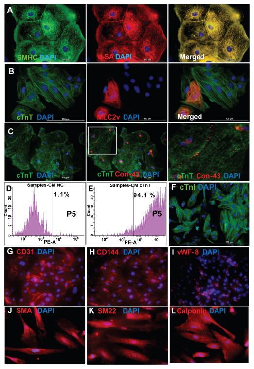
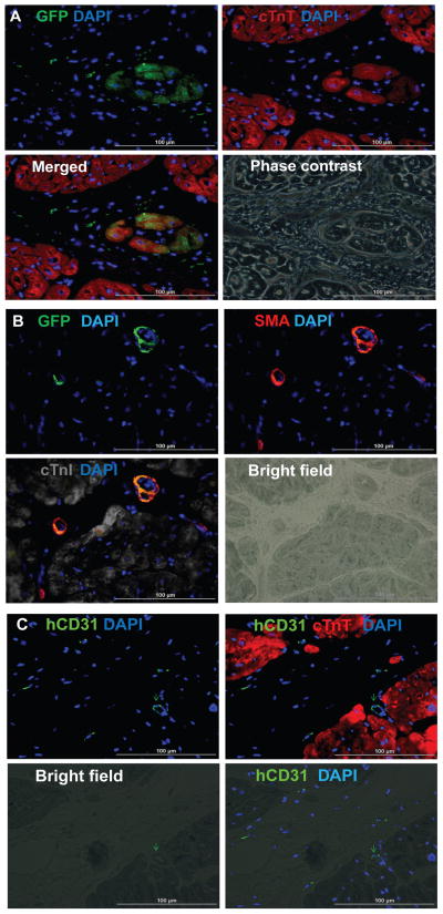
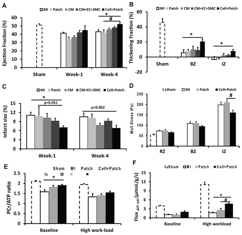
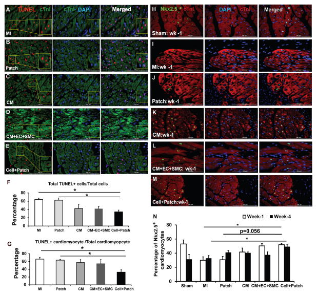
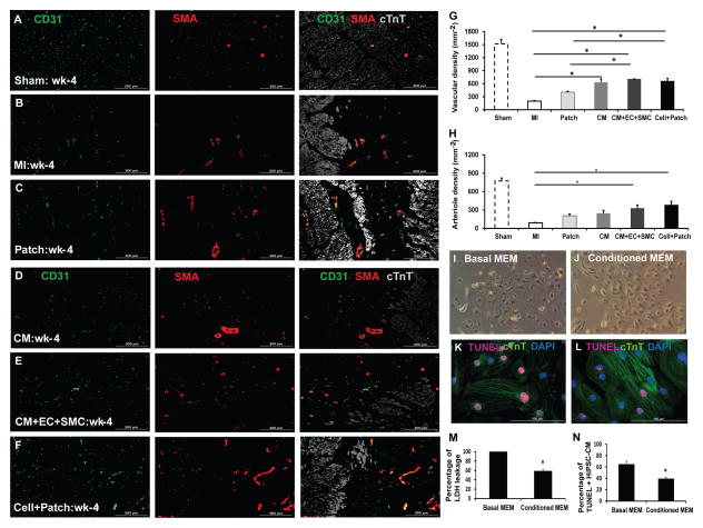
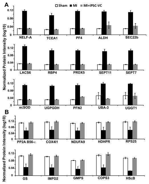
Human induced pluripotent stem cells (hiPSCs) hold promise for myocardial repair following injury, but preclinical studies in large animal models are required to determine optimal cell preparation and delivery strategies to maximize functional benefits and to evaluate safety. Here, we utilized a porcine model of acute myocardial infarction (MI) to investigate the functional impact of intramyocardial transplantation of hiPSC-derived cardiomyocytes, endothelial cells, and smooth muscle cells, in combination with a 3D fibrin patch loaded with insulin growth factor (IGF)-encapsulated microspheres. hiPSC-derived cardiomyocytes integrated into host myocardium and generated organized sarcomeric structures, and endothelial and smooth muscle cells contributed to host vasculature. Trilineage cell transplantation significantly improved left ventricular function, myocardial metabolism, and arteriole density, while reducing infarct size, ventricular wall stress, and apoptosis without inducing ventricular arrhythmias. These findings in a large animal MI model highlight the potential of utilizing hiPSC-derived cells for cardiac repair.


Conflict of interest statement

CONFLICT OF INTERESTS: NA

Figures

Figure 1
Figure 1. Differentiation of human iPSCs into cardiomyocytes, endothelial cells, and smooth muscle cells
hiPSCs were differentiated into CMs via the Sandwich method (Zhang et al., 2012), and the lineage of the differentiated hiPSC-CMs was confirmed via the expression of (A) slow myosin heavy chain (SMHC) and α-sarcomeric actin (α-SA); (B) cardiac troponin T (cTnT) and the ventricular- specific cardiomyocyte protein myosin light chain 2v (MLC2v); and (C) cTnT and the gap-junction protein connexin-43 (Con-43); nuclei were counterstained with DAPI. The boxed region in the 2nd panel of C is shown at higher magnification. (D–F) The purity of the hiPSC-CM population was evaluated via flow cytometry analysis of cTnT expression in (D) isotype controls and (E) purified hiPSC-CMs, and by (F) immunofluorescence analysis of cardiac troponin I (cTnI) expression; nuclei were counterstained with DAPI. Bar: 100 μm in A–B, 200 μm in C and F. (See also Movies S1, S2, and S3). hiPSCs were differentiated into ECs and SMCs as described previously (Hill et al., 2010; Woll et al., 2008). (G–I) The lineage of the differentiated hiPSC-ECs was confirmed via the expression of (G) CD31, (H) CD144, and (I) vWF-8; and (J–L) the lineage of the differentiated hiPSC-SMCs was confirmed via the expression of (J) smooth-muscle actin (SMA), (K) SM22, and (L) calponin. Nuclei were counterstained with DAPI. (Bar: 100 μm in A–B, 200 μm in C and F. Magnification G–L=200x). (See also Figure S1).
Figure 2
Figure 2. hiPSC-derived cardiac cells engraft and survive after transplantation into the hearts of swine with MI
(A) Engraftment of the injected cells was evaluated in sections stained for the presence of GFP; muscle fibers were visualized via fluorescent immunostaining for cTnT, and nuclei were counterstained with DAPI. The sections displayed in the first three panels of A were imaged with a phase-contrast microscope. (B) Engrafted cells were identified in arterioles via immunofluorescent staining for the co-expression of GFP and SMA; muscle fibers were visualized via immunofluorescent staining for cTnI and nuclei were counterstained with DAPI. (C) Engrafted cells were identified in blood vessels (i.e., capillaries and arterioles) via immunofluorescent staining for the human-specific isoform of CD31; muscle fibers were visualized via cTnT staining and nuclei were counterstained with DAPI. (Bar=100 μm). (See also Figure S3).
Figure 3
Figure 3. Transplanted hiPSC-derived cardiac lineage cells improve cardiac function
(A) LV ejection fractions were evaluated at Week 1 and Week 4 after MI injury and treatment. (B) LV wall systolic thickening fractions in the infarct zone (IZ) and at the border zone (BZ) of ischemia were evaluated at Week 4. (C) Infarct sizes were evaluated at Week 1 and Week 4 and expressed as a percentage of the LV surface area. (D) LV wall stress in the IZ, in the BZ, and in uninjured regions of the myocardium (i.e., the remote zone [RZ]) was evaluated at Week 4. Four weeks after MI injury and treatment, (E) PCr/ATP ratios and (F) the ATP hydrolysis rate were determined in the BZ under both baseline conditions and after a high cardiac workload was induced via catecholamine administration; measurements were obtained via a double-saturation 31P MRS-MST protocol. For panels B, D, E, and F, measurements in Sham animals were performed in regions that corresponded to the site of injury in the other experimental groups. *p<0.05 vs MI; #p<0.05 vs Patch. (See also Figures S2&S4, and Table S1).
Figure 4
Figure 4. Transplantation of hiPSC-derived cardiac cells reduces cardiomyocyte apoptosis and enhances endogenous cell survival
(A–G) Apoptotic cells were identified in sections from the border zone of infarct in hearts from animals in the (A) MI, (B) Patch, (C) CM, (D) CM+EC+SMC, and (E) Cell+Patch groups with the TUNEL assay. Muscle fibers were visualized via fluorescent immunostaining for cTnI, and nuclei were counterstained with DAPI; the boxed regions toward the left of panels A, B, C, D, and E are displayed at higher magnification in the boxes at the right of the images. (F) Apoptosis was quantified as the percentage of cells that were positive for TUNEL staining. (G) Cardiomyocyte apoptosis was quantified as the percentage of cTnI-positive cells that were also positive for TUNEL staining. (H–N) NkX2.5 expression was evaluated in sections from the border zone of infarct hearts that compared to Sham operated normal hearts: (H) Sham, (I) MI, (J) Patch, (K) CM, (L) CM+EC+SMC and (M) Cell+Patch. The immunofluorescent staining positives with anti-Nkx2.5 antibody are shown green; muscle fibers were visualized via fluorescent immunostaining for cTnI, and nuclei were counterstained with DAPI. (N) The percentage of cardiomyocytes that expressed Nkx2.5 was determined at Week 1 and Week 4 after injury. (*p<0.05; bar=100 μm). (See also Table S2)
Figure 5
Figure 5. The hiPSC-derived cardiac cells enhance the angiogenic response, and inhibit apoptosis
Vascular density and arteriole density at Week 4 after MI were evaluated in sections from the border zone of infarct in the hearts of animals from the (A) Sham, (B) MI, (C) Patch, (D) CM, (E) CM+EC+SMC and (F) Cell+Patch groups via immunofluorescent staining for CD31 and SMA; muscle fibers were visualized via cTnT staining. (G) Vascular density was determined by counting CD31+ vascular structures, and (H) arteriole density was determined by counting vascular structures that expressed both CD31 and SMA. (*p<0.05; bar=200μm). (I–N) hiPSC-CMs were cultured under hypoxic conditions in (I) basal media (Basal MEM) or (J) media collected from the hiPSC-derived vascular cells (Conditioned MEM) for 48 hours; then, (K–L) apoptotic cells were identified via the TUNEL assay, (M) cytotoxicity was quantified via the intensity of lactate dehydrogenase fluorescence observed in the media, and (N) apoptosis was quantified as the percentage of cells that were positive for TUNEL staining. *p<0.05 vs basal MEM; magnification: 100x for I&J; bar=100 μm. (See also Figure S5 & Table S2).
Figure 6
Figure 6. Protein expression levels are significantly altered in MI and partially restored by cell transplantation
Myocardial protein expression profiles were evaluated in animals that had been treated with (MI+iPSC-VC) or without (MI) hiPSC-VC transplantation after experimentally induced MI; control assessments were performed in animals that underwent all surgical procedures for the induction of MI except for the ligation step (SHAM); results are displayed for 25 proteins whose expression levels (A) increased or (B) decreased after MI and were restored to normal levels by cell therapy. NELF-A, negative elongation factor A; TCEA1, transcription elongation factor A protein 1; PF4, platelet factor 4; ALDH, aldehyde dehydrogenase [mitochondrial]; SEC22b, vesicle-trafficking protein SEC22b; LACS6, long-chain-fatty-acid-CoA ligase 6; RBP4, retinol-binding protein 4; PRDX5, peroxiredoxin-5 [mitochondrial]; SEPT11, septin-11; SEPT7, septin-7; ecSOD, extracellular superoxide dismutase [Cu-Zn]; UGPGDH, UDP-glucose-6-dehydrogenase; PFN2, profiling-2; UBA-3, NEDD8-activating enzyme E1 catalytic subunit; UGGT1, UDP-glucose:glycoprotein glucosyltransferase 1; PP2A B56-ε, serine/threonine-protein phosphatase 2A 56 kDa regulatory subunit ε isoform; COX4I1, cytochrome C oxidase subunit 4 isoform 1, mitochondrial; NDUFA9, NADH dehydrogenase [ubiquinone] 1 α subcomplex subunit 2; HDHPR, dihydropteridine reductase; RPS25, 40S ribosomal protein S25; GS, glutamine synthetase; IMPD2, inosine-5′-monophosphate dehydrogenase 2; GMPS, GMP synthase [glutamine-hydrolyzing]; COPS3, COP9 signlosome complex subunit 3; HScB, iron-sulfur cluster co-chaperone protein HScB [mitochondrial]. (See also Figure S6&S7).

Comment in

References

    1. Beauchamp JR, Morgan JE, Pagel CN, Partridge TA. Dynamics of myoblast transplantation reveal a discrete minority of precursors with stem cell-like properties as the myogenic source. The Journal of cell biology. 1999;144:1113–1122. - PMC - PubMed
    1. Brutsaert DL. Cardiac endothelial-myocardial signaling: its role in cardiac growth, contractile performance, and rhythmicity. Physiological reviews. 2003;83:59–115. - PubMed
    1. Burridge PW, Keller G, Gold JD, Wu JC. Production of de novo cardiomyocytes: human pluripotent stem cell differentiation and direct reprogramming. Cell stem cell. 2012;10:16–28. - PMC - PubMed
    1. Caspi O, Huber I, Kehat I, Habib M, Arbel G, Gepstein A, Yankelson L, Aronson D, Beyar R, Gepstein L. Transplantation of human embryonic stem cell-derived cardiomyocytes improves myocardial performance in infarcted rat hearts. Journal of the American College of Cardiology. 2007;50:1884–1893. - PubMed
    1. Chen D, Dorling A. Critical roles for thrombin in acute and chronic inflammation. Journal of thrombosis and haemostasis: JTH. 2009;7(Suppl 1):122–126. - PubMed

Publication types

MeSH terms