Review
. 2015 Feb;25(2):74-81.
doi: 10.1016/j.tcb.2014.10.003. Epub 2014 Dec 2.

Merkel cells and neurons keep in touch

Affiliations
Review

Merkel cells and neurons keep in touch

Seung-Hyun Woo et al. Trends Cell Biol. 2015 Feb.

Abstract

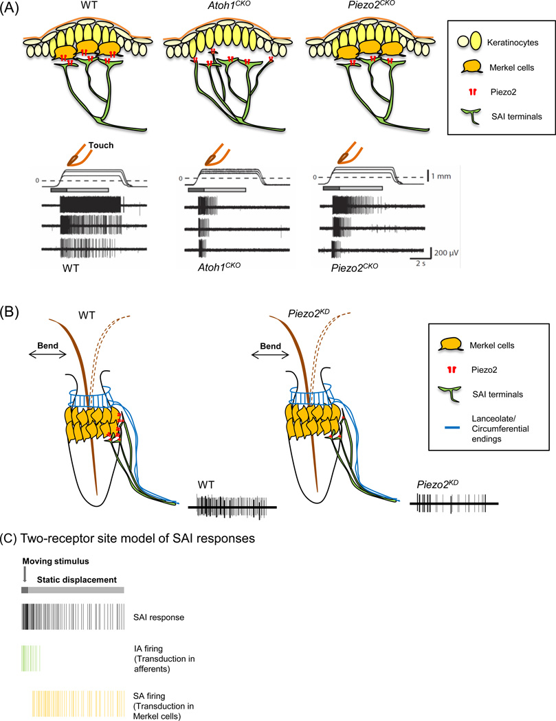
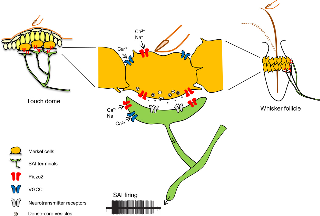
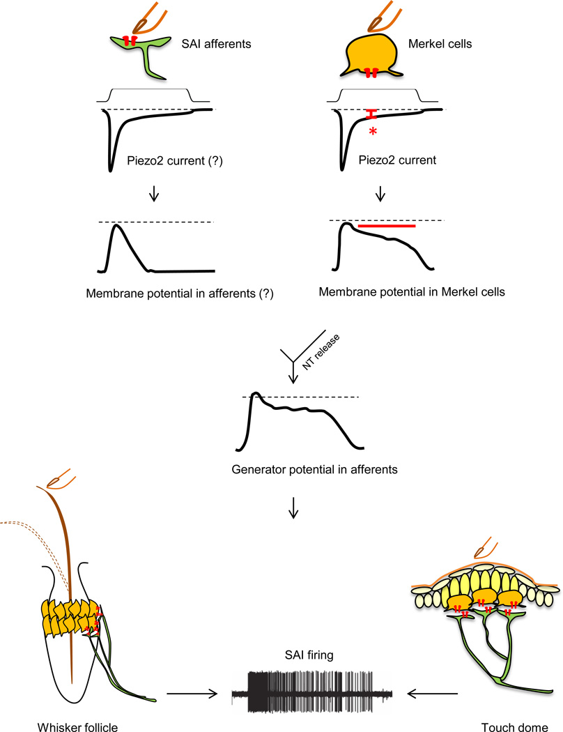
The Merkel cell-neurite complex is a unique vertebrate touch receptor comprising two distinct cell types in the skin. Its presence in touch-sensitive skin areas was recognized more than a century ago, but the functions of each cell type in sensory transduction have been unclear. Three recent studies demonstrate that Merkel cells are mechanosensitive cells that function in touch transduction via Piezo2. One study concludes that Merkel cells, rather than sensory neurons, are principal sites of mechanotransduction, whereas two other studies report that both Merkel cells and neurons encode mechanical inputs. Together, these studies settle a long-standing debate on whether or not Merkel cells are mechanosensory cells, and enable future investigations of how these skin cells communicate with neurons.

Keywords: Piezo; mechanoreceptor; mechanosensory cells; somatosensory; touch dome.


Figures

Figure 1
Figure 1. SA firing responses of Merkel cell-neurite complexes in touch domes and whisker follicles
(A) Mechanically evoked responses from afferents innervating touch domes in wild-type (WT) (left), Atoh1CKO (middle) and Piezo2CKO (right) mice. Displacement applied to a touch dome causes action potential firing in SAI afferents. Moving displacements (dark gray bar) evoke firing in all three genotypes. In wild-type mice, static displacement (light gray bar) evokes slowly adapting (SA) firing; however, firing during static displacement is truncated to intermediately adapting (IA) firing in both Atoh1CKO and Piezo2CKO touch domes. (B) Mechanically evoked responses from sensory afferent bundles innervating WT whisker follicle (left) and the whisker follicle with Piezo2 knockdown (Piezo2KD) (right). Piezo2 knockdown in Merkel cells causes action potentials to be reduced (right). (C) A two-receptor site model of SAI responses. SAI afferents transduce moving stimuli, and Merkel cells mediate sustained firing during static displacement. All traces have been recreated from [–50].
Figure 2
Figure 2. A model of touch transduction in the Merkel cell-neurite complex
1) Gentle pressure on the skin or hair deformation of the whisker opens mechanotransduction channels, hypothesized to be Piezo2, in SAI afferents to initiate SAI action potential firing. 2) Simultaneously, it opens Piezo2 channels in Merkel cells, which causes Merkel cell depolarization. 3) Voltage-gated calcium channels (VGCC) in Merkel cells are subsequently activated, and 4) neurotransmitters are released as a result and contribute to SAI firing. Adapted from [49, 50, 74].
Figure 3
Figure 3. A model of mechanotransduction in each component of the Merkel cell-neurite complex
Left: Skin indentation by touch opens rapidly inactivating mechanotransduction channels, hypothesized to be Piezo2, in SAI afferents and causes a rapid depolarization. Right: Simultaneously, it opens Piezo2 channels in Merkel cells. A Piezo2-dependent small, long-lasting current (red asterisk) induces a sustained depolarization in Merkel cells (red bar) due to the high membrane resistance of Merkel cells. Merkel cell depolarization causes consequent neurotransmitter (NT) release from Merkel cells to SAI afferents. Combined generator potential changes from the afferents and Merkel cells contribute to slowly adapting action potential firing in SAI afferents.

References

    1. Rice FL, Albrecht PJ. Cutaneous Mechanisms of Tactile Perception: Morphological and Chemical Organization of the Innervation to the Skin. In: Kaas JH, Gardner EP, editors. The Senses: A Comprehensive Reference. Academic Press; 2008. pp. 1–31.
    1. Gardner EP, Martin JH, Jessell TM. The bodily senses. In: Kandel ER, Schwartz JH, Jessell TM, editors. Principles of Neural Science. McGraw-Hill; 2000. pp. 430–449.
    1. Brown AG, Iggo A. A quantitative study of cutaneous receptors and afferent fibres in the cat and rabbit. The Journal of physiology. 1967;193:707–733. - PMC - PubMed
    1. Abraira VE, Ginty DD. The sensory neurons of touch. Neuron. 2013;79:618–639. - PMC - PubMed
    1. Takahashi-Iwanaga H. Three-dimensional microanatomy of longitudinal lanceolate endings in rat vibrissae. The Journal of comparative neurology. 2000;426:259–269. - PubMed