. 2014 Dec 4;159(6):1277-89.
doi: 10.1016/j.cell.2014.10.053.

Gut microbiota elicits a protective immune response against malaria transmission

Affiliations

Gut microbiota elicits a protective immune response against malaria transmission

Bahtiyar Yilmaz et al. Cell. .

Abstract

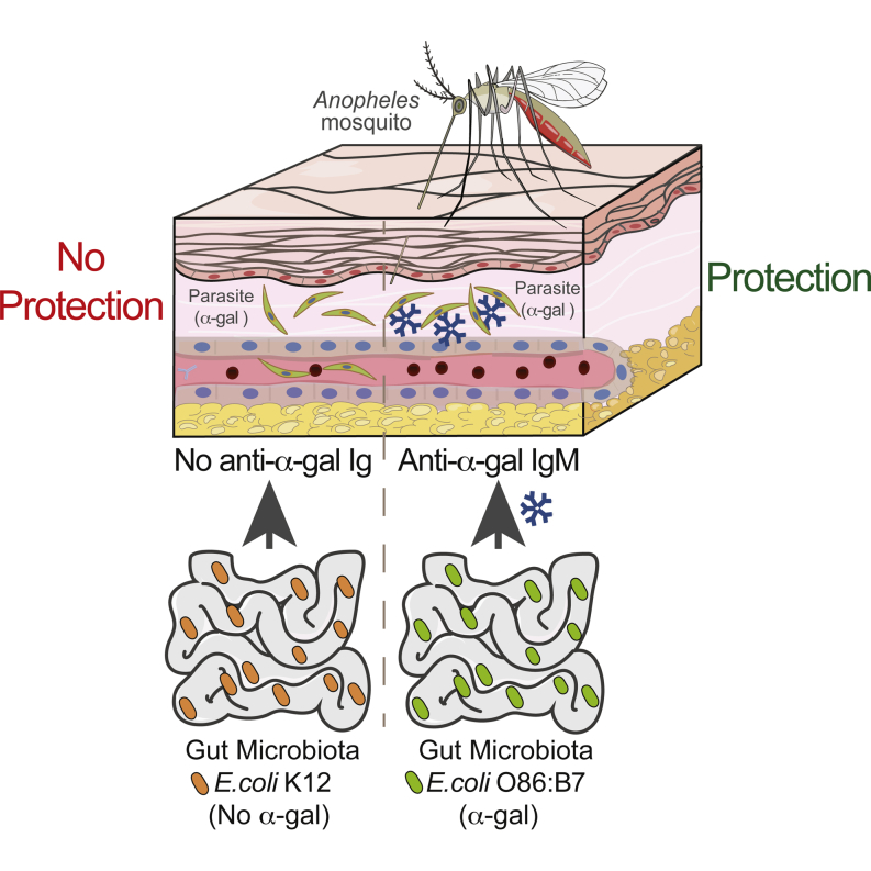
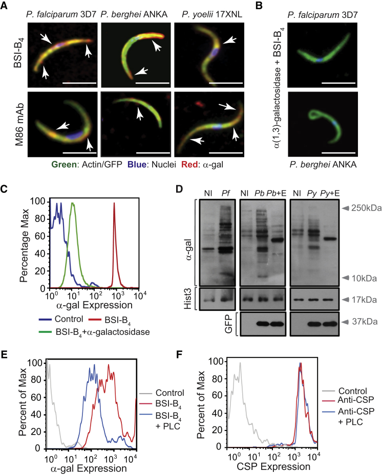
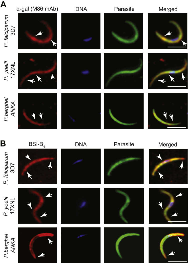
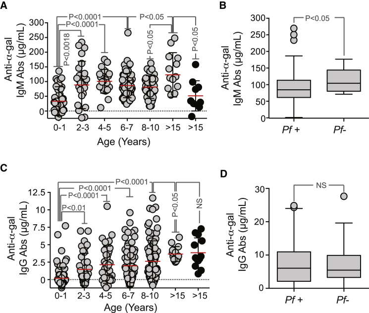
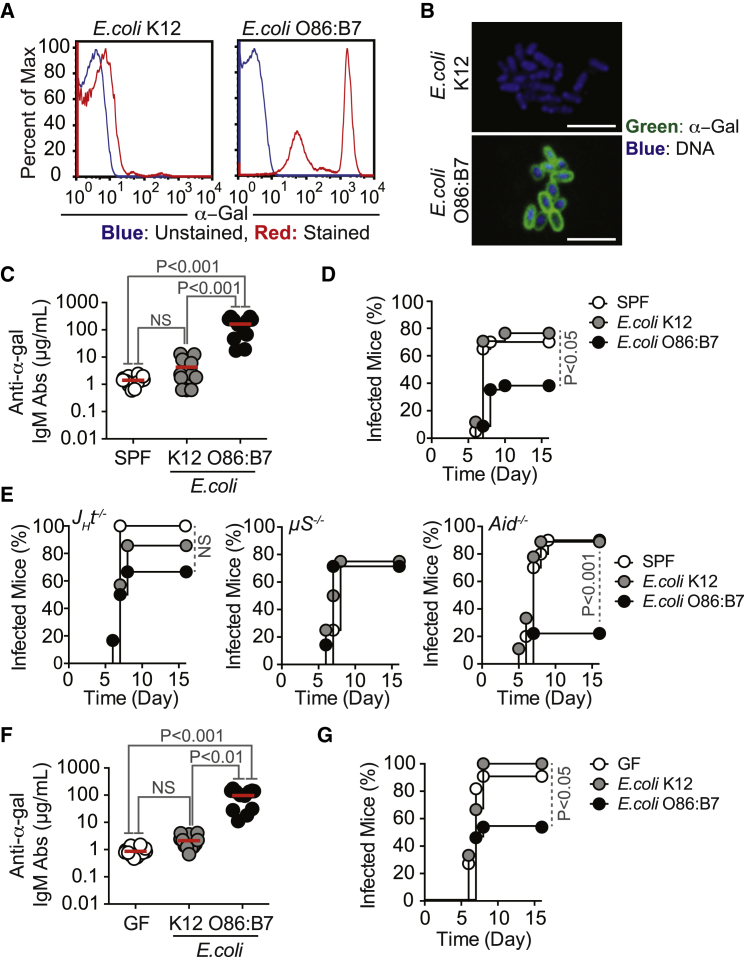
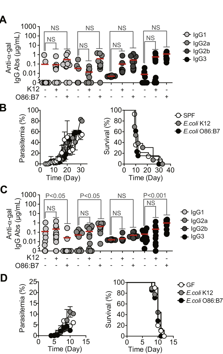
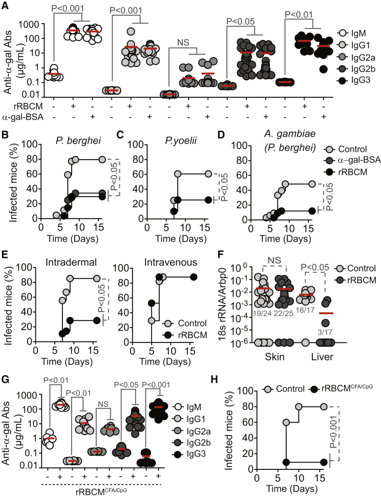
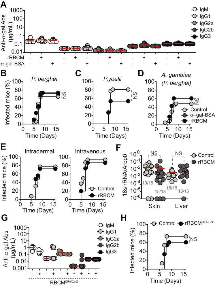
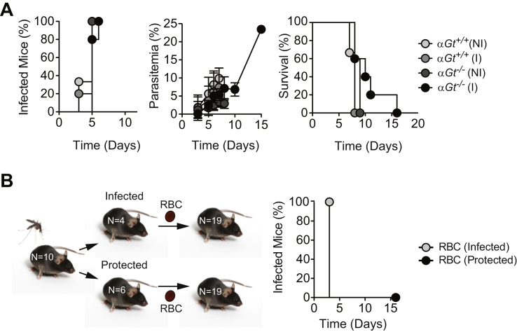
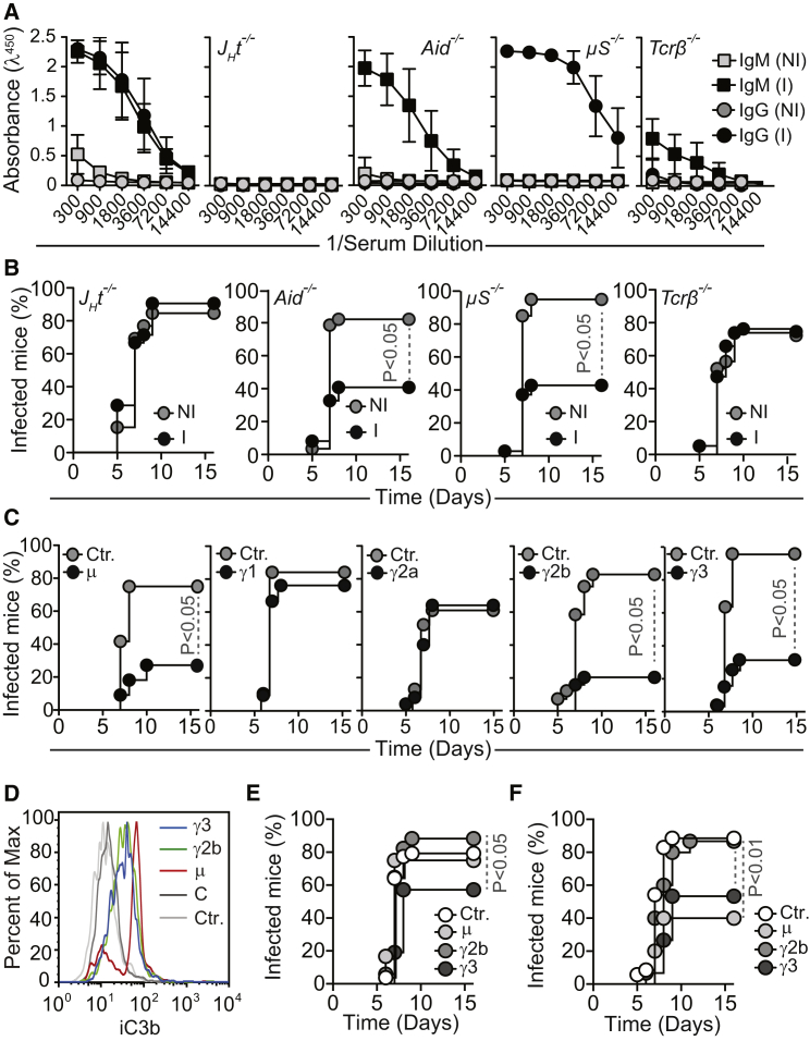
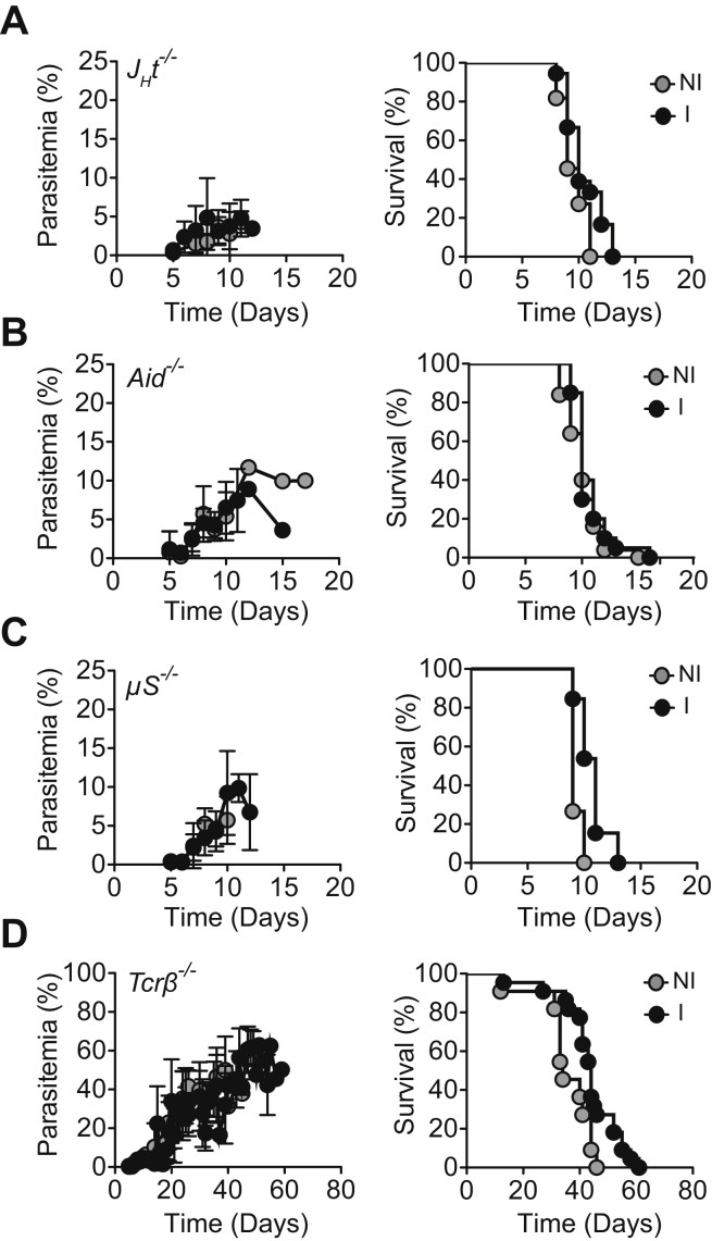
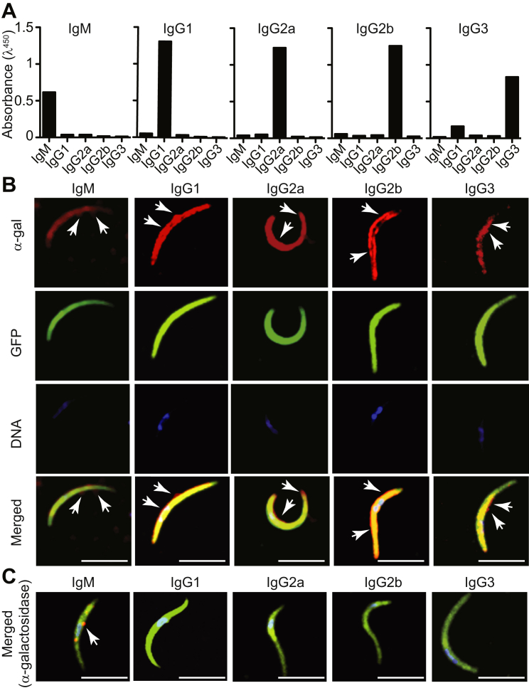
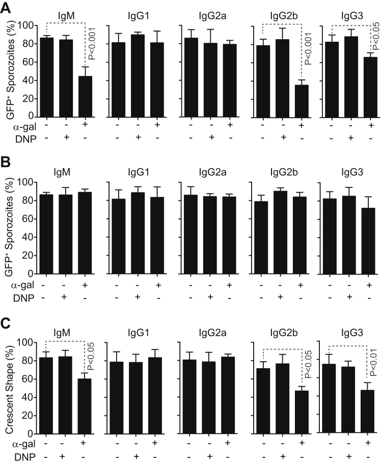
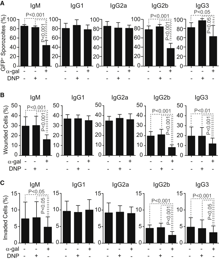
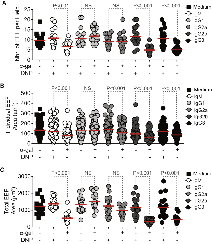
Glycosylation processes are under high natural selection pressure, presumably because these can modulate resistance to infection. Here, we asked whether inactivation of the UDP-galactose:β-galactoside-α1-3-galactosyltransferase (α1,3GT) gene, which ablated the expression of the Galα1-3Galβ1-4GlcNAc-R (α-gal) glycan and allowed for the production of anti-α-gal antibodies (Abs) in humans, confers protection against Plasmodium spp. infection, the causative agent of malaria and a major driving force in human evolution. We demonstrate that both Plasmodium spp. and the human gut pathobiont E. coli O86:B7 express α-gal and that anti-α-gal Abs are associated with protection against malaria transmission in humans as well as in α1,3GT-deficient mice, which produce protective anti-α-gal Abs when colonized by E. coli O86:B7. Anti-α-gal Abs target Plasmodium sporozoites for complement-mediated cytotoxicity in the skin, immediately after inoculation by Anopheles mosquitoes. Vaccination against α-gal confers sterile protection against malaria in mice, suggesting that a similar approach may reduce malaria transmission in humans.


Figures

None
Graphical abstract
Figure 1
Figure 1
Detection of α-Gal in Plasmodium Sporozoites (A) Composite images of GFP/actin (green), α-gal (red; white arrows), and DNA (blue) in Plasmodium sporozoites. (B) Same staining as (A), after removal of α-gal by α-galactosidase. Images are representative of 2–3 independent experiments. Scale bar, 5 μm. (C) Detection of α-gal in PbAHsp70-GFP sporozoites by flow cytometry, representative of three independent experiments. (D) Detection of α-gal in proteins extracted from salivary glands of noninfected (NI), P. falciparum 3D7 (Pf), PbAHsp70-GFP (Pb), or P. yoelii 17XNL (Py)-infected A. mosquitoes. Histone H3 (Hist3) and GFP were detected as loading controls. When indicated, α-gal was digested using α-galactosidase (E). (E and F) Detection of α-gal (E) and CSP (F) in PbAHsp70-GFP sporozoites treated or not with phospholipase C (+PLC). Control is not stained. Data representative of 2–4 independent experiments. See also Figure S1.
Figure S1
Figure S1
Detection of α-Gal in Plasmodium Sporozoites, Related to Figure 1 (A and B) Expression of GFP (P. berghei ANKA) and actin (P. falciparum 3D7 and P. yoelli 17XNL) shown in green, α-gal shown in red and DNA (DAPI) shown in blue. The α-gal epitope was detected with (A) anti-α-gal mAb (M86) or (B) the BSI-B4 lectin. Representative of 2-3 experiments. Arrows indicate α-gal staining. Scale bar, 5 μm.
Figure 2
Figure 2
Anti-α-Gal IgM Abs Are Associated with Protection against Malaria Transmission in Individuals from a Malaria Endemic Region (A) Anti-α-gal IgM Abs in individuals from a malaria endemic region in Mali (gray dots) or from the United States (black dots). Mean (red bars) ± SD. (B) Levels of anti-α-gal IgM Abs in P. falciparum-infected (Pf+) versus noninfected (Pf−) children >4 years of age are shown as box plots in the same population as in (A). (C) Anti-α-gal IgG Abs in individuals from a malaria endemic region in Mali (gray dots) or from the United States (black dots). Mean (red bars) ± SD. (D) Levels of anti-α-gal IgG Abs in P. falciparum-infected (Pf+) versus noninfected (Pf−) children >4 years of age are shown as box plots in the same population as in (C).
Figure 3
Figure 3
Gut Colonization by E. coli Expressing α-Gal Protects against Plasmodium Infection (A and B) Detection of α-gal in E. coli strains by (A) flow cytometry and (B) immunofluorescence. Representative of 2–3 independent experiments. Composite images in (B), i.e., α-gal (green) and DNA (blue) at 100× magnification. Scale bar, 10 μm. (C and D) α1,3Gt−/− mice maintained under SPF were treated with streptomycin for 7 days. (C) Anti-α-gal IgM Abs levels were measured in α1,3Gt−/− mice not colonized (SPF), colonized with E. coli K12, or colonized with O86.B7 strains (2–3 experiments; n = 12). (D) Incidence of blood stage of Plasmodium infection (%) in mice colonized as in (C) and exposed to PbAEEF1a-GFP-infected A. stephensi mosquitoes (four experiments; n = 17–34). (E) Incidence of blood stage of Plasmodium infection (%) in α1,3Gt−/−JHT−/−, α1,3Gt−/−Aid−/−, and α1,3Gt−/−μS−/− mice colonized as in (C) and exposed to PbAHsp70-GFP-infected A. stephensi mosquitoes (1–2 experiments; n = 4–10). (F) Anti-α-gal IgM Abs were measured in GF α1,3Gt−/− mice not colonized (GF), colonized with E. coli K12, or colonized with O86.B7 strains (2–3 experiments; n = 12). (G) Incidence of blood stage of Plasmodium infection (%) in mice colonized as in (F) and exposed to PbAEEF1a-GFP-infected A. stephensi mosquitoes (four experiments; n = 9–13). Mean (red bars). See also Figure S2.
Figure S2
Figure S2
Anti-α-Gal IgG Abs in Gut-Colonized α1,3Gt−/− Mice and Disease Assessment after Exposure to Infected Mosquitoes, Related to Figure 3 (A) Anti-α-gal IgG subclass Ab levels in α1,3Gt−/− mice not-colonized (SPF), colonized with E. coli K12 or O86.B7 strains after streptomycin treatment (2-3 experiments; n = 12). (B) Parasitemia (%) and survival (%) in same mice as (A) after exposure to PbAEEF1a-GFP infected A. stephensi mosquitoes (4 experiments; n = 17-34). (C) Anti-α-gal IgG subclass Ab levels in GF α1,3Gt−/− mice not colonized (GF), colonized with E. coli K12 or O86.B7 strains (2-4 experiments; n = 12). (D) Parasitemia (%) and survival (%) in same mice as (C) after exposure to PbAEEF1a-GFP infected A. stephensi mosquitoes (4 experiments; n = 7-10 per group). Mean (red bars).
Figure 4
Figure 4
Protective Effect of α-Gal Immunization (A) Anti-α-gal Abs in the serum of control (−) versus rRBCM (+) or α-gal-BSA (+) immunized α1,3Gt−/− mice (2–3 experiments; n = 12–29). (B–D) Incidence of blood stage of infection (%) in α1,3Gt−/− mice treated as in (A) and exposed to (B) PbAEEF1a-GFP-infected A. stephensi mosquitoes (seven experiments; n = 27–44), (C) P. yoelii 17XNL-infected A. stephensi mosquitoes (five experiments; n = 28–39), or (D) PbAEEF1a-GFP-infected A. gambiae mosquitoes (four experiments; n = 27–34). (E) Incidence of blood stage of infection (%) in nonimmunized (control) versus immunized (rRBCM) α1,3Gt−/− mice receiving PbAEEF1a-GFP sporozoites (3–4 experiments; n = 17–28). (F) Plasmodium 18 s rRNA/Arbp0 mRNA in skin and liver of nonimmunized (control) versus immunized (rRBCM) α1,3Gt−/− mice exposed to PbAEEF1a-GFP-infected A. stephensi mosquitoes (3–5 experiments). Infected/total mice (gray nbrs). (G) Same as (A) in control (−) versus immunized (+; rRBCM emulsified in CFA+CpG) α1,3Gt−/− mice (two experiments; n = 6–23). (H) Incidence of blood stage of infection (%) in α1,3Gt−/− mice treated as in (G) and infected as in (B) (three experiments; n = 16–19). In (A), (F), and (G), dots are individual mice and mean (red bars). See also Figures S3 and S4.
Figure S3
Figure S3
α1,3Gt+/+ Mice Are Not Protected against Malaria Transmission, Related to Figure 4 (A) Anti-α-gal antibodies in the serum of control (-) versus rRBCM (+) or α-gal-BSA (+) immunized α1,3Gt+/+ mice (2-3 experiments; n = 12-16). (B–D) Incidence of blood stage of infection (%) in α1,3Gt+/+ mice treated as in (A) and exposed to (B) PbAEEF1a-GFP infected A. stephensi mosquitoes (7 experiments; n = 19-48), (C) P. yoelii 17XNL infected A. stephensi mosquitoes (5 experiments; n = 26-30) or (D) PbAEEF1a-GFP infected A. gambiae mosquitoes (4 experiments; n = 27-28). (E) Incidence of blood stage of infection (%) in nonimmunized (Control) versus immunized (rRBCM) α1,3Gt+/+ mice receiving PbAEEF1a-GFP sporozoites (3-4 experiments; n = 15-26). (F) Plasmodium 18 s rRNA/Arbp0 mRNA in skin and liver of nonimmunized (Control) versus immunized (rRBCM) α1,3Gt+/+ mice exposed to PbAEEF1a-GFP infected Anopheles stephensi mosquitoes (3-5 experiments). Infected/total mice (gray Nbrs). (G) Same as (A) in control (-) versus immunized (+; rRBCM emulsified in CFA plus CpG) α1,3Gt+/+ mice (2 experiments; n = 5). (H) Same as (B) in mice treated as in (G) (3 experiments; n = 15). In (A, F and G) dots are individual mice and mean (red bars).
Figure S4
Figure S4
Immunization against α-Gal Is Ineffective in Protecting against Blood Stage of Infection but Confers Sterile Protection against Malaria Transmission, Related to Figure 4 (A) Incidence of blood stage of infection (%; left panel), parasitemia (%; middle panel) and survival (%; right panel) upon inoculation of PbAEEF1a-GFP infected RBC. Mice were either immunized (I) with rRBCM or not immunized (NI) (One experiment; n = 3-5). (B) Schematic representation of infection protocol (left panel) and incidence of blood stage of infection (%; right panel) in α1,3Gt−/− mice transferred with RBC from α1,3Gt−/− mice infected PbAEEF1a-GFP or protected from transmission of PbAEEF1a-GFP infection by A. stephensi mosquitoes (2 experiments; n = 19).
Figure 5
Figure 5
Protective Effect of Anti-α-Gal Abs (A) Relative absorbance of anti-α-gal Abs (Mean ± SD) in serial serum dilutions from nonimmunized (NI) or rRBCM-immunized (I) α1,3Gt−/− mice (two experiments; n = 10). (B) Incidence of blood stage infection (%) in specific immune component-deleted α1,3Gt−/− mice immunized (I) or not (NI) as in (A) and exposed to PbAEEF1a-GFP-infected mosquitoes (3–7 experiments; n = 13–41). (C) Incidence of blood stage of infection (%) in α1,3Gt−/− mice after passive transfer of anti-α-gal Abs versus controls (no passive transfer; ctr.) exposed to PbAEEF1a-GFP-infected mosquitoes (4–7 experiments; n = 19–32). (D) C3 deposition in PbAHsp70-GFP sporozoites not exposed (ctr.) or exposed to anti-α-gal Abs plus mouse complement (C). Representative of three independent experiments. (E) Incidence of blood stage of infection (%) in α1,3Gt−/−C3−/− mice after passive transfer of anti-α-gal IgM (μ), IgG2b (γ2b), or IgG3 (γ3) Abs versus controls (ctr.; no passive transfer) not receiving Abs, exposed to PbAEEF1a-GFP-infected mosquitoes (four experiments; n = 21–37). (F) Same as (E) in PMN-depleted α1,3Gt−/− mice (four experiments; n = 15–25). See also Figures S5 and S6.
Figure S5
Figure S5
Blood Stage Infection and Lethality, Related to Figure 5 (A–D) Parasitemia (%; left panels) and survival (%; right panels) are shown for infected (A) α1,3Gt−/−Jht−/−, (B) α1,3Gt−/−Aid−/−, (C) α1,3Gt−/−μS−/− and (D) α1,3Gt−/−Tcrβ−/− mice immunized with rRBCM (I) vs. control nonimmunized (NI) (3-7 experiment; n = 11-22).
Figure S6
Figure S6
Specificity of Anti-α-Gal mAbs, Related to Figure 5 (A) Comparison of relative binding of anti-α-gal mAbs (125 ng/ml of mAb) determined by ELISA using α-gal-BSA as a solid phase antigen. (B) Comparison of relative binding capacity of anti-α-gal mAbs (50 μg/ml of mAb) determined by immunofluorescence using PbAEEF1a-GFP sporozoites as an antigen. mAb (red), GFP (green) and DNA (blue) staining. Merged images show composite of the three staining. Representative of 2-3 experiments. (C) Similar staining as in (B) for PbAEEF1a-GFP sporozoites treated with α-galactosidase. Composite images are shown with mAb (red), GFP (green) and DNA (blue) staining. White arrows in (B) and (C) indicate binding of different mAb to the α-gal epitope. Scale bar, 5 μm.
Figure S7
Figure S7
Cytotoxic Effect of Anti-α-Gal Antibodies, Related to Figure 7 (A) Mean percentage (%) of viable GFP+PbAHsp70-GFP sporozoites ± STD (3-4 experiments) after in vitro exposure to anti-α-gal (α-gal) or anti-DNP mAbs in the presence of rabbit complement. (B) Mean percentage (%) of viable GFP+PbAHsp70-GFP sporozoites ± STD (3-4 experiments) after in vitro exposure to anti-α-gal (α-gal) or anti-DNP mAbs in the absence of complement. (C) Mean percentage (%) of viable crescent shaped PbAEEF1a-GFP sporozoites ± STD (3 experiments) after in vitro exposure to anti-α-gal (α-gal) or anti-DNP (DNP) mAb in the presence of mouse complement.
Figure 6
Figure 6
Protective Effect of Anti-α-Gal Abs against Hepatocyte Infection (A) Mean percentage (%) of viable GFP+PbAHsp70-GFP sporozoites ± STD (3–4 experiments) after exposure in vitro to anti-α-gal or control anti-DNP mAbs in the presence of mouse complement. (B and C) Mean percentage (%) of HepG2 cells (B) wounded (Dextran-Red+) or (C) invaded (GFP+) by PbAHsp70-GFP sporozoites treated as in (A) ± SD (six experiments).
Figure 7
Figure 7
Protective Effect of Anti-α-Gal Abs against Plasmodium Maturation in Hepatocytes (A) Number of EEF per field (dots; 20–23 fields). (B) Area of individual EEF (dots) (n = 111–256 EEFs counted in 20–23 fields). (C) Total area of EEF (dots) per field (20–23 fields). HepG2 cells were incubated with PbAHsp70-GFP sporozoites, previously exposed to anti-α-gal or control anti-DNP mAbs in the presence of complement (A–C). See also Figure S7.

Comment in

References

    1. Avila J.L., Rojas M., Galili U. Immunogenic Gal alpha 1----3Gal carbohydrate epitopes are present on pathogenic American Trypanosoma and Leishmania. J. Immunol. 1989;142:2828–2834. - PubMed
    1. Avila J.L., Rojas M., Velazquez-Avila G. Characterization of a natural human antibody with anti-galactosyl(alpha 1-2)galactose specificity that is present at high titers in chronic Trypanosoma cruzi infection. Am. J. Trop. Med. Hyg. 1992;47:413–421. - PubMed
    1. Belkaid Y., Hand T.W. Role of the microbiota in immunity and inflammation. Cell. 2014;157:121–141. - PMC - PubMed
    1. Belnoue E., Kayibanda M., Vigario A.M., Deschemin J.C., van Rooijen N., Viguier M., Snounou G., Rénia L. On the pathogenic role of brain-sequestered alphabeta CD8+ T cells in experimental cerebral malaria. J. Immunol. 2002;169:6369–6375. - PubMed
    1. Benatuil L., Kaye J., Rich R.F., Fishman J.A., Green W.R., Iacomini J. The influence of natural antibody specificity on antigen immunogenicity. Eur. J. Immunol. 2005;35:2638–2647. - PubMed

Supplemental References

    1. Circolo A., Garnier G., Fukuda W., Wang X., Hidvegi T., Szalai A.J., Briles D.E., Volanakis J.E., Wetsel R.A., Colten H.R. Genetic disruption of the murine complement C3 promoter region generates deficient mice with extrahepatic expression of C3 mRNA. Immunopharmacology. 1999;42:135–149. - PubMed
    1. Eto T., Ichikawa Y., Nishimura K., Ando S., Yamakawa T. Chemistry of lipid of the posthemyolytic residue or stroma of erythrocytes. XVI. Occurrence of ceramide pentasaccharide in the membrane of erythrocytes and reticulocytes of rabbit. J. Biochem. 1968;64:205–213. - PubMed
    1. García-González M., Bettinger S., Ott S., Olivier P., Kadouche J., Pouletty P. Purification of murine IgG3 and IgM monoclonal antibodies by euglobulin precipitation. J. Immunol. Methods. 1988;111:17–23. - PubMed
    1. Kisailus E.C., Kabat E.A. A study of the specificity of Bandeiraea simplicifolia lectin I by competitive-binding assay with blood-group substances and with blood-group A and B active and other oligosaccharides. Carbohydr. Res. 1978;67:243–255. - PubMed
    1. LaTemple D.C., Galili U. Adult and neonatal anti-Gal response in knock-out mice for alpha1,3galactosyltransferase. Xenotransplantation. 1998;5:191–196. - PubMed

Publication types

MeSH terms