. 2014 Dec 4;159(6):1290-9.
doi: 10.1016/j.cell.2014.10.057.

Host adaptation of a bacterial toxin from the human pathogen Salmonella Typhi

Affiliations

Host adaptation of a bacterial toxin from the human pathogen Salmonella Typhi

Lingquan Deng et al. Cell. .

Abstract

Salmonella Typhi is an exclusive human pathogen that causes typhoid fever. Typhoid toxin is a S. Typhi virulence factor that can reproduce most of the typhoid fever symptoms in experimental animals. Toxicity depends on toxin binding to terminally sialylated glycans on surface glycoproteins. Human glycans are unusual because of the lack of CMAH, which in other mammals converts N-acetylneuraminic acid (Neu5Ac) to N-glycolylneuraminic acid (Neu5Gc). Here, we report that typhoid toxin binds to and is toxic toward cells expressing glycans terminated in Neu5Ac (expressed by humans) over glycans terminated in Neu5Gc (expressed by other mammals). Mice constitutively expressing CMAH thus displaying Neu5Gc in all tissues are resistant to typhoid toxin. The atomic structure of typhoid toxin bound to Neu5Ac reveals the structural bases for its binding specificity. These findings provide insight into the molecular bases for Salmonella Typhi's host specificity and may help the development of therapies for typhoid fever.


Figures

Figure 1
Figure 1. Comparison of typhoid toxin binding to paired Neu5Ac- and Neu5Gc-terminated glycans by a customized microarray
Chemical structures of Neu5Ac and Neu5Gc are shown. The two molecules differ by only one single oxygen atom. Vertical axis values represent the normalized average of relative fluorescence units and horizontal axis indicates the glycan number in the array. See also Fig. S1, Table 1, and Table S1.
Figure 2
Figure 2. Typhoid toxin binding to red blood cells and lymphocytes from humans and chimpanzees
(A) Relative levels of Neu5Ac/Neu5Gc in human and chimpanzee red blood cells (RBCs). (B) Binding of different amounts of typhoid toxin to human and chimpanzee RBCs. (C) Relative levels of Neu5Ac/Neu5Gc in human and chimpanzee lymphocytes. (D) Binding of typhoid toxin to human and chimpanzee lymphocytes. Similar results were obtained in several independent repetitions of the experiments. RFI: relative fluorescence intensity. PBS: phosphate buffered saline.
Figure 3
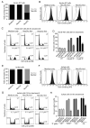
Figure 3. Typhoid toxin binds to and is cytotoxic towards cells displaying Neu5Ac-but not to those displaying Neu5Gc-terminated glycans
Human intestinal epithelial Henle-407 cells (A-D) and human T lymphocyte Jurkat cells (E-H) were left untreated (medium only) or fed Neu5Ac or Neu5Gc in a culture medium for 4 days. Cells were then analyzed by HPLC to examine their relative sialic acid composition (A and E), or used in typhoid toxin binding (B and F) and toxicity assays by examining the cell cycle profile of toxin-treated cells (C, D, G, and H). Data in D and H are the mean ± SEM; ***P<0.0001, compared to the percent of control (medium-treated) cells in G2/M in the same group. See also Fig. S2.
Figure 4
Figure 4. Mice engineered to constitutively express CMAH, resulting in elevated levels of Neu5Gc in all tissues, are resistant to typhoid toxin
Purified preparations of wild type typhoid toxin or a binding-defective mutant (PltBS35A) were systemically administered into mice defective in (Cmah−/−) or constitutively expressing CMAH (Cmahtg), or control (C57BL/6) mice. Four days after treatment their total weight (A) and the total number of white cells (WBC) (B, top panel) or neutrophils (B, bottom panel) were measured as indicated in Materials and Methods. Black circles represent the percentage of the weight of an animal relative to its weight immediately before treatment (A). Circulating white blood cells were counted in a hematology analyzer (B). Alternatively, peripheral blood cells from animals that had received the indicated treatments were stained with an antibody directed to the neutrophil cell marker Gr1 and the number of stained cells was determined by flow cytometry (C). The histograms shown are from ungated samples. Similar results were obtained in several independent repetitions of the experiment. RFI: relative fluorescence intensity. TT: typhoid toxin. WT: wild type. Data in B are the mean ± SEM; ***P<0.0001, **P< 0.002 (relative to the buffer control in the same group). (D) Survival of mice after administration of different amounts of typhoid toxin. PBS: phosphate buffered saline. The difference in the survival curves of PBS vs toxin treated (all concentrations) control and Cmah−/− animals was statistically significant (P<0.001; log-rank Mantel-Cox test). The difference between the survival curves in control and Cmah−/− mice after administration of 2 μg of toxin was statistically significant (P<0.001). However, after administration of 10 μg of toxin the difference between the survival curves in control and Cmah−/− mice was not statistically significant (P<0.6). See also Fig. S3.
Figure 5
Figure 5. Typhoid toxin does not bind to chimpanzee tissues
Frozen sections of small intestine from humans or chimpanzees were stained with fluorescently labeled typhoid toxin or its binding-defective PltBS35A mutant (red) and counterstained with Hoescht (blue). Scale bar: 100 μm.
Figure 6
Figure 6. Crystal structure of typhoid toxin B subunit PltB bound to its sialic acid ligand
(A) The atomic structure of the PltB pentamer in complex with the GalNAcβ1-4(Neu5Acα2-8Neu5Acα2-3)Galβ1-4Glc oligosaccharide is shown as a ribbon cartoon with each protomer depicted in a different color. In the PltB pentamer, only partial oligosaccharide density (Neu5Ac-Neu5Ac-Gal) is seen in Chain C (purple) and E (yellow). Cyan sticks represent the sugar carbon atoms, blue sticks represent nitrogen atoms, and red sticks represent oxygen atoms. (B) Surface charge distribution of the PltB pentamer structure and sugar-binding pockets. (C) Close-up views of Neu5Ac-Neu5Ac-Gal and its composite annealed omit difference density map. PltB chain E and its key residue Ser35 are shown in yellow. Green mesh represents the sugar difference density map contoured at 2.5σ. (D) Interactions between PltB and Neu5Ac. Chain E of PltB is shown as a yellow colored ribbon cartoon, the amino acids interacting with the sugar are shown as sticks, and the direct interactions are shown in black dash. Water is shown as gray balls and water-mediated interactions are shown as purple dashes. (E) Structure/function analysis of the PltB glycan-binding site. Typhoid holotoxin toxin preparations containing the indicated PltB mutants were tested for their ability to intoxicate cultured Henle-407 cells. Toxicity was evaluated by determining the percentage of cells arrested at the G2/M phase of the cell cycle, which is a measure of typhoid toxin’s CdtB activity. Data are the mean ± SEM; ***P<0.0001, compared to the percent cells treated with wild type toxin that are in G2/M. (F) Comparison of the sugar binding sites of PltB and SubB bound to Neu5Ac and Neu5Gc, respectively. Critical residues that differ between SubB (Tyr78) and PltB (Val103) are highlighted as sticks. Other interacting amino acids and sugars are shown in lines. PltB is shown in yellow, Neu5Ac in Cyan, SubB in Green and Neu5Gc in light purple. See also Fig. S4-Fig. S7 and Table S2.

References

    1. Adams P, Afonine P, Bunkóczi G, Chen V, Davis I, Echols N, Headd J, Hung L-W, Kapral G, Grosse-Kunstleve R, et al. PHENIX: a comprehensive Python-based system for macromolecular structure solution. Acta Cryst. 2010;D66:213–221. - PMC - PubMed
    1. Beddoe T, Paton A, Le Nours J, Rossjohn J, Paton J. Structure, biological functions and applications of the AB5 toxins. Trends Biochem Sci. 2010;35:411–418. - PMC - PubMed
    1. Byres E, Paton A, Paton J, Löfling J, Smith D, Wilce M, Talbot U, Chong D, Yu H, Huang S, et al. Incorporation of a non-human glycan mediates human susceptibility to a bacterial toxin. Nature. 2008;456:648–652. - PMC - PubMed
    1. Chou H, Hayakawa T, Diaz S, Krings M, Indriati E, Leakey M, Paabo S, Satta Y, Takahata N, Varki A. Inactivation of CMP-N-acetylneuraminic acid hydroxylase occurred prior to brain expansion during human evolution. Proc Natl Acad Sci U S A. 2002;99:11736–11741. - PMC - PubMed
    1. Crump J, Mintz E. Global trends in typhoid and paratyphoid Fever. Clin Infect Dis. 2010;50:241–246. - PMC - PubMed

Publication types

MeSH terms