. 2014 Dec 4;159(6):1300-11.
doi: 10.1016/j.cell.2014.11.017.

Beta-lactam antibiotics induce a lethal malfunctioning of the bacterial cell wall synthesis machinery

Affiliations

Beta-lactam antibiotics induce a lethal malfunctioning of the bacterial cell wall synthesis machinery

Hongbaek Cho et al. Cell. .

Abstract

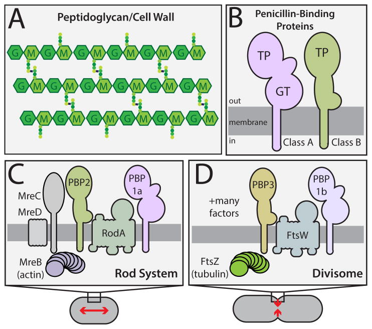
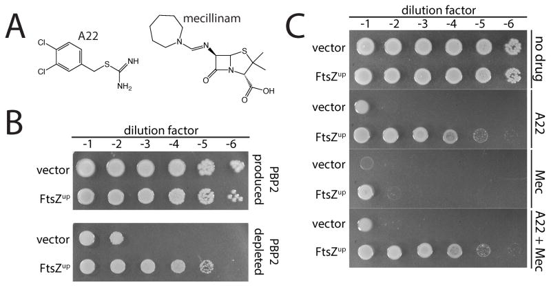
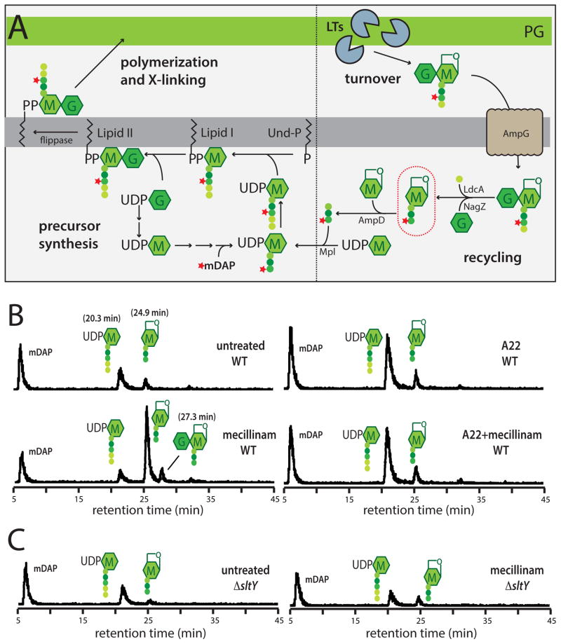
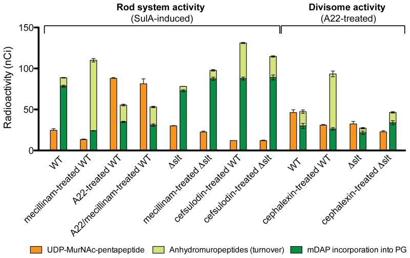
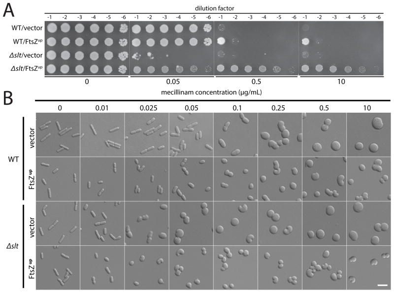
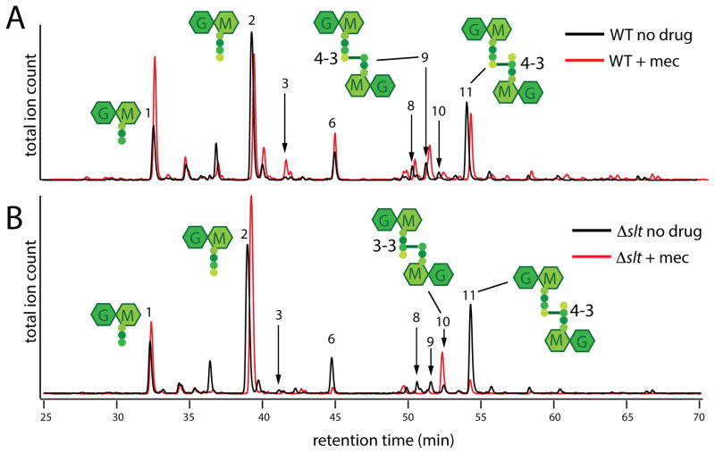
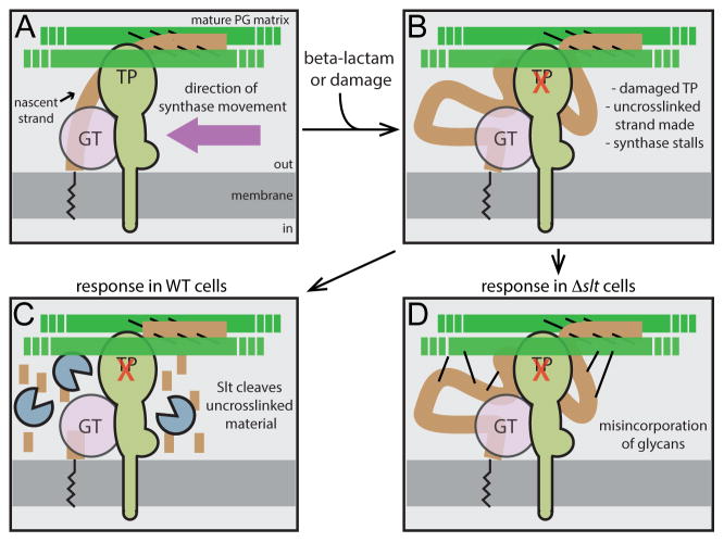
Penicillin and related beta-lactams comprise one of our oldest and most widely used antibiotic therapies. These drugs have long been known to target enzymes called penicillin-binding proteins (PBPs) that build the bacterial cell wall. Investigating the downstream consequences of target inhibition and how they contribute to the lethal action of these important drugs, we demonstrate that beta-lactams do more than just inhibit the PBPs as is commonly believed. Rather, they induce a toxic malfunctioning of their target biosynthetic machinery involving a futile cycle of cell wall synthesis and degradation, thereby depleting cellular resources and bolstering their killing activity. Characterization of this mode of action additionally revealed a quality control function for enzymes that cleave bonds in the cell wall matrix. The results thus provide insight into the mechanism of cell wall assembly and suggest how best to interfere with the process for future antibiotic development.


Figures

Figure 1
Figure 1. Peptidoglycan structure and the machines that synthesize it
A. The PG matrix consists of glycan chains with the repeating unit of N-acetylmuramic acid (MurNAc, M) and N-acetylglucosamine (GlcNAc, G). Attached to the MurNAc sugars are peptides (colored circles) used to form crosslinks between adjacent glycans. B. Domain structure of the PG synthases. Both classes of PBPs have a single transmembrane domain with a large catalytic domain in the periplasm. C–D. Schematics highlighting the components of the two main PG synthetic systems in rod-shaped bacteria. Both systems require a dedicated bPBP (PBP2 or PBP3) and a SEDS (shape/elongation/division/sporulation) family protein (RodA or FtsW) for proper function. See text for details.
Figure 2
Figure 2. Mecillnam induces a lethal malfunctioning of the Rod system
A. Shown are the chemical structures of A22 and mecillinam. B. Conditional essentiality of PBP2. Cultures of strain HC439/pHC817 [ΔpbpA Plac::pbpA] harboring either a vector control (pSC101, vector) or a derivative (pTB63, FtsZup) were serially diluted and spotted on LB agar with either 1 mM IPTG (PBP2 replete) or agar without inducer (PBP2 depletion). Plates were incubated overnight at 30°C and photographed. C. Cultures of strain MG1655 [WT] harboring either pSC101 or pTB63 serially diluted and spotted on LB agar as in part B. Agar was supplemented with A22 (10 μg/ml), mecillinam (1 μg/ml), or both drugs as indicated.
Figure 3
Figure 3. Measurement of PG synthesis and turnover following mecillinam treatment
A. Schematic summarizes the PG synthesis and recycling pathways. Sugars and peptides are represented as in Figure 1A. The composition of the pentapeptide of the cytoplasmic precursors is L-Ala-gamma-D-Glu-meso-diaminopimelic acid (mDAP)-D-Ala-D-Ala. UDP-sugars are first made in the cytoplasm before being transferred to the lipid carrier undecaprenol-phosphate (Und-P). The final precursor lipid II is the substrate used by the PBPs to form PG. As a result of the crosslinking mechanism and the activity of carboxypeptidase enzymes that remove the terminal D-Ala, the PG layer consists primarily of glycans with tetrapeptides. The turnover product circled in red will accumulate in a ΔampD strain if nascent PG is degraded during a pulse labeling experiment. See text for details. B–C. Measurement of PG synthesis and turnover by the Rod system. Cells of TU278 [ΔlysA ΔampD] (B) or its Δslt derivative (HC419) (C) producing SulA to block cell division were pulse labeled with [3H]-mDAP following treatment with the indicated drug(s) (10 μg/ml each when added). Soluble metabolites were separated by HPLC and detected using an in-line radiodetector. The resulting chromatograms are shown. Peaks are labeled with schematics of the corresponding compounds. The identity of each peak is based on the results shown in Figure S3. See text for details.
Figure 4
Figure 4. Quantification of PG synthesis and turnover following beta-lactam treatment
Cells of TU278 [ΔlysA ΔampD] or its Δslt derivative (HC419) were grown, radiolabeled, and analyzed as in Figure 3. The amount of UDP-MurNAc-pentapeptide and total anhydromuropeptides produced were quantified from the area under the peaks in HPLC chromatograms. Radiolabel incorporation into PG was determined by quantifying the amount of label released from cells by lysozyme. Antibiotic concentrations used were: mecillinam (10 μg/ml), A22 (10 μg/ml), cephalexin (10 μg/ml), and cefsulodin (100 μg/ml). Results are the average of three independent experiments with the error bars representing the standard deviation. The drop in precursor levels between untreated and mecillinam-treated WT cells is significant (p < 0.005). See text for details.
Figure 5
Figure 5. Effect of mecillinam on the growth and morphology of cells defective for Slt
A. Cells of MG1655 [WT] or HC408 [Δslt] harboring either pSC101 (vector) or pTB63 (FtsZup) were grown overnight, diluted, and spotted on agar containing the indicated concentration of mecillinam as described for Figure 2B. B. The same cells were grown to exponential phase and diluted to an OD600 of 0.025 into LB medium containing the indicated concentration of mecillinam. Growth was continued for an additional 3 hours and the cells were fixed. The fixed cells were then imaged on agarose pads using DIC optics. Bar equals 4 microns.
Figure 6
Figure 6. Muropeptide composition of PG following mecillinam treatment
Cells of MG1655 [WT] (A) or HC408 [Δslt] (B) were grown to an OD600 of approximately 0.5 and diluted 1:20 into fresh LB medium with or without mecillinam (10 μg/ml) as indicated. Growth was continued for an additional 3 hours and PG sacculi were prepared from cells of each culture. The purified PG was then digested with the muramidase mutanolysin and the resulting muropeptides were reduced and analyzed by LC/MS. Total ion count chromatograms are shown with the chromatograms of the mecillinam-treated samples off-set for clarity. Note that the scales of each chromatogram are not identical. They were scaled to show the relative muropeptide composition rather than the total amount of material. For example, the total peak area in the chromatogram from mecillinam-treated WT cells is one third that of the corresponding untreated sample even though an equivalent of twice the volume was injected. Schematics of several major muropeptide products are shown near their corresponding peaks with the numbers referring to the type of crosslink (4-3 v.s. 3-3). The identities for all labeled peaks and the quantification of their relative amounts are listed in Table S1. The results were reproducible over two biological replicates of each sample and three technical replicates for each biological replicate.
Figure 7
Figure 7. Role of Slt in maintaining coordination between GT and TP activity during PG biogenesis
Shown is a schematic highlighting the role of Slt in PG biogenesis. Panel (A) shows the normal synthetic process with properly coupled GT and TP activities. The new glycan strands (brown) are polymerized from lipid-linked precursors (black zig-zag line) by an enzyme with GT activity (pink) that may either be the canonical domain of an aPBP or an as yet unidentified GT enzyme. The newly polymerized glycans are rapidly crosslinked into the mature matrix (green) by an associated TP activity shown here as a bPBP, but it could also be the TP domain of an aPBP. Other components of the putative synthetic complex including cytoskeletal elements were omitted for simplicity. When the TP active site is damaged or targeted by a beta-lactam (panel B), GT activity continues to produce glycan chains that are no longer crosslinked into the matrix. In WT cells (panel C), such strands are targeted for destruction by Slt. Conversely, in cells inactivated for Slt (panel D), the uncrosslinked glycans produced by the damaged/targeted machinery are not degraded, but are instead aberrantly incorporated into the matrix by an alternative crosslinking enzyme. In cells with normal FtsZ levels, the resulting morphological changes are lethal, leading to beta-lactam hypersensitivity of Slt-defective cells.

Comment in

  • Antimicrobials: New tricks for old drugs.
    Nunes-Alves C. Nunes-Alves C. Nat Rev Microbiol. 2015 Feb;13(2):68. doi: 10.1038/nrmicro3421. Epub 2014 Dec 22. Nat Rev Microbiol. 2015. PMID: 25534806 No abstract available.

References

    1. Banzhaf M, van den Berg van Saparoea B, Terrak M, Fraipont C, Egan A, Philippe J, Zapun A, Breukink E, Nguyen-Distèche M, den Blaauwen T, et al. Cooperativity of peptidoglycan synthases active in bacterial cell elongation. Mol Microbiol. 2012;85:179–194. - PubMed
    1. Beachey EH, Keck W, de Pedro MA, Schwarz U. Exoenzymatic activity of transglycosylase isolated from Escherichia coli. Eur J Biochem. 1981;116:355–358. - PubMed
    1. Begg K, Nikolaichik Y, Crossland N, Donachie WD. Roles of FtsA and FtsZ in activation of division sites. J Bacteriol. 1998;180:881–884. - PMC - PubMed
    1. Bendezú FO, de Boer PAJ. Conditional lethality, division defects, membrane involution, and endocytosis in mre and mrd shape mutants of Escherichia coli. J Bacteriol. 2008;190:1792–1811. - PMC - PubMed
    1. Bernhardt TG, de Boer PAJ. SlmA, a nucleoid-associated, FtsZ binding protein required for blocking septal ring assembly over Chromosomes in E. coli. Mol Cell. 2005;18:555–564. - PMC - PubMed

Publication types

MeSH terms