Review
. 2014 Dec;26(12):4568-83.
doi: 10.1105/tpc.114.131623. Epub 2014 Dec 5.

Phytochromes: an atomic perspective on photoactivation and signaling

Affiliations
Review

Phytochromes: an atomic perspective on photoactivation and signaling

E Sethe Burgie et al. Plant Cell. 2014 Dec.

Abstract

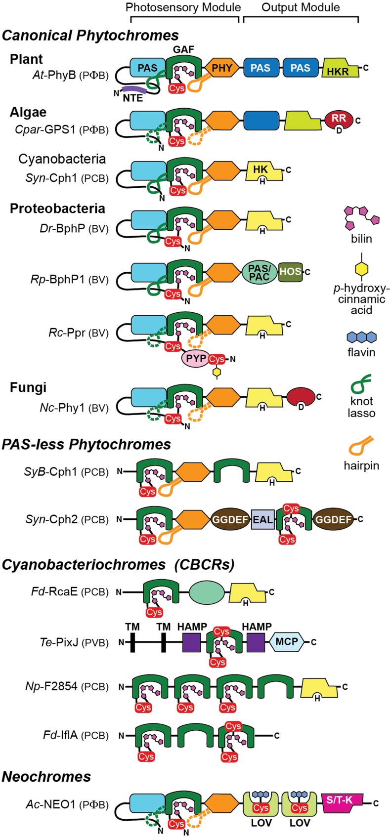
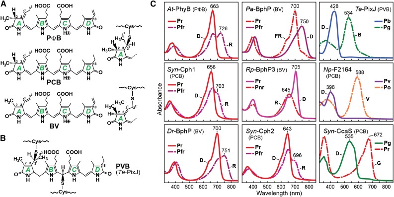
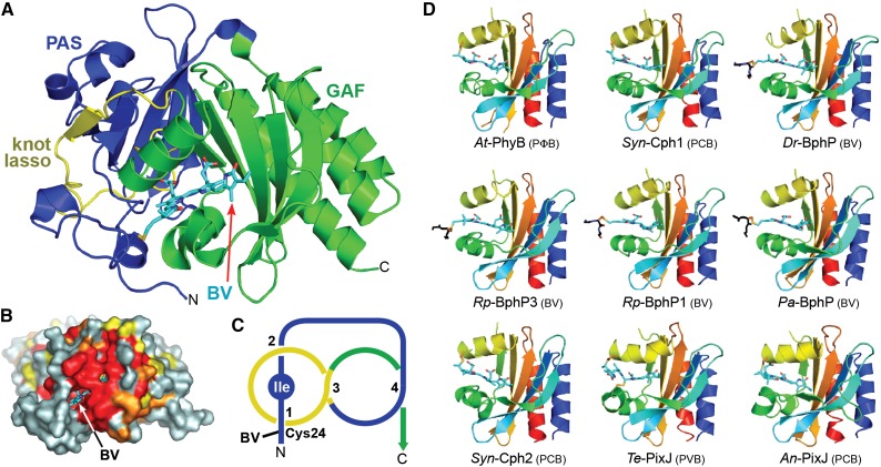
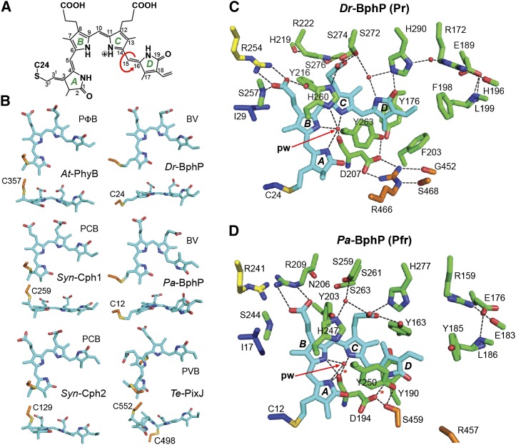
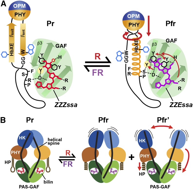
The superfamily of phytochrome (Phy) photoreceptors regulates a wide array of light responses in plants and microorganisms through their unique ability to reversibly switch between stable dark-adapted and photoactivated end states. Whereas the downstream signaling cascades and biological consequences have been described, the initial events that underpin photochemistry of the coupled bilin chromophore and the ensuing conformational changes needed to propagate the light signal are only now being understood. Especially informative has been the rapidly expanding collection of 3D models developed by x-ray crystallographic, NMR, and single-particle electron microscopic methods from a remarkably diverse array of bacterial Phys. These structures have revealed how the modular architecture of these dimeric photoreceptors engages the buried chromophore through distinctive knot, hairpin, and helical spine features. When collectively viewed, these 3D structures reveal complex structural alterations whereby photoisomerization of the bilin drives nanometer-scale movements within the Phy dimer through bilin sliding, hairpin reconfiguration, and spine deformation that ultimately impinge upon the paired signal output domains. When integrated with the recently described structure of the photosensory module from Arabidopsis thaliana PhyB, new opportunities emerge for the rational redesign of plant Phys with novel photochemistries and signaling properties potentially beneficial to agriculture and their exploitation as optogenetic reagents.


Figures

Figure 1.
Figure 1.
Architecture of Representative Members of the Phy Superfamily within Seed and Seedless Plants (Neochromes), Algae (Glaucophyte), Proteobacteria, and Cyanobacteria (PAS-Less and CBCRs). Shown are Arabidopsis (At) PhyB, Cyanophora paradoxa CCMP329 (glaucophyte, Cpar) GPS1, Synechocystis PCC6803 (Syn) Cph1 and Cph2, D. radiodurans (Dr) BphP, R. palustris (Rp) BphP1, R. centenum (Rc) Ppr, Neurospora crassa (Nc) Phy1, Synechococcus OSB’ (SyB) Cph1, T. elongatus (Te) PixJ, N. punctiforme (Np) F2854, F. diplosiphon (Fd) RcaE and IflA, and A. capillus-veneris (Ac) neochrome NEO1. The known or likely bilin type for each Phy is indicated in parenthesis. Domains include conserved residues in diguanylate phosphodiesterase (EAL), cGMP phosphodiesterase/adenylyl cyclase/FhlA (GAF), conserved residues in diguanylate cyclase (GGDEF), histidine kinase/adenylyl cyclase/methyl binding protein/phosphatase (HAMP), histidine kinase (HK), histidine-kinase-related (HKR), 2-helix output sensor (HOS), light/oxygen/voltage (LOV), methyl-accepting chemotaxis protein (MCP), Phy N-terminal extension (NTE), Period/Arnt/Single-Minded (PAS), PAS domain followed by C-terminal motif similar to PAS domain (PAS/PAC), Phy-specific (PHY), photoactive yellow protein (PYP), response regulator (RR), serine/threonine-kinase (S/T-K), and predicted transmembrane (TM). The knot lasso and hairpin motifs are indicated by the green and orange loops, respectively. Confirmed and predicted lassos/hairpins are in solid and dashed lines, respectively. Cys, cysteine residue that covalently binds the chromophores: bilin, flavin, or p-hydroxycinnamic acid. H, phosphoacceptor histidine in HK domains; D, phosphoacceptor aspartate in RR domains; C, C terminus; N, N terminus.
Figure 2.
Figure 2.
Bilin Chromophores and Absorption Spectra of Representative Phys within the Superfamily. (A) Chemical structure and apoprotein thioether linkage position for various bilins used in Phys. The four pyrrole rings (A-D) and important carbon positions in the bilin are indicated. (B) Chemical structure and dual thioether linkage specific to CBCRs with blue/green photocycles. (C) Absorption spectra of the dark-adapted (D) and photoactivated states after blue (B), far-red (FR), green (G), red (R), and violet (V) excitation for canonical Phys with Pr dark-adapted states (At-PhyB, Syn-Cph1, and Dr-BphP), a bathy-Phy (Pa-BphP), a Pnr Phy (Rp-BphP3), a PAS-less Phy (Syn-Cph2), and three CBCRs (Te-PixJ, Np-F2164, and Syn-CcaS). Dashed lines denote the photoactivated state. The bilin type is indicated in parenthesis for each Phy. Absorption maxima are indicated.
Figure 3.
Figure 3.
3D Structures of the Bilin Binding Region in Phys. (A) Ribbon diagram of the PAS-GAF region of Dr-BphP (see Protein Databank [PDB] codes 1ZTU, 2O9C, and 4Q0H). Shown are the PAS (blue) and GAF (green) domains and the knot lasso (yellow). BV (arrow) is displayed in cyan with the nitrogens and oxygens colored in blue and red, respectively. N, N terminus; C, C terminus. The sulfur moiety in Cys-24 that forms the thioether linkage with BV is in yellow. (B) Surface view of the PAS-GAF region of Dr-BphP showing the buried chromophore. Residues with 90, 75, and 60% identity within the Phy superfamily are colored in red, orange, and yellow, respectively. BV (arrow) is displayed in cyan. (C) Diagram of the figure-of-eight knot connecting the PAS (blue) and GAF domains (yellow) in Dr-BphP. The isoleucine at the nexus of the knot is labeled. (D) Ribbon diagrams of GAF domains in their dark-adapted states from representative canonical Phys (At-PhyB, PDB code 4OUR; Syn-Cph1, 2VEA; and Dr-BphP, 2O9C), bathy-Phys (Pa-BphP, 3C2W; Rp-BphP1, 4GW9), a Pnr Phy (Rp-BphP3, 2OOL), a PAS-less Phy (Syn-Cph2, 4BWI), and CBCRs (Te-PixJ, 4GLQ; An-PixJ, 3W2Z). Polypeptide chains are displayed in rainbow with the N-terminal ends in blue and the C-terminal ends in red. Bilins are colored in cyan, and the cysteine(s) forming the thioether linkage(s) are in orange with the sulfur atoms in yellow. A portion of the polypeptide for proteobacterial Phys that adjoins the site of thioether linkage is included and shown in black. ([A] and [B] are adapted from Wagner et al. [2005], Figures 1B and 4A.)
Figure 4.
Figure 4.
Structure of the Bilin and Bilin Binding Pocket in Representative Phys. (A) Chemical diagram of BV bound to Dr-BphP. The pyrrole rings (A-D), carbon positions, and location of the Cys-24 linkage are labeled. Arrow shows the location of the prototypic Z to E isomerization of the bilin during photoconversion. (B) Conformation in side and top views of the bilin bound to representative Phys in their dark-adapted states. The bilin type is indicated for each Phy. The cysteine(s) that links the bilin via a thioether bond is shown. (C) and (D) Bilin and surrounding amino acids in the canonical Phy Dr-BphP (C) and the bathy-Phy Pa-BphP (D) in their dark-adapted Pr and Pfr end states, respectively (PDB codes 4Q0J and 3C2W). Amino acids in the PAS domain and upstream region, GAF domain, PHY domain, and the knot lasso are indicated in blue, green, orange, and yellow, respectively. The sulfur moiety in the cysteine forming the thioether linkage with the bilin is in yellow. Fixed waters are in red spheres. pw, pyrrole water. Dashed lines indicate hydrogen bonds.
Figure 5.
Figure 5.
Structure of the PSM from Representative Phys in Their Dark-Adapted States. (A) Ribbon diagrams of PSMs from Syn-Cph1 (PDB code 2VEA), Pa-BphP (3C2W), and Dr-BphP (4Q0J) in their dark-adapted states. Helical spine is highlighted by the dashed red lines. Hairpin is located by the bracket. The paired subunits of the Pa-BphP and Dr-BphP dimers are in gray. Other coloring is as in Figure 3. The spectral state of each structure is indicated in parenthesis. (B) SPEM model of the full-length Dr-BphP dimer. Only a portion of the histidine kinase domain was imaged due to conformational flexibility. The crystal structure of the PSM is included for one subunit with the space-filling model of the bilin shown. ([A] is adapted from data presented in Essen et al. [2008], Figure 1B; Yang et al. [2008], Figure 1A; and Burgie et al. [2014b], Figure 5A.)
Figure 6.
Figure 6.
Conformation of the PHY-Domain Hairpin from Representative Phys and Its Interaction with the GAF Domain. Ribbon diagrams of crystal structures were drawn from Dr-BphP as Pr (PDB ID code 4Q0J) and a mixed Pr/Pfr state (4O01), Syn-Cph1 as Pr (2VEA), Syn-Cph2 as Pr (4BWI), At-PhyB as Pr (4OUR), Pa-BphP as Pfr (3C2W), and Rp-BphP1 (4GW9) as Pfr. The bilin type (arrow) and the spectral state of each parent model are indicated. The coloring is as in Figure 3. Side chains are shown for relevant amino acids. Dashed lines highlight hydrogen bond contacts between the DIP (Asp-Ile-Pro) motif aspartate in the GAF domain and either the conserved arginine or serine residues in the PRXSF motif from the hairpin stem. The distance separating the GAF and PHY domain globular regions is shown; it was measured from the loop separating the β1 and β2 strands of the GAF domain and the α carbon of a conserved tryptophan (Trp-483 in Dr-BphP) just proximal to the exiting α-helix of the PHY domain. βent and βexit label the entrance (N-terminal) and exit (C-terminal) β-strands in the hairpin. (Adapted from Burgie et al. [2014b], Figure 6.)
Figure 7.
Figure 7.
Conformational Changes Associated with Phy Photoconversion. (A) Photoconversion of Te-PixJ from Pb to Pg drives bilin sliding within the GAF pocket. Shown are portions of the GAF domain β-sheet that impinges upon the bilin. Selected residues that contact the bilin directly are labeled. Hydrogen bonds are indicated with dashed lines. Pb and Pg carbons are colored white and pink, respectively, with the exception that cysteine carbons of the thioether linkages are colored orange. Carbons of Pb and Pg state bilins are colored gray and magenta, respectively. Oxygens, red; nitrogens, cyan; sulfurs, yellow. Asterisk identifies the light-labile thioether linkage. (B) Paired Pr (4Q0J) and Pr/Pfr mixed structures (4O01) of the PSM from Dr-BphP showing the PAS (blue), GAF (green), and PHY (orange) domains. (C) to (E) Negative-staining SPEM images of full-length Dr-BphP in its dark-adapted Pr and photoactivated Pfr end states. The crystal structures of the corresponding Pr (PDB ID code 4Q0J) and red-light-treated states (4O01) were superposed on the Pr (C) and Pfr/Pfr’ models ([D] and [E]). Each micrograph is shown in two orientations, and height and width of the respective species are indicated. Domain coloring of the crystal structures is the same as in (B). The positions of the PHY domains in (D) and (E) were adjusted to better fit the SPEM density (magenta). (F) Superimposed SPEM density of the Pr versus the Pfr (left) and Pfr’ conformers (right). The position of the histidine kinase domain is indicated (HK). Arrows highlight the direction of displacement of the PHY or HK domains from the Pr state to the Pfr or Pfr’ states. ([A] is adapted from Cornilescu et al. [2014], Figure 4; [B] from Takala et al. [2014], Figure 2A, and Burgie et al. [2014b], Figure 5A; and [C] to [F] from Burgie et al. [2014b], Figures 7C to 7F.)
Figure 8.
Figure 8.
3D Crystal Structure of the PSM from Arabidopsis PhyB. (A) Ribbon diagram of the PSM with the PAS (blue), GAF (green), and PHY (orange) domains indicated. The knot-lasso is colored yellow, the helical spine is indicated with a dashed red line, and the hairpin is delineated with a bracket. Connectivity of the PHY domain in the disordered regions is illustrated with dashed black lines. The second subunit of the dimer is colored gray. PΦB is colored cyan. N, N terminus. (B) Bilin and surrounding amino acids. NTE, GAF domain, knot lasso, hairpin, and PΦB carbons are colored blue, green, yellow, orange, and cyan, respectively. Amino acid side chains are labeled. Hydrogen bonds are indicated with black dashed lines. Oxygens, red; nitrogens, blue; and sulfurs, gold. pw, pyrrole water. (Adapted from Burgie et al. [2014a], Figure 2C.)
Figure 9.
Figure 9.
Toggle Model for Photoconversion of Canonical Phys That Translates Light into a Conformational Signal. (A) Conformational changes within the GAF domain and hairpin associated with bilin photoisomerization and sliding and hairpin deformation using At-PhyB(Pr) and Pa-BphP(Pfr) as the examples. Upon light-induced rotation of the D pyrrole ring, the bilin breaks its D-ring/His-403 connection and slides (see arrow) within the GAF domain crevice to form a new contact between the D-ring and Asp-307 and the C-ring propionate and His-403. The tryptophan pair, Tyr-276 and Tyr-303 adjacent to the D-ring, rotate in directions opposite to the D-ring rotation. Together, the effects initiate a collision of Tyr-361 with Phe-585, breakage of the Asp-307/Arg-582 contact, and other changes that collectively release the hairpin stem from the GAF domain. The freed hairpin stem becomes helical, swivels, reforms a new contact between Asp-307 and Ser-584 in the PRXSF motif, and swaps the βent for a βexit/Phe-588 connection with the GAF-domain surface. The rotation and helical conversion of strand βexit presumably reorients the PHY domain relative to the GAF domain and/or tugs on the helical spine connecting the PHY domain to OPM (not shown) to eventually actuate signaling changes in the OPM. The conformations of the bilin in the two end states are indicated. (B) Global conformational changes within the Dr-BphP Phy dimer. The PHY domains adjust their positions relative to their GAF domains through the light-induced conformational changes in the PHY domain hairpin stem from β-strand to helical followed by straightening of the bowed helical spines. Ultimately, the position and/or mobility of the paired OPMs are impacted to alter their signaling potential, in this case phosphotransferase activity by a histidine kinase domain (HK). HP, hairpin. Pfr and Pfr' represent the two photoactivated states observed by SPEM. ([A] is adapted from Burgie et al. [2014a], Figure 5; and [B] from Burgie et al. [2014b], Figure 11.)

Similar articles

Cited by

References

    1. Ádám É., Hussong A., Bindics J., Wüst F., Viczián A., Essing M., Medzihradszky M., Kircher S., Schäfer E., Nagy F. (2011). Altered dark- and photoconversion of phytochrome B mediate extreme light sensitivity and loss of photoreversibility of the phyB-401 mutant. PLoS ONE 6: e27250. - PMC - PubMed
    1. Al-Sady B., Ni W., Kircher S., Schäfer E., Quail P.H. (2006). Photoactivated phytochrome induces rapid PIF3 phosphorylation prior to proteasome-mediated degradation. Mol. Cell 23: 439–446. - PubMed
    1. Anders K., Gutt A., Gärtner W., Essen L.O. (2014). Phototransformation of the red light sensor cyanobacterial phytochrome 2 from Synechocystis species depends on its tongue motifs. J. Biol. Chem. 289: 25590–25600. - PMC - PubMed
    1. Anders K., Daminelli-Widany G., Mroginski M.A., von Stetten D., Essen L.O. (2013). Structure of the cyanobacterial phytochrome 2 photosensor implies a tryptophan switch for phytochrome signaling. J. Biol. Chem. 288: 35714–35725. - PMC - PubMed
    1. Auldridge M.E., Forest K.T. (2011). Bacterial phytochromes: more than meets the light. Crit. Rev. Biochem. Mol. Biol. 46: 67–88. - PubMed

Publication types

LinkOut - more resources