. 2015 Apr;29(4):1258-68.
doi: 10.1096/fj.14-262527. Epub 2014 Dec 5.

Tumor suppressor ataxia telangiectasia mutated functions downstream of TGF-β1 in orchestrating profibrotic responses

Affiliations

Tumor suppressor ataxia telangiectasia mutated functions downstream of TGF-β1 in orchestrating profibrotic responses

Jessica M Overstreet et al. FASEB J. 2015 Apr.

Abstract

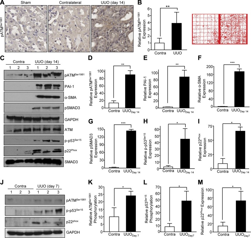
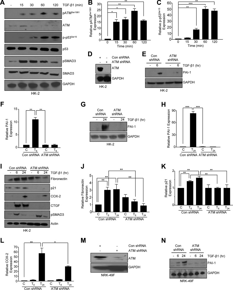
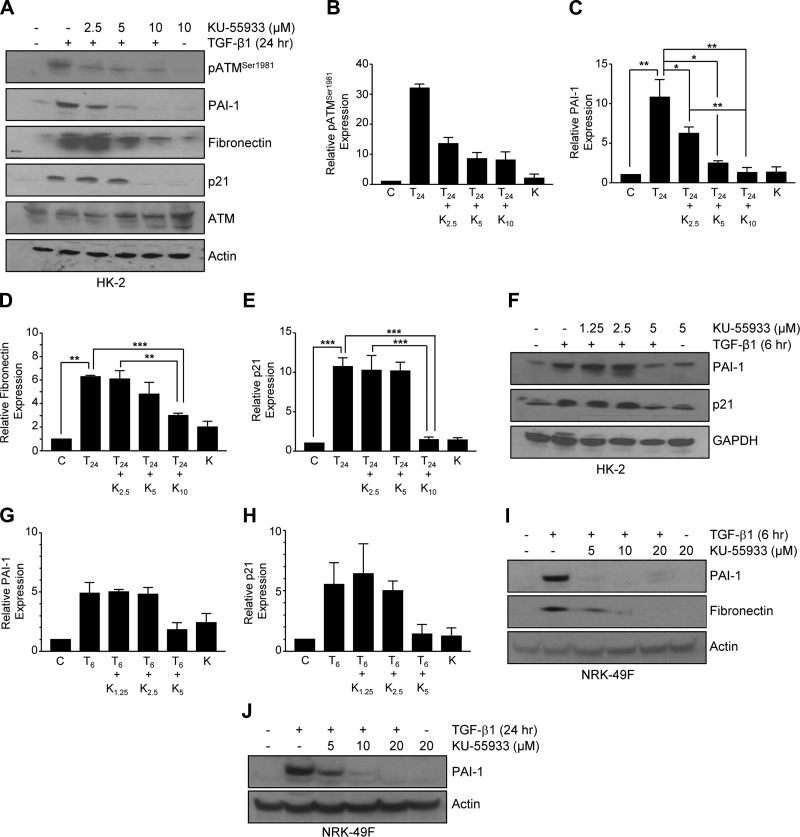
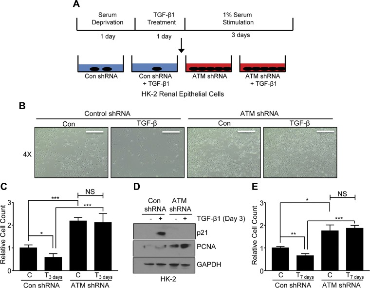
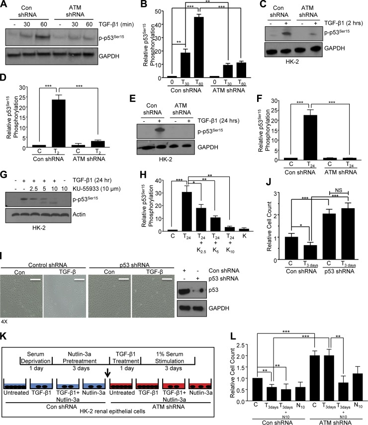
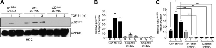
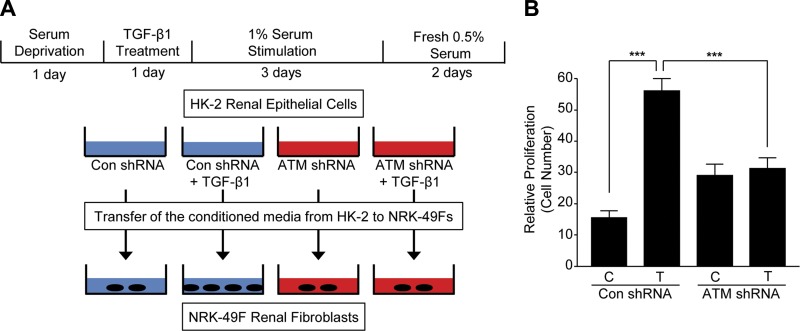
Effective therapy to prevent organ fibrosis, which is associated with more than half of all mortalities, remains elusive. Involvement of tumor suppressor ataxia telangiectasia mutated (ATM) in the TGF-β1 pathway related to renal fibrosis is largely unknown. ATM activation (pATM(Ser1981)) increased 4-fold in the tubulointerstitial region of the unilateral ureteral obstruction-injured kidney in mice correlating with SMAD3 and p53(Ser15) phosphorylation and elevated levels of p22(phox) subunit of the NADPH oxidases (NOXs), and fibrotic markers, plasminogen activator inhibitor-1 (PAI-1), and fibronectin, when compared to contralateral (contra) or sham controls. In fact, ATM is rapidly phosphorylated at Ser(1981) by TGF-β1 stimulation. Stable silencing and pharmacologic inhibition of ATM ablated TGF-β1-induced p53 activation (>95%) and subsequent PAI-1, fibronectin, connective tissue growth factor, and p21 expression in human kidney 2 (HK-2) tubular epithelial cells and normal rat kidney-49 fibroblasts (NRK-49F). ATM or p53 depletion in HK-2 cells, moreover, bypassed TGF-β1-mediated cytostasis evident in control short hairpin RNA-expressing HK-2 cells. Interestingly, stable silencing of NOX subunits, p22(phox) and p47(phox), in HK-2 cells blocked TGF-β1-induced pATM(Ser1981) (>90%) and target gene induction via p53-dependent mechanisms. Furthermore, NRK-49F fibroblast proliferation triggered by conditioned media from TGF-β1-stimulated, control vector-transfected HK-2 cells decreased (∼ 50%) when exposed to conditioned media from ATM-deficient, TGF-β1-treated HK-2 cells. Thus, TGF-β1 promotes NOX-dependent ATM activation leading to p53-mediated fibrotic gene reprogramming and growth arrest in HK-2 cells. Furthermore, TGF-β1/ATM-initiated paracrine factor secretion by dysfunctional renal epithelium promotes interstitial fibroblast growth, suggesting a role of tubular ATM in mediating epithelial-mesenchymal cross-talk highlighting the translational benefit of targeting the NOX/ATM/p53 axis in renal fibrosis.

Keywords: NOX; ROS; kidney fibrosis; p53.


Figures

Figure 1.
Figure 1.
Increased activation of ATM in the fibrotic kidney following UUO in mice. A) Obstructed (UUO) and contra control kidneys were removed from mice following 14 d of experimental UUO. Paraffin kidney sections were stained with phospho-ATMSer1981 (pATMSer1981). Sham kidneys derived from mice receiving a surgical incision without kidney manipulation served as an additional control. Scale bars, 60 μm. B) Histogram depicts the expression level of pATMSer1981 quantified from 56 adjacent areas (see red inset; 7 × 8 grid, 100 μm per square) of the contra and UUO mouse kidneys. Statistical significance was determined using the Student’s t test; **P < 0.01. C) Western blot analysis for pATMSer1981, total ATM, pSMAD3, total SMAD3, p-p53Ser15, p22phox expression, and profibrotic markers (α-SMA and PAI-1) in 3 individual kidney lysates from Contra and UUO-injured mice following 14 d of ureteral ligation. GAPDH is a loading control. DI) Western blot analysis (C) quantified in graph by calculating the mean ± sd for protein levels in obstructed compared to Contra kidney in 3 individual mice (n = 3). J) Immunoblotting of pATMSer1981, p-p53Ser15, and p22phox expression in kidney lysates from Contra and obstructed (UUO) mouse kidneys after 7 d of ureteral ligation. KM) Graphs depict the mean ± sd of protein expression patterns from (J). *P < 0.05; **P < 0.01; ***P < 0.001.
Figure 2.
Figure 2.
Activation and requirement of ATM in TGF-β–induced gene expression in renal tubular epithelial cells and fibroblasts. A) Immunoblotting for pATMSer1981, phospho-p53Ser15 (p-p53Ser15), and pSMAD3 provided an assessment of activation kinetics following a time course of TGF-β1 stimulation (0–120 min). Histograms in (B) and (C) measure pATMSer1981 and p-p53Ser15, respectively, determined by the mean ± sd. *P < 0.05, **P < 0.01, and ***P < 0.001 vs. untreated controls. DH) Con shRNA-expressing or ATM shRNA–expressing HK-2 renal epithelial cells remained untreated or treated with TGF-β1 for 6 (E) or 24 h (G) and immunoblotted for PAI-1. Knockdown efficiency of ATM was confirmed in HK-2 cells (D). F, H) Plotted data of (E) and (G) represent the mean ± sd for TGF-β1–induced PAI-1 expression arbitrarily setting levels in untreated cells with con shRNA as 1. **P < 0.01; ***P < 0.001. IL) Western blot analysis of TGF-β1–dependent profibrotic gene expression in ATM-depleted cells compared to mock controls. TGF-β1–mediated SMAD3 activation is evident in both con shRNA and ATM shRNA-expressing cells. Graphs depict the mean ± sd of fibronectin (I, J), p21 (I, K), and COX-2 (I, L) in ATM knockdown cells compared to their control counterparts where untreated, con shRNA was arbitrarily set as 1. *P < 0.05; **P < 0.01. M, N) Expression of PAI-1 protein following stable generation of NRK-49F rat renal fibroblasts expressing control or ATM shRNA in response to TGF-β1 treatment for 6 or 24 h. Efficiency of ATM knockdown in NRK-49Fs was measured by blotting for ATM (M). GAPDH (A, D, E, G, M, N) and actin (I) provided loading controls.
Figure 3.
Figure 3.
Pharmacologic inhibition of ATM blocks TGF-β–stimulated PAI-1 induction in renal tubular epithelial cells and fibroblasts. AH) HK-2 cells were pretreated with KU-55933 at the indicated doses followed by TGF-β1 exposure for 6 (F) or 24 h (A). Western blotting assessed PAI-1, fibronectin, and p21 expression. Inhibitor function was confirmed using pATMSer1981 as a readout. BE, G, H) Histograms depict the expression of PAI-1 (C, G), fibronectin (D), and p21 (E, H) as the mean ± sd in triplicate experiments. *P < 0.05; **P < 0.01; ***P < 0.001. I, J) Effect of TGF-β1–mediated PAI-1 and fibronectin protein levels following pretreatment with KU-55933 in NRK-49F fibroblasts at 6 (I) and 24 h (J). GAPDH (F) and actin (A, I, J) provided the loading controls. In all histograms, untreated HK-2 or NRK-49F controls were arbitrarily set as 1.
Figure 4.
Figure 4.
ATM is a mediator of TGF-β–induced growth arrest in renal tubular epithelial cells. A) Schematic of experimental plan for growth arrest studies in HK-2 cells. Serum-starved (1 d) subconfluent (30%) HK-2 cells were treated with TGF-β for 1 d followed by 1% serum for 3 or 7 d to stimulate proliferation. B) Phase-contrast images of con shRNA– and ATM shRNA–expressing HK-2 cells stimulated with or without TGF-β1 followed by 3 d of growth in 1% serum. C, E) Relative cell count of epithelial cells stimulated with TGF-β for 3 (C) or 7 d (E) was depicted in histograms (mean ± sd) setting relative cell count in untreated con shRNA as 1 at both time points. *P < 0.05; **P < 0.01; ***P < 0.001. D) Immunoblotting of p21 and PCNA expression in HK-2 cells following TGF-β stimulation for 3 d with or without ATM depletion. GAPDH is a loading control. N.S., not significant.
Figure 5.
Figure 5.
ATM is required for p53 phosphorylation downstream of TGF-β in renal epithelial cells. AF) ATM knockdown in HK-2 cells eliminated p53Ser15 phosphorylation in response to TGF-β stimulation at 0.5, 1 (A), 2 (C), or 24 h (E) compared to con shRNA cultures. Graphs (mean ± sd) in (B), (D), and (F) represent the quantification of (A), (C), and (E), respectively, arbitrarily setting p-p53Ser15 levels in untreated con shRNA cells as 1 for both experiments (n = 3). G, H) The effect of KU-55933 pretreatment on TGF-β1–induced p53Ser15 phosphorylation. H) Histogram reflects the dose-dependent reduction in p-p53Ser15 by TGF-β quantified by the mean ± sd of 3 independent experiments setting p-p53Ser15 levels in untreated controls as 1. I) Phase-contrast images of con shRNA and p53 shRNA–expressing HK-2 cells treated with or without TGF-β followed by a 3 d stimulation of 1% serum to promote growth. Western blot analysis confirmed the knockdown efficiency of p53. J) Graphs depict the relative cell count of above cultures (mean ± sd) setting relative cell count in untreated con shRNA as 1. K) Schematic of experimental plan involving the p53 activator Nutlin-3A. Sparsely confluent (30%) HK-2 cells were serum deprived for 1 d, pretreated with Nutlin-3a for 3 d, followed by TGF-β and 1% serum treatment for 3 d. L) Plotted cell count is the mean ± sd of 3 experiments arbitrarily setting cell count in unstimulated, con shRNA–expressing HK-2 cells as 1. GAPDH (A, C, E, I) and actin (G) confirmed equal loading. *P < 0.05; **P < 0.01; ***P < 0.001. N.S., not significant.
Figure 6.
Figure 6.
NOX subunits, p47phox and p22phox, are necessary for TGF-β1–dependent profibrotic gene induction. A) Knockdown efficiency of the p22phox and p47phox subunits was confirmed by Western blot analysis. B) TGF-β1 treatment for 6 or 24 h promoted the expression of PAI-1 in the control cells, but not p22phox-depleted renal epithelial cells, whereas HK-2 cells with p47phox silencing have reduced PAI-1 expression compared to con shRNA in response to TGF-β1. C) PAI-1 expression is shown in the graph (mean ± sd) setting protein levels in untreated, con shRNA as 1. *P < 0.05; **P < 0.01; ***P < 0.001. D) TGF-β1–dependent p21 and fibronectin were ablated in p47phox and p22phox knockdown HK-2 cells. GAPDH (A), actin (B), and ERK2 (D) acted as loading controls.
Figure 7.
Figure 7.
TGF-β1–mediated ATM activation (pATMSer1981) and p53 phosphorylation (p-p53Ser15) require NOX1/NOX2/NOX4. A) Western blot analysis of TGF-β1–mediated p53Ser15 phosphorylation following genetic silencing of p47phox and p22phox in renal epithelial cells. GAPDH (A) confirmed equal loading. B, C) Histograms (mean ± sd) illustrate relative p-p53Ser15 and pATMSer1981 levels, respectively, in p47phox or p22phox-depleted HK-2 cells following TGF-β1 exposure. *P < 0.05; **P < 0.01; ***P < 0.001.
Figure 8.
Figure 8.
Increased renal fibroblast proliferation by conditioned media derived from TGF-β1–stimulated con shRNA-, but not ATM shRNA-, expressing HK-2 cells. A) Schematic of epithelial-fibroblast cross-talk. Con shRNA or ATM-depleted HK-2 cells were treated with TGF-β1 for 1 d followed by additon of 1% serum for 3 d to promote growth arrest, washed with PBS to remove any residual TGF-β1 prior to addition of fresh 0.5% FBS/DMEM for 48 h. Conditioned media isolated from epithelial cells were directly added to semiconfluent NRK-49F fibroblasts at similar cell density for 2 d. B) Plots (mean ± sd) represent relative NRK-49F cell counts of over 100 (2 × 2 mm) fields for each experimental condition. ***P < 0.001.
Figure 9.
Figure 9.
Proposed model of ATM activation downstream of TGF-β signaling in the context of renal biology. TGF-β ligand binding initiates ALK5-dependent signal transduction resulting in the activation of the SMAD2/SMAD3/SMAD4. In a collateral pathway, TGF-β utilizes the p47phox and p22phox-dependent NOXs to generate ROS necessary for ATM activation as well as its downstream substrate p53. p53 and SMAD, in turn, form a complex that binds to TGF-β target gene promoters (e.g., PAI-1) stimulating optimal gene expression. Other ROS-dependent non-SMAD elements, including c-src/EGFR/MAPK, function to recruit additional transcriptional elements as part of a highly interactive canonical and noncanonical TGF-β transcriptional complex.

Similar articles

Cited by

References

    1. Wynn T. A. (2007) Common and unique mechanisms regulate fibrosis in various fibroproliferative diseases. J. Clin. Invest. 117, 524–529 - PMC - PubMed
    1. Friedman S. L., Sheppard D., Duffield J. S., Violette S. (2013) Therapy for fibrotic diseases: nearing the starting line. Sci. Transl. Med. 5, 167sr1. - PubMed
    1. Liu Y. (2011) Cellular and molecular mechanisms of renal fibrosis. Nat. Rev. Nephrol. 7, 684–696 - PMC - PubMed
    1. Kisseleva T., Brenner D. A. (2007) Role of hepatic stellate cells in fibrogenesis and the reversal of fibrosis. J. Gastroenterol. Hepatol. 22(Suppl 1), S73–S78 - PubMed
    1. Lee C. G., Kang H. R., Homer R. J., Chupp G., Elias J. A. (2006) Transgenic modeling of transforming growth factor-beta(1): role of apoptosis in fibrosis and alveolar remodeling. Proc. Am. Thorac. Soc. 3, 418–423 - PMC - PubMed

Publication types

MeSH terms

Substances