. 2015 Mar;100(3):336-44.
doi: 10.3324/haematol.2014.112748. Epub 2014 Dec 5.

Anti-CD33 chimeric antigen receptor targeting of acute myeloid leukemia

Affiliations

Anti-CD33 chimeric antigen receptor targeting of acute myeloid leukemia

Carol O'Hear et al. Haematologica. 2015 Mar.

Abstract

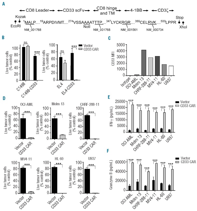
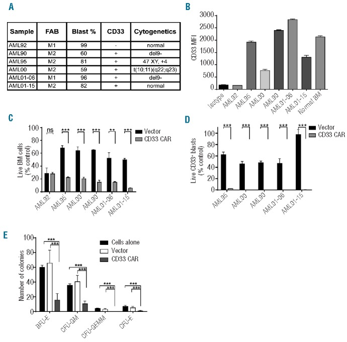
Current therapies for acute myeloid leukemia are associated with high failure and relapse rates. Adoptive immunotherapies, which have shown promise in the treatment of hematologic malignancies, have the potential to target acute myeloid leukemia through pathways that are distinct and complementary to current approaches. Here, we describe the development of a novel adoptive immunotherapy specific for this disease. We generated a second generation CD33-specific chimeric antigen receptor capable of redirecting cytolytic effector T cells against leukemic cells. CD33 is expressed in approximately 90% of acute myeloid leukemia cases and has demonstrated utility as a target of therapeutic antibodies. Chimeric antigen receptor-modified T cells efficiently killed leukemia cell lines and primary tumor cells in vitro. The anti-leukemia effect was CD33-specific, mediated through T-cell effector functions, and displayed tumor lysis at effector:target ratios as low as 1:20. Furthermore, the CD33-redirected T cells were effective in vivo, preventing the development of leukemia after prophylactic administration and delaying the progression of established disease in mice. These data provide pre-clinical validation of the effectiveness of a second-generation anti-CD33 chimeric antigen receptor therapy for acute myeloid leukemia, and support its continued development as a clinical therapeutic.


Figures

Figure 1.
Figure 1.
Anti-CD33 CAR-modified T cells kill tumor cells in vitro. (A) Schematic representation of the anti-CD33 CAR showing the CD8 leader, CD33 single chain variable fragment (scFv), CD8 hinge and transmembrane domains, 4-1BB co-stimulatory domain, and CD3ζ intracellular signaling domain. Restriction sites added during subcloning and Genbank accession numbers for sequence information are shown. (B) Specific cytotoxicity of anti-CD33 CAR or vector-transduced T cells against C1498-CD33 and EL4-CD33 tumor cell lines or their parental CD33-negative controls. Quantitative flow cytometry was performed after co-culture for 24 h at an E:T ratio of 1:2. The percentage of live tumor cells compared with cultures lacking added T cells is plotted. (C) The difference in mean fluorescence intensity (MFI) of studied AML cell lines compared with isotype control staining is plotted. (D) The indicated AML cell lines were co-cultured with anti-CD33 CAR or vector-transduced T cells at an E:T ratio of 1:2 for 24 h and analyzed as in (B). Supernatants from co-cultures of AML cell lines with either vector-transduced or anti-CD33 CAR-transduced T cells were analyzed by enzyme-linked immunosorbent assay for interferon-γ (E) and granzyme B (F). Means + 1 SD are plotted. Data shown are representative of three or more experiments. ***P<0.001.
Figure 2.
Figure 2.
Anti-CD33 CAR- modified T cells mediate tumor cell killing at low E:T ratios. Molm-13 (A) and Mv4-11 (B) cell lines were incubated with either anti-CD33 CAR T cells or vector-transduced T cells at the indicated E:T ratios for 24 or 48 h. Residual viable tumor cells were quantified by flow cytometry and the results expressed as percentage of live tumor cells identified in cultures without added T cells. Means of triplicate wells ± 1 SD are shown. Data are representative of three experiments.
Figure 3.
Figure 3.
Anti-CD33 CAR-modified T cells target primary AML blasts and normal CD33+ cord blood cells in vitro. (A) French-American-British classification, blast percentage, CD33 status, and cytogenetics of primary AML bone marrow samples are shown. (B) The mean fluorescent intensity (MFI) of studied primary AML samples and normal bone marrow stained for CD33 is plotted. Primary tumor samples listed in (A) were cultured alone or with anti-CD33 CAR-modified or vector-transduced T cells at a 1:2 E:T ratio for 24 h. Residual live bone marrow (BM) cells (C) and CD33+ blasts (D) were quantified by flow cytometry and results expressed as percentage of identically gated cells from cultures without T cells. Results represent the mean of triplicate wells + 1 SD (E) CD34+ cord blood cells were incubated alone or with an equivalent number of vector-transduced or anti-CD33 CAR T cells for 24 h. The mixture was plated on methylcellulose medium and colonies were enumerated at 14 days. Results represent the mean of three experiments, each performed in duplicate (E). ***P<0.001.
Figure 4.
Figure 4.
Anti-CD33 CAR-modified T cells prevent AML development in NSG mice. NSG mice received 1×10⁶ Molm-13-luc cells intravenously followed by retro-orbital transfer of saline, 10×106 anti-CD33 CAR-modified or vector-transduced T cells. Mice were sacrificed on day 20 and infiltrating YFP+CD33+ tumor cells determined by flow cytometry. (A) Number of tumor cells in the blood, bone marrow, liver, and spleen in mice treated with saline, vector-transduced T cells, or anti-CD33 CAR T cells. Each open circle represents one animal and means are indicated by horizontal lines. Results are pooled from two experiments. (B) Representative flow cytometry plots demonstrating CD33+ tumor presence in the indicated tissues. **P<0.01, ***P<0.001.
Figure 5.
Figure 5.
Anti-CD33 CAR-modified T cells exhibit anti-leukemic effects in an AML treatment model. On day 0, NSG mice were inoculated intravenously with 1×10⁶ Molm-13-luc cells. Four days later, at which time substantial tumor burden was evident by bioluminescence imaging, 10×106 anti-CD33 CAR T cells, vector-transduced T cells, or saline was administered. (A) Images of mice from one experiment are shown. (B) Kaplan-Meier analysis of each treatment group. (C) Bioluminescent signal intensities are plotted. Data (B, C) are pooled from two experiments and (C) mean values from each group ± 1 SD are shown. ***P<0.001.

References

    1. Dohner H, Estey EH, Amadori S, et al. Diagnosis and management of acute myeloid leukemia in adults: recommendations from an international expert panel, on behalf of the European LeukemiaNet. Blood. 2010;115(3):453–474. - PubMed
    1. Woods WG, Neudorf S, Gold S, et al. A comparison of allogeneic bone marrow transplantation, autologous bone marrow transplantation, and aggressive chemotherapy in children with acute myeloid leukemia in remission. Blood. 2001;97(1):56–62. - PubMed
    1. Rubnitz JE, Lensing S, Zhou Y, et al. Death during induction therapy and first remission of acute leukemia in childhood: the St. Jude experience. Cancer. 2004;101(7):1677–1684. - PubMed
    1. Jin L, Hope KJ, Zhai Q, Smadja-Joffe F, Dick JE. Targeting of CD44 eradicates human acute myeloid leukemic stem cells. Nat Med. 2006;12(10):1167–1174. - PubMed
    1. Majeti R, Chao MP, Alizadeh AA, et al. CD47 is an adverse prognostic factor and therapeutic antibody target on human acute myeloid leukemia stem cells. Cell. 2009; 138(2):286–299. - PMC - PubMed

Publication types

MeSH terms

Substances