. 2015 Jan 15;194(2):606-14.
doi: 10.4049/jimmunol.1402383. Epub 2014 Dec 5.

Natural anti-intestinal goblet cell autoantibody production from marginal zone B cells

Affiliations

Natural anti-intestinal goblet cell autoantibody production from marginal zone B cells

Daiju Ichikawa et al. J Immunol. .

Abstract

Expression of a germline VH3609/D/JH2 IgH in mice results in the generation of B1 B cells with anti-thymocyte/Thy-1 glycoprotein autoreactivity by coexpression of Vk21-5/Jk2 L chain leading to production of serum IgM natural autoantibody. In these same mice, the marginal zone (MZ) B cell subset in spleen shows biased usage of a set of Ig L chains different from B1 B cells, with 30% having an identical Vk19-17/Jk1 L chain rearrangement. This VH3609/Vk19-17 IgM is reactive with intestinal goblet cell granules, binding to the intact large polymatrix form of mucin 2 glycoprotein secreted by goblet cells. Analysis of a μκ B cell AgR (BCR) transgenic (Tg) mouse with this anti-goblet cell/mucin2 autoreactive (AGcA) specificity demonstrates that immature B cells expressing the Tg BCR become MZ B cells in spleen by T cell-independent BCR signaling. These Tg B cells produce AGcA as the predominant serum IgM, but without enteropathy. Without the transgene, AGcA autoreactivity is low but detectable in the serum of BALB/c and C.B17 mice, and this autoantibody is specifically produced by the MZ B cell subset. Thus, our findings reveal that AGcA is a natural autoantibody associated with MZ B cells.


Figures

FIGURE 1.
FIGURE 1.
MZ B cell–associated BCR, VH3609/Vk19-17. (A) Analysis of B cell subsets (gated rectangles color coded on plots) in the peritoneal cavity washout cells (PerC) and spleen from a VH3609t mouse. The VH3609id+ cell gate is marked by a vertical line. The majority of 13H8khi/med Igκ B cells are also VH3609id+ in the MZ B subset (arrow). (B) Igκ single-cell sequence data from MZ B and FO B subsets of four individual VH3609t mice. Percentages are from a total of 25–36 samples per mouse B cell subset gated as VH3609id(10C7)+Igλ. Three Igκs enriched in the MZ B cell subset, in contrast with FO B cells (empty bars), are shown. Vk19-17/Jk1 Ig L chains used by individual MZ B cells were all identical. (C) Selected Igκ-chain usage data comparing the immature, FO B, and MZ B cells in VH3609μTg mice (all data are provided in Supplemental Table I). Confirmation of highest usage of identical Vk19-17/Jk1 Ig L chains by MZ B cells (in CDR3, the Vk-Jk junction encodes a proline [P] residue).
FIGURE 2.
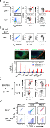
FIGURE 2.
Btk-dependent MZ B cell development by VH3609/Vk19-17 BCR B cells. (A) Spleen B cell FACS analysis of the VH3609/Vk19-17 μκTg mouse line, EP67, at 3 wk of age. The CD21hi B cell area is marked by red dotted line. Predominance of CD21hi pre-MZ B and MZ B cells in Tg+ mice (starred) relative to Tg littermate. (B) FACS analysis of 2 mo adult EP67 spleen B cells. Tgμκ+ B cells, the predominant population, comprise 40–50% in spleen, and half become either FO B or MZ B cells. MZ B cell phenotype cells are colocalized in the MZ with SIGNR1+ macrophages (ER-TR9+), and FO B cell phenotype cells are present in B220+ follicles with CD21hi FO DCs (20× objective lens). Quantitative PCR analysis of selected genes was compared in sorted VH3609id+ MZ B and FO B cells from EP67 mice (n = 3 each, mean and SE) with wild type C.B17 mouse FO B cells. (C) Mixed cell transfer of adult mouse BM hematopoietic stem cell–enriched fractions using a 1:10 Tgμκ EP67/non-Tg ratio. Six weeks after transfer, spleen B cell FACS analysis was performed, gating for VH3609id+ (Tg+, IgMa+) cells (9% of total B cells) and VH3609id (Tg, IgMb+) B cells. Representative data of four recipients (n = 4) from two separate transfer experiments are shown. All mice showed predominant MZ B cell generation by the Tg+ B cells. Transfer of EP67 BM alone generated both MZ B and FO B cells, as found in intact adult EP67 mice (data not shown). (D) Analysis of BM AA4+ immature B and spleen B cells in 2-mo-old EP67 μκTg mice without or with Xid (Btk mutant) background. In contrast with BM, in spleen there is a reduction of total B cell numbers (2-fold lower in Xid), together with reduction of μκTg B cell frequency, and an absence of VH3609/Vk19-17 MZ B cells (red ellipse region) in EP67.Xid mice. Representative data are shown from analyses of four mice.
FIGURE 3.
FIGURE 3.
AGcA by MZ B cell–associated VH3609/Vk19-17 IgM (MK19). (A) Colon mucosal cell suspension was stained by MK19, in contrast with thymus cell suspension that was stained by MK21. (B) Cryosection staining of colon. Goblet cells and contiguous luminal mucus were stained with MK19, but not MK21. IB4 (BSI-B4) using a 10× objective lens. (C) MK19 staining of transverse sections of intestines and stomach using a 20× objective lens. (D) Colon goblet cells (horizontal section) were shown to be PAS+ (paraffin section staining). MK19-stained granules in the goblet cells, which were distinct from PNA and IB4 bound supranuclear areas and nuclei stained by DAPI, as assessed using a 20× objective lens. (E) Apical goblet cell staining by MK19 was similar to DBA lectin binding in colon. MK19 staining (bottom middle) was merged with PNA and WGA lectins (top middle). (F) MK19 staining of Muc1- and Muc2-deficient mouse colon demonstrates predominantly Muc2-dependent binding of MK19. (G) Absence of MK19 staining of rat colon goblet cells. (E)–(G) using a 10× objective lens.
FIGURE 4.
FIGURE 4.
Predominant MK19 AGcA binding to intact Muc2 glycoprotein. (A) Western blot of colon NP-40 lysate. Mucus released from crypts by centrifugation was suspended in 1% NP-40 and applied to 3.3% SDS-PAGE with a 2.5% stacking gel for detection of glycosylated colon mucin(s). Comparison of wild-type B6 versus Muc2KO.B6. Anti-Muc2 (N terminus peptide). (B) Colonic crypts from Rag1 null mice were incubated with MK19 (or MK21), cross-linked (CL) with DSP, then suspended in NP-40 buffer for SDS-PAGE. Left set is Western blot of IgM H chain band using soluble and insoluble lysate, showing the presence of MK19 IgM CL colon granules detected by anti-IgM. Right set of the same lysates preincubated with MK19 shows copresence of Muc2. MK19 IgM and Muc2 were both predominantly in the insoluble fraction. (C) Soluble material from MK19 IgM prebound (MK21 as a control) colon granules, followed by cross-linking, was incubated with anti-IgM–bound protein G Dynabeads for 18 h, then washed, eluted, and applied to SDS-PAGE analysis. Presence of MK19 bound to ∼600 kDa DBA+Muc2 is shown. IB, immunoblot; LB, lectin blot.
FIGURE 5.
FIGURE 5.
Natural AGcA IgM production from MZ B cells. (A) The presence of AGcA PCs (high IgMa+ and CD19) in EP67 mouse spleen. CD19+ B cells are also stained with IgMa+ at a lower level than PCs. (B) Predominant AGcA IgM in serum of EP67 mice was independent of T cells (EP67.RagKO). μκTg IgM is IgMa on a C.B17 (IgMb) mouse background. n = 9 each. (C) Colon and thymus cryosection staining comparison between EP67 AGcA μκTg mouse and 3369 ATA μκTg mouse sera, both with Tg IgMa predominance in serum. (D) AGcA reactivity by IgM produced from normal mouse MZ B cells. Colon from Rag1 null mice stained by LPS-stimulated B cell culture supernatant IgM (adjusted to 2–10 μg/ml) from BALB/c mice. B1 (B1a) B cells in the peritoneal cavity and MZ B cells, and FO B cells in spleen were purified from the same mouse. Representative of three experiments (two with BALB/c, one with C.B17). (A), (C), and (D) using a 20× objective lens. RP, red pulp; WP, white pulp.
FIGURE 6.
FIGURE 6.
Natural AGcA autoantibody in intestine. (A) The presence of AGcA B cells (IgMa+) in isolated lymphoid follicles in EP67 mouse colon, and absence of goblet cell prebound AGcA IgM (upper panel), unless the tissue section is first incubated with serum from EP67 μκTg mice containing AGcA autoantibody (lower panel). (B) Analysis of DSS-treated (8 d after ceasing DSS treatment) EP67 and 3369 mouse intestine. AGcA (but not ATA) plasma cells are increased in intestinal lamina propria (selected area, enlarged at right). (C) DSS-treated EP67 colon section staining (8 d after ceasing DSS treatment). AGcA B cell follicle (iLF, right side of the IgMa-stained figure) and AGcA PC infiltration into the damaged region, in comparison with the PNA+ undamaged goblet cell area. (A) and (C) using a 20× objective lens. (B) using a 10× and 20× objective lens.

References

    1. Boyden S. V. 1966. Natural antibodies and the immune response. Adv. Immunol. 5: 1–28. - PubMed
    1. Avrameas S. 1991. Natural autoantibodies: from ‘horror autotoxicus’ to ‘gnothi seauton’. Immunol. Today 12: 154–159. - PubMed
    1. Coutinho A., Kazatchkine M. D., Avrameas S. 1995. Natural autoantibodies. Curr. Opin. Immunol. 7: 812–818. - PubMed
    1. Merbl Y., Zucker-Toledano M., Quintana F. J., Cohen I. R. 2007. Newborn humans manifest autoantibodies to defined self molecules detected by antigen microarray informatics. J. Clin. Invest. 117: 712–718. - PMC - PubMed
    1. Ehrenstein M. R., Notley C. A. 2010. The importance of natural IgM: scavenger, protector and regulator. Nat. Rev. Immunol. 10: 778–786. - PubMed

Publication types

MeSH terms

LinkOut - more resources