. 2015 Jan 1;194(1):68-75.
doi: 10.4049/jimmunol.1401653. Epub 2014 Dec 5.

Th1/Th17 plasticity is a marker of advanced β cell autoimmunity and impaired glucose tolerance in humans

Collaborators, Affiliations

Th1/Th17 plasticity is a marker of advanced β cell autoimmunity and impaired glucose tolerance in humans

Linnea Reinert-Hartwall et al. J Immunol. .

Abstract

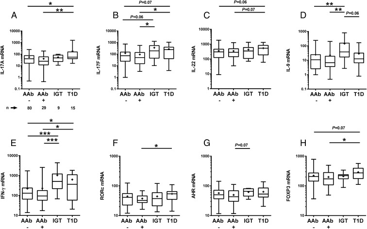
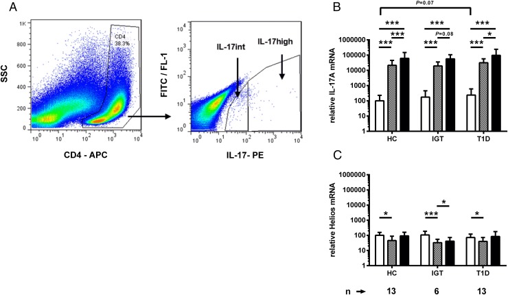
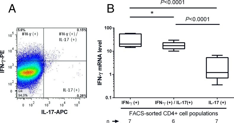
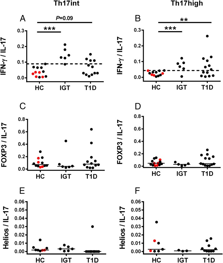
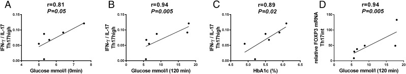
Upregulation of IL-17 immunity and detrimental effects of IL-17 on human islets have been implicated in human type 1 diabetes. In animal models, the plasticity of Th1/Th17 cells contributes to the development of autoimmune diabetes. In this study, we demonstrate that the upregulation of the IL-17 pathway and Th1/Th17 plasticity in peripheral blood are markers of advanced β cell autoimmunity and impaired β cell function in human type 1 diabetes. Activated Th17 immunity was observed in the late stage of preclinical diabetes in children with β cell autoimmunity and impaired glucose tolerance, but not in children with early β cell autoimmunity. We found an increased ratio of IFN-γ/IL-17 expression in Th17 cells in children with advanced β cell autoimmunity, which correlated with HbA1c and plasma glucose concentrations in an oral glucose tolerance test, and thus impaired β cell function. Low expression of Helios was seen in Th17 cells, suggesting that Th1/Th17 cells are not converted thymus-derived regulatory T cells. Our results suggest that the development of Th1/Th17 plasticity may serve as a biomarker of disease progression from β cell autoantibody positivity to type 1 diabetes. These data in human type 1 diabetes emphasize the role of Th1/Th17 plasticity as a potential contributor to tissue destruction in autoimmune conditions.


Figures

FIGURE 1.
FIGURE 1.
Gene expression levels in anti-CD3– and anti-CD28–stimulated PBMCs in the autoantibody-negative children (AAb, n = 80), the children with early β cell autoimmunity (AAb+, n = 29), children with advanced β cell autoimmunity (IGT, n = 9), and in children with type 1 diabetes (n = 15). Relative mRNA levels of IL-17A (A), IL-17F (B), IL-22 (C), IL-9 (D), IFN-γ (E), RORc (F), AHR (G), and FOXP3 (H). The boxes represent lower and higher quartiles. Horizontal line in the box represents the median value and whiskers represent minimum and maximum values. Plus sign in the box represents the mean value of the group. The p values were calculated with the Student t test for unpaired samples. Gene expression levels were analyzed with RT-qPCR. Relative gene expression was assessed for each marker by comparing ΔCt value of each sample to the ΔCt value of the calibrator. *p < 0.05, **p < 0.01, ***p < 0.001.
FIGURE 2.
FIGURE 2.
(A) Gating strategy for FACS sorting of Th17 cells. IL-17–secreting CD4+ cells were stained with a cytokine capture assay and identified based on the IL-17–PE intensity of the CD4+ gated lymphocyte population and sorted into Th17high and Th17int cells. Dead autofluorescent cells were excluded from the sorting based on autofluorescence signal detected on FITC/FL-1 channel. (B) IL-17A mRNA expression is remarkably higher in Th17int (gray bars) and Th17high (black bars) cell fractions in comparison with anti-CD3– and anti-CD28–stimulated PBMCs (white bars). (C) mRNA of Helios, the marker of Tregs, was not enriched in sorted Th17 cells. Bars represent mean and whiskers SD. Relative gene expression of the markers was calculated by comparing ΔCt values of IL-17A and Helios to the average ΔCt value of IL-17A in the stimulated PBMCs of healthy controls. Comparison of different cell populations within the study group was performed with the Student t test for paired samples. Differences between the study groups within a cell population were compared with the Student t test for unpaired samples. *p < 0.05, ***p < 0.001.
FIGURE 3.
FIGURE 3.
IFN-γ mRNA expression level in the IFN-γ– and/or IL-17–secreting CD4+ cells sorted from anti-CD3– and anti-CD28–stimulated PBMCs. (A) Frozen PBMCs were thawed and stimulated with anti-CD3 and anti-CD28 for 72 h and sorted with FACS into CD4+IFN-γ+, CD4+IFN-γ+IL-17+, and CD4+IL-17+ cells. IFN-γ mRNA expression in sorted cell fractions was analyzed with RT-qPCR. A representative FACS plot of IFN-γ and IL-17 protein expression in CD4+ cells of a child with advanced β cell autoimmunity and IGT is shown. (B) The IFN-γ mRNA expression was highest in the FACS-sorted CD4+ cells secreting IFN-γ alone (n = 7), being somewhat lower in CD4+ cells cosecreting of IFN-γ and IL-17 (n = 6) and lowest in the CD4+ cells secreting IL-17 alone (n = 7). Dead cells were excluded from the analysis by 7-aminoactinomycin D staining of the dual-labeled cells. The p values were calculated with the Student t test for paired samples. *p < 0.05.
FIGURE 4.
FIGURE 4.
The plasticity of sorted Th17 cells was evaluated by calculating the ratio of IFN-γ/IL-17A, FOXP3/IL-17A, and Helios/IL-17A mRNA in Th17int (left column) and Th17high cells (right column) in healthy children (HC), children with advanced β cell autoimmunity and IGT, and in children with type 1 diabetes. Red symbols in the HC group denote the subjects with early β cell autoimmunity. (A and B) The highest IFN-γ/IL-17 ratio in Th17 cells of the healthy children was used as a cutoff for increased Th17 plasticity (dashed line). An increased ratio of IFN-γ/IL-17 in Th17high and Th17int cells was seen in the children with advanced β cell autoimmunity and in the children with type 1 diabetes in comparison with HC. (CF) FOXP3/IL-17 and Helios/IL-17 ratios in sorted Th17 cells were comparable between the study groups. Statistical significance was calculated with the Fisher exact test. Horizontal lines represent the median ratio. **p < 0.01, ***p < 0.001.
FIGURE 5.
FIGURE 5.
Children with advanced β cell autoimmunity and IGT have an increased IFN-γ/IL-17 ratio in Th17‑ cells, which correlates with the parameters reflecting β cell function (AC). (A) Fasting plasma glucose concentration correlated with IFN-γ/IL-17 ratio in Th17high cells in children with advanced β cell autoimmunity. (B) IFN-γ/IL-17 ratio in Th17high cells correlated with plasma glucose concentrations at 120 min in the OGTT in children with advanced β cell autoimmunity. (C) HbA1c levels correlated with the IFN-γ/IL-17 ratio in Th17high cells in children with advanced β cell autoimmunity. (D) Statistically significant correlation between the expression of FOXP3 mRNA in Th17int cells and the 120 min glucose concentration in the OGTT was observed in children with advanced β cell autoimmunity. Correlations were calculated with a nonparametric Spearman’s test.

References

    1. Bottazzo G. F., Florin-Christensen A., Doniach D. 1974. Islet-cell antibodies in diabetes mellitus with autoimmune polyendocrine deficiencies. Lancet 304: 1279–1283. - PubMed
    1. MacCuish A. C., Irvine W. J., Barnes E. W., Duncan L. J. 1974. Antibodies to pancreatic islet cells in insulin-dependent diabetics with coexistent autoimmune disease. Lancet 304: 1529–1531. - PubMed
    1. Foulis A. K., McGill M., Farquharson M. A. 1991. Insulitis in type 1 (insulin-dependent) diabetes mellitus in man—macrophages, lymphocytes, and interferon-gamma containing cells. J. Pathol. 165: 97–103. - PubMed
    1. Kallmann B. A., Hüther M., Tubes M., Feldkamp J., Bertrams J., Gries F. A., Lampeter E. F., Kolb H. 1997. Systemic bias of cytokine production toward cell-mediated immune regulation in IDDM and toward humoral immunity in Graves’ disease. Diabetes 46: 237–243. - PubMed
    1. Emamaullee J. A., Davis J., Merani S., Toso C., Elliott J. F., Thiesen A., Shapiro A. M. 2009. Inhibition of Th17 cells regulates autoimmune diabetes in NOD mice. Diabetes 58: 1302–1311. - PMC - PubMed

Publication types

MeSH terms