. 2015 Jan 30;290(5):2659-69.
doi: 10.1074/jbc.M114.610824. Epub 2014 Dec 5.

The TRPM8 protein is a testosterone receptor: I. Biochemical evidence for direct TRPM8-testosterone interactions

Affiliations

The TRPM8 protein is a testosterone receptor: I. Biochemical evidence for direct TRPM8-testosterone interactions

Swapna Asuthkar et al. J Biol Chem. .

Abstract

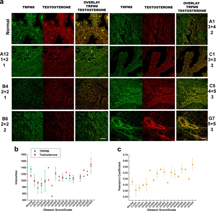
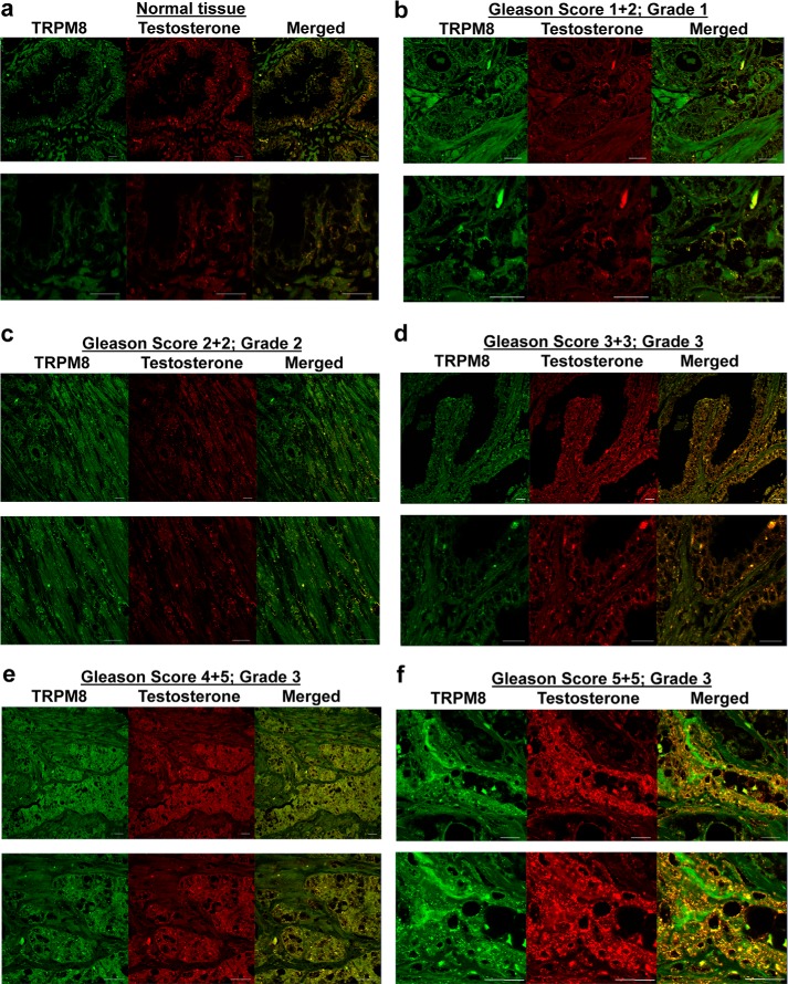
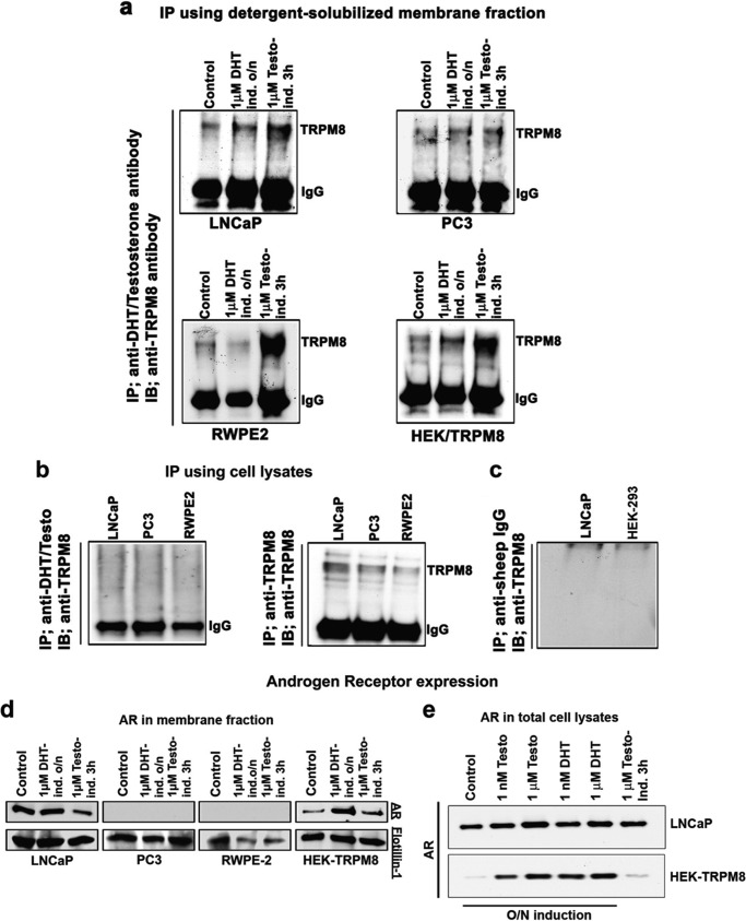
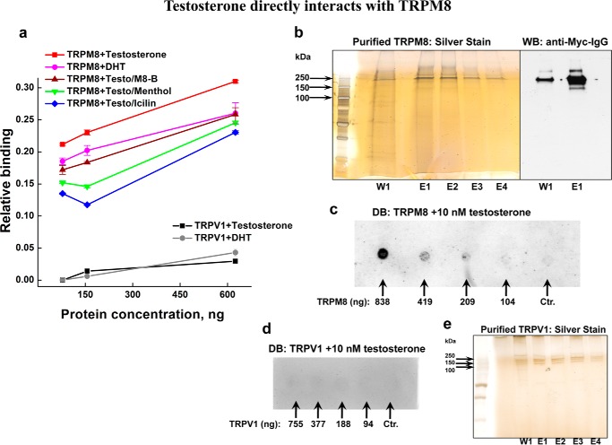
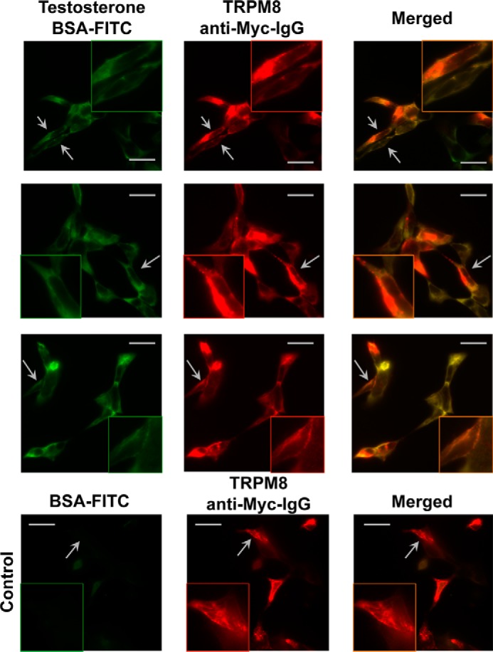
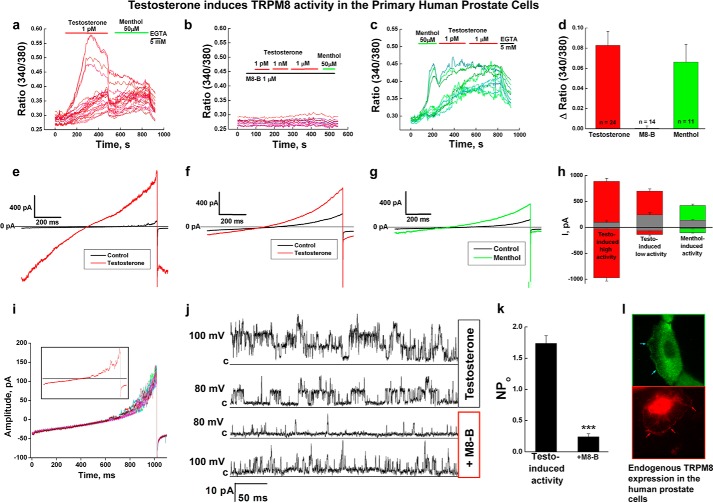
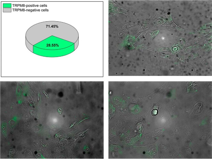
The transient receptor potential ion channel of the melastatin subfamily, TRPM8, is a major cold receptor in the peripheral nervous system. Along with the sensory neurons, the TRPM8 protein is highly expressed in the prostate epithelial cells, and this expression is regulated by androgens. Here we investigated the expression and intracellular localization of the TRPM8 channel in relationship to androgens. We performed experiments using human prostate tissues obtained from healthy individuals and patients with prostate cancer at various stages of the disease as well as in cultured cells. Using an immunohistochemistry approach, we detected an intensive colocalization pattern of the TRPM8 protein with endogenous androgens in all tissues tested, suggesting possible interactions. Co-immunoprecipitation experiments performed using cultured prostate epithelial cells, prostate cancer cells, and HEK-293 cells stably expressing TRPM8 further confirmed direct binding of the steroid hormone, testosterone, to the TRPM8 protein. Applications of picomolar concentrations of testosterone to the primary human prostate cells, endogenously expressing TRPM8, elicited Ca(2+) responses and channel currents, and those were inhibited in the presence of TRPM8 antagonist, N-(2-aminoethyl)-N-(4-(benzyloxy)-3-methoxybenzyl)thiophene-2-carboxamide hydrochloride. These results indicate that the TRPM8 channel is physically associated with testosterone and suggest that, in addition to a genomic role, testosterone plays a role in direct regulation of the TRPM8 channel function.

Keywords: Androgen; Calcium Channel; Cold and Menthol Receptor TRPM8; Ion Channel; Testosterone; Transient Receptor Potential Channels (TRP Channels); Transient Receptor Potential Melastatin 8 Channel.


Figures

FIGURE 1.
FIGURE 1.
Testosterone colocalization with the TRPM8 protein in human prostate tissues. Shown is immunohistochemistry analysis of a prostate cancer tissue microarray containing 60 cases (180 cores) of prostate adenocarcinoma (grades 1–4) and nine cases (27 cores) of normal prostate tissues using Alexa Fluor anti-TRPM8 (green) and anti-testosterone/DHT (red) secondary antibodies, as described under “Experimental Procedures.” Microscopic examination was performed (Zeiss LSM 510 upright confocal microscope) at ×40 magnification. a, immunohistochemical detection of the TRPM8 protein and testosterone obtained from the tissue array. b, relative intensities obtained for the TRPM8 protein (green) and testosterone/DHT (red). The values were averaged from the samples obtained from the same patients and from several regions within one sample because prostate cancer tissues are highly heterologous. The Pearson coefficients are plotted against the Gleason score/grade in c. The Pearson coefficient was calculated in ImageJ using the colocalization finder plugin to evaluate the relative colocalization pattern between TRPM8 and testosterone. Scale bar, 20 μm. Error bars, S.E.
FIGURE 2.
FIGURE 2.
Testosterone colocalization with TRPM8 in human prostate tissues. Images additional to and enlarged from those presented in Fig. 1 show immunohistochemistry analysis of the prostate cancer tissue microarray (US Biomax) containing 60 cases (180 cores) of prostate adenocarcinoma (grades 1–4) and nine cases (27 cores) of normal prostate tissues using Alexa Fluor anti-TRPM8 (green) and anti-testosterone/DHT (red) secondary antibodies as described under “Experimental Procedures.” Microscopic examination was done (Zeiss LSM 510 upright confocal microscope) at ×40 magnification. a, representative images obtained from normal prostate; b, prostate cancer tissues (Gleason score 1 + 2, grade 1); c, Gleason score 2 + 2, grade 2; d, Gleason score 3 + 3, grade 3; e, Gleason score 4 + 5, grade 3; f, Gleason score 5 + 5, grade 3. The bottom panels are enlarged images of the top panels. Scale bars, 20 μm.
FIGURE 3.
FIGURE 3.
Testosterone directly binds to the TRPM8 protein. Shown are IP and immunoblot (IB) analysis using the membrane extracts of LNCaP, PC3, RWPE-2, and HEK-TRPM8 control and treated (DHT and testosterone) cells. a, IP was performed using the detergent-solubilized fractions, containing 0.1% Nonidet P-40 and 0.5% dodecylmaltoside. The membrane fractions were obtained from the epithelial prostate cells RWPE-2, prostate cancer cells PC3 and LNCaP, and HEK-293 stably expressing TRPM8. To extract protein into detergent micelles, the membrane fractions were incubated overnight at 4 °C on a shaker. IP was performed using a sheep anti-testosterone/DHT antibody (Fisher), and the eluate was immunoprobed using a rabbit anti-TRPM8 antibody to show the association of testosterone/DHT with TRPM8 as described under “Experimental Procedures.” The protein band that corresponds to TRPM8 monomer was detected at ∼130 kDa. IgG denotes the anti-IgG heavy chain antibody control in the IP assays. b, IP performed on the total cell lysates did not result in detectable levels of TRPM8 with testosterone. c, IP was performed using negative control anti-sheep IgG, and the immunoprecipitated proteins were immunoblotted against anti-TRPM8 IgG with the secondary anti-rabbit antibody. d, the immunoblots from the membrane fractions were developed using antibodies specific for the AR; flotillin was used as a loading control for detergent-solubilized membrane fractions. e, AR in total cell lysates of LNCaP and HEK-293 cells after androgen treatment: Western blot analysis for the AR protein using 40 μg of total cell lysates from control-, testosterone-, and DHT-treated LNCaP and HEK-293 cells stably expressing TRPM8. The blots are representatives from three independent experiments. O/N, overnight.
FIGURE 4.
FIGURE 4.
Direct interaction between the purified TRPM8 protein and testosterone in the presence or absence of the TRPM8-specific inhibitor M8-B and the TRPM8 agonists menthol and icilin. a, ELISA plates coated with TRPM8 or TRPV1 (78, 156, or 625 ng/well) were incubated with various TRPM8 agonist/antagonists (200 nm testosterone, 200 nm DHT, 1 μm M8-B, 50 μm menthol, or 10 μm icilin) at various dilutions in PBS as mentioned under “Experimental Procedures.” After washing, binding of testosterone or DHT to immobilized TRPM8 or TRPV1 was detected with a sheep polyclonal anti-testosterone/DHT antibody. The binding of testosterone/DHT to TRPM8 and TRPV1 is represented graphically. The negative control anti-IgG did not show any binding to immobilized TRPM8 and TRPV1. The OD values were deducted from the test values. b, silver staining and Western blot (WB) of the purified TRPM8 samples. TRPM8 protein was eluted with NCB buffer containing Myc peptide (50 μg/ml), 0.1% Nonidet P-40, and 0.03% LMNG. TRPM8 shows high stability in the presence of LMNG, and even after incubating with the SDS-loading buffer for 10 min at 95 °C, it runs on the SDS-gel preferably in the form of dimer. The silver-stained gel demonstrates wash 1 (W1), done with NCB/Nonidet P-40/LMNG buffer; eluates 1–4 (E1–E4) were obtained with the NCB/Nonidet P-40/LMNG elution buffer containing Myc peptide. c, dot blot (DB) was done on the purified TRPM8 protein treated with 10 nm testosterone. 2 μl of TRPM8 (in various concentrations, indicated on the blot) diluted with the NCB elution buffer containing the same detergent concentration were applied onto the nitrocellulose membrane, dried for 30 min at 37 °C, and then incubated with 10 nm testosterone in 3% BSA-PBS buffer for 1 h at 37 °C. The blot was then developed with anti-DHT/testosterone IgG. The control sample contained NCB elution buffer, with all of the additives and detergents described in b. d, dot blot performed on the purified TRPV1 protein treated with 10 nm testosterone. The conditions are the same as described in c. 2 μl of TRPV1 were used in various dilutions done with the NCB elution buffer. The protein concentrations are indicated on the blot. The control sample represents NCB elution buffer with the same detergent composition. e, silver staining of TRPV1, obtained under the same purification conditions used for TRPM8, those described in b. Error bars, S.E.
FIGURE 5.
FIGURE 5.
Immunocytochemistry experiments demonstrate TRPM8-testosterone interaction on HEK-293 cells. The top three rows show HEK-293 cells transiently expressing Myc-tagged TRPM8 treated with testosterone/BSA/FITC (testosterone, 10 nm; BSA/FITC, 1 nm). Cells expressing TRPM8 also show staining corresponding to testosterone/BSA/FITC and suggest TRPM8-testosterone interaction. BSA/FITC (1 nm) (bottom row) was used as a control and demonstrates no staining on TRPM8. The images were obtained with an Olympus BX61 confocal microscope (Minneapolis, MN) (×60 objective). Scale bars, 10 μm.
FIGURE 6.
FIGURE 6.
TRPM8 channel activity in the primary human prostate epithelial cells. a, testosterone-induced (1 pm) Ca2+ uptake observed in the primary human prostate cells (n = 5). b, preapplication of the TRPM8 antagonist M8-B (1 μm) inhibits testosterone- and menthol-induced activation (n = 8). c, menthol-induced (50 μm) Ca2+ uptake. d, summary of the testosterone- and menthol-induced responses of endogenous TRPM8 channels in the prostate cells. e–i, whole-cell patch clamp experiments. e, testosterone-induced (50 nm) high amplitude channel activity, voltage ramp from −100 to 100 mV (n = 3); f, testosterone-induced (50 nm) low amplitude channel activity, voltage ramp from −100 to 100 mV (n = 4); g, menthol-induced (500 μm) channel activity, voltage ramp from −100 to 100 mV (n = 3); h, bar graph summary of the testosterone- and menthol-induced responses (values of currents are taken at −100 and 100 mV); i, basal channel activity observed in the voltage ramp recordings obtained from −100 to 100 mV. j and k, whole-cell configuration recording where single channel events were observed in the presence of testosterone (50 nm) at 100 and 80 mV. The subsequent addition of TRPM8 antagonist M8-B (8 μm) inhibited the channel openings. j, representative current traces; k, summary of the NPo before (number of events analyzed = 17,456) and after the addition of M8-B (number of events analyzed = 5204) (error bars, S.E.). l, immunocytochemistry experiments show endogenous TRPM8 channel expression on the plasma and endoplasmic reticulum membranes. The protein was detected with TRPM8 antibody and with secondary antibody Alexa-488 (green) or Alexa-594 (red) to choose better resolution. The images were obtained using an Olympus-BX61 confocal microscope with a ×60 objective. Error bars, S.E.
FIGURE 7.
FIGURE 7.
Endogenous TRPM8 expression in the primary human prostate cells. Immunocytochemistry experiments demonstrate expression of the endogenous TRPM8 protein in about 29% of the cells in the second week of cultivation after the recovery from cryopreservation. The protein was detected with anti-TRPM8 antibody.

References

    1. Bautista D. M., Siemens J., Glazer J. M., Tsuruda P. R., Basbaum A. I., Stucky C. L., Jordt S.-E., Julius D. (2007) The menthol receptor TRPM8 is the principal detector of environmental cold. Nature 448, 204–208 - PubMed
    1. Dhaka A., Murray A. N., Mathur J., Earley T. J., Petrus M. J., Patapoutian A. (2007) TRPM8 is required for cold sensation in mice. Neuron 54, 371–378 - PubMed
    1. Colburn R. W., Lubin M. L., Stone D. J., Jr., Wang Y., Lawrence D., D'Andrea M. R., Brandt M. R., Liu Y., Flores C. M., Qin N. (2007) Attenuated cold sensitivity in TRPM8 null mice. Neuron 54, 379–386 - PubMed
    1. Behrendt H. J., Germann T., Gillen C., Hatt H., Jostock R. (2004) Characterization of the mouse cold-menthol receptor TRPM8 and vanilloid receptor type-1 VR1 using a fluorometric imaging plate reader (FLIPR) assay. Br. J. Pharmacol. 141, 737–745 - PMC - PubMed
    1. McKemy D. D., Neuhausser W. M., Julius D. (2002) Identification of a cold receptor reveals a general role for TRP channels in thermosensation. Nature 416, 52–58 - PubMed

Publication types

LinkOut - more resources