. 2015 Mar 1;24(5):1441-56.
doi: 10.1093/hmg/ddu561. Epub 2014 Dec 5.

Epigenetic dysregulation of hairy and enhancer of split 4 (HES4) is associated with striatal degeneration in postmortem Huntington brains

Affiliations

Epigenetic dysregulation of hairy and enhancer of split 4 (HES4) is associated with striatal degeneration in postmortem Huntington brains

Guang Bai et al. Hum Mol Genet. .

Abstract

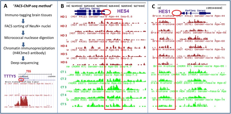
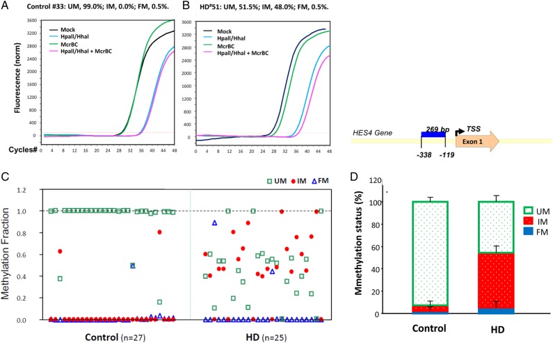
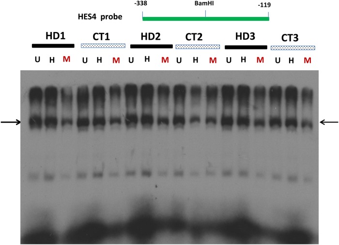
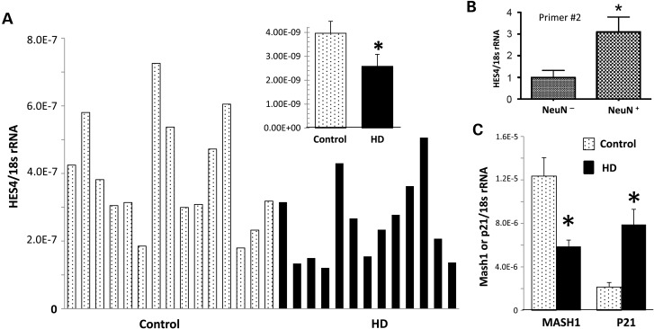
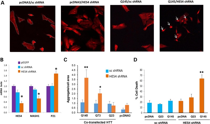
To investigate epigenetic contributions to Huntington's disease (HD) pathogenesis, we carried out genome-wide mapping of the transcriptional mark, trimethyl-histone H3-lysine 4 (H3K4me3) in neuronal nuclei extracted from prefrontal cortex of HD cases and controls using chromatin immunoprecipitation followed by deep-sequencing. Neuron-specific mapping of the genome-wide distribution of H3K4me3 revealed 136 differentially enriched loci associated with genes implicated in neuronal development and neurodegeneration, including GPR3, TMEM106B, PDIA6 and the Notch signaling genes hairy and enhancer of split 4 (HES4) and JAGGED2, supporting the view that the neuronal epigenome is affected in HD. Importantly, loss of H3K4me3 at CpG-rich sequences on the HES4 promoter was associated with excessive DNA methylation, reduced binding of nuclear proteins to the methylated region and altered expression of HES4 and HES4 targeted genes MASH1 and P21 involved in striatal development. Moreover, hypermethylation of HES4 promoter sequences was strikingly correlated with measures of striatal degeneration and age-of-onset in a cohort of 25 HD brains (r = 0.56, P = 0.006). Lastly, shRNA knockdown of HES4 in human neuroblastoma cells altered MASH1 and P21 mRNA expression and markedly increased mutated HTT-induced aggregates and cell death. These findings, taken together, suggest that epigenetic dysregulation of HES4 could play a critical role in modifying HD disease pathogenesis and severity.


Figures

Figure 1.
Figure 1.
Detection and distribution of H3K4me3 peaks surrounding the HES4 and HES1 genes in HD and control subjects. (A) Flow chart of the FACS-ChIP-seq procedure as described in the Materials and Methods section for detecting genome-wide distribution of H3K4me3 marks from NeuN+ cortical nuclei of six HD and five control subjects. Low panel: detection of H3K4me3 peak signal for Y chromosome gene TTTY5 by FACS-ChIP-seq H3K4me3 peaks are distributed in punctuated pattern and highly enriched in TSS of TTTY5 gene (as indicated by red circle). H3K4me3 peaks surround TTS of TTTY5 were absent in a female subject (first line) but present in a male sample (second line), confirming specificity of the H3K4me3 peaks detected by FACS-ChIP-seq. (B) H3K4me3 peaks were detected by FACS-ChIP-seq in NeuN+ cortical nuclei from six HD and five control subjects as described in the Materials and Methods section. H3K4me3 peaks are clustered around TSS of the HES4 gene (as indicated red circle). Moreover, the H3K4me3 peak (tag) densities (ad indicated by red long square/box) in HD were lower, compared with controls. (C) In contrast, the H3K4me3 peak densities around HES1 gene were indistinguishable between HD and control subjects.
Figure 2.
Figure 2.
DNA methylation of the HES4 promoter of HD and control cortex. DNA methylation states of a 269-bp fragment of the HES4 promoter in the PFC of 27 controls and 25 HD were measured by Methyl-Profiler as described in the Materials and Methods section. (Left) Examples of qPCR curves of all four reactions in one control and in one HD for the HES4 gene. DNA methylation states for the HES4 promoter are expressed as fractions of unmethylated (UM), intermediate-methylated (IM) or fully methylated (FM) DNA. (Right) IM was robustly increased from 5% of total input DNA in control to 49% in HD while UM fraction in the HES4 promoter was reduced in HD. In contrast, FM of the HES4 gene did not exhibit significant change.
Figure 3.
Figure 3.
The binding of nuclear proteins to the HES4 promoter is reduced after DNA hypermethylation in vitro. Binding of nuclear proteins from HD and control cortex to the 269-bp fragment of the HES4 promoter with in vitro DNA methylation by EMSA as described in the Materials and Methods section. This 269-bp fragment of the HES4 promoter was first digested BamHI into two identical DNA fragments and in vitro methylated and then re-annealed unmethylated (U), fully methylated (M) and hemi-methylated (H) double strand DNA probes for EMSA. Note that nuclear protein binding (indicated by arrows) was reduced and shifted to high molecular weight band at the fully methylated HES4 promoter compared with the un-methylated or hemi-methylated HES4 promoter.
Figure 4.
Figure 4.
The mRNA levels for HES4 as well as two down-stream target genes, MASH1 and P21, are reduced in the cortex of HD compared with controls. mRNA levels for HES4 (A) and its down-stream target MASH1 and P21 (C) in total cortex tissues were detected by qPCR analysis as described in the Materials and Methods section. We also measured relative heterogenous nuclear HES4 mRNA levels in NeuN and NeuN+ nuclei FACS sorted from human brains using two different primer sets [primer #2, (B) and primer #3 (data not shown]. (A) HES4 mRNA is reduced ∼40% in HD cortex compared with control. (B) HES4 mRNA is enriched in human neuronal nuclei (N = 3, P = 0.05, Mann–Whitney, one-tailed). (C) MASH1 mRNA in HD cortex compared with the control while P21 mRNA was increased in the cortex of HD compared with control. *P < 0.05 (n = 14, t-test). All data are presented as relative ratio of targeted mRNA over 18s rRNA, and presented as mean ± SEM.
Figure 5.
Figure 5.
shRNA knockdown of HES4 in human neuroblastoma cells alters mRNA levels of HES4, MASH1 and P21, and markedly increases mutant HTT-induced aggregates and cell death. HTB-11 cells were transfected with shRNA-HES4-1 or sc shRNA with or without different (23, 73 and 145) CAG repeats (HTT-Q23, HTT-Q73 and HTT-Q145), and qRT–PCR analysis, immunohistochemistry and cell death assays were performed as described in the Materials and Methods. (A) Ubiquitin immunohistochemistry showed that co-transfection of shRNA-HES4-1 and mutant HTT-Q145 for 24 h produced highly dense protein aggregates (aggregates = white arrow, cellular area = dash line) with no effect of shRNA-HES4-1 or HTT-Q145 alone compared with the cells transfected with shNRA-control/pcDNA3. (B) Quantitative analyses of HES4, MASH1 and P21 mRNAs in HBT-11 cells transfected with EGFP, sc shRNA or HES4-1 shRNA. Data are presented as mean values (plus s.e. from six transfection) of fold change relative to cells receiving EGFP after normalized to 18 s rRNA. HES4 shRNA decreased HES4 mRNA by ∼70%, MASH1 mRNA by ∼50% and increased P21 mRNA by ∼40%, compared with the sc shRNA. (C) Exacerbated HTT aggregates by shRNA knockdown of HES4 mRNA in HTB-11 cells. Cells were ubiquitin stained to detect the protein aggregation which was measured using automated image analysis as described in the Materials and Methods. Co-transfection of HES4 shRNA exacerbated HTT Q145-, Q73- (but not Q23-) induced cellular aggregates, as evident by increased relative size of cellular aggregates, in HTB-11 cells. (D) Trypan blue exclusion assay showed that shRNA-HES4-1 or HTT-Q145 alone did not have significant effect on cell death, but co-transfection of shRNA-HES4-1 and Q145 increased cell death rates compared with shRNA-control. **P < 0.01, Student's t-test or One-way ANOVA, post-hoc Dunn's test (B–D).

References

    1. Martin J.B., Gusella J.F. Huntington's disease. Pathogenesis and management. N. Engl. J. Med. 1986;315:1267–1276. - PubMed
    1. Vonsattel J.P., DiFiglia M. Huntington disease. J. Neuropathol. Exp. Neurol. 1998;57:369–384. - PubMed
    1. Group, T.H.s.D.C.R. A novel gene containing a trinucleotide repeat that is expanded and unstable on Huntington's disease chromosomes. The Huntington's Disease Collaborative Research Group. Cell. 1993;72:971–983. - PubMed
    1. MacDonald M.E., Gines S., Gusella J.F., Wheeler V.C. Huntington's disease. Neuromol. Med. 2003;4:7–20. - PubMed
    1. Gusella J.F., MacDonald M.E. Huntington's disease: the case for genetic modifiers. Genome Med. 2009;1:80. - PMC - PubMed

Publication types

MeSH terms