. 2015 Jan 15;75(2):405-14.
doi: 10.1158/0008-5472.CAN-14-2475. Epub 2014 Dec 5.

Kinome-wide functional screen identifies role of PLK1 in hormone-independent, ER-positive breast cancer

Affiliations

Kinome-wide functional screen identifies role of PLK1 in hormone-independent, ER-positive breast cancer

Neil E Bhola et al. Cancer Res. .

Erratum in

Abstract

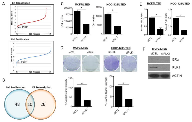
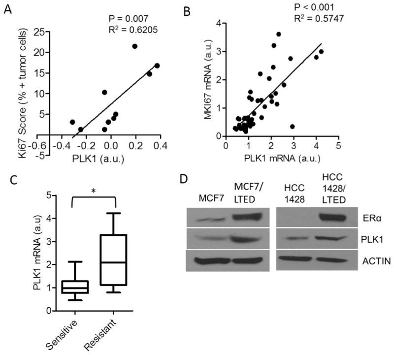
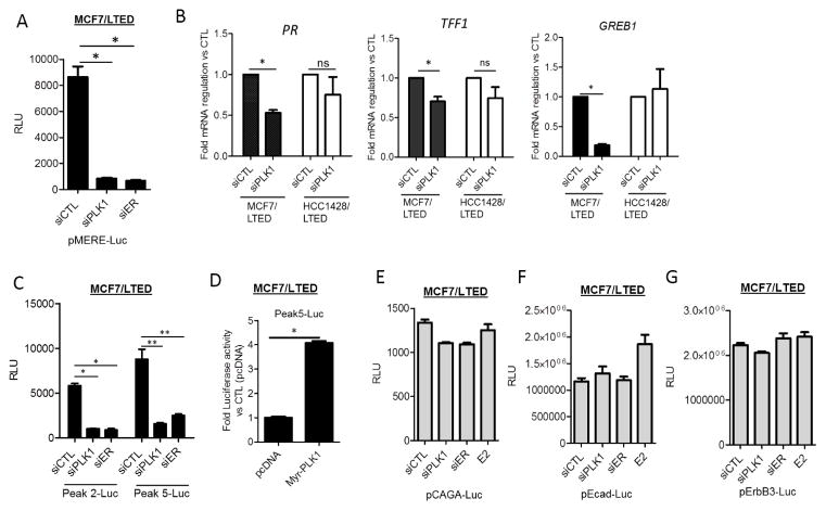
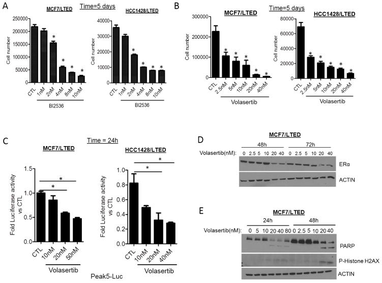
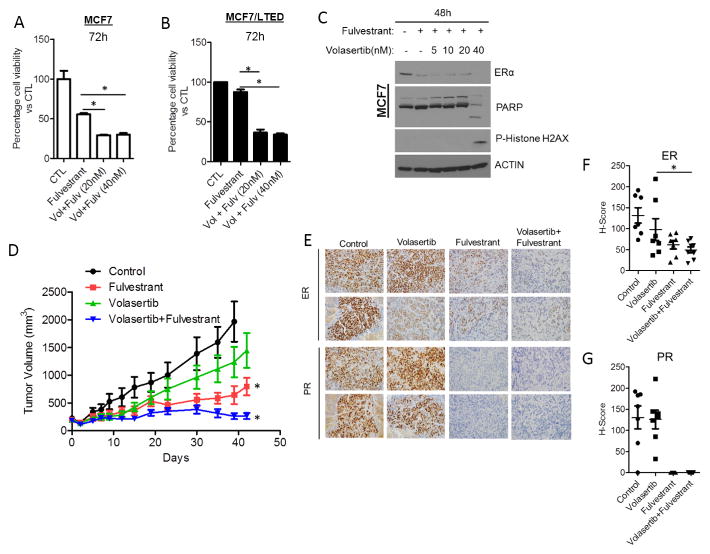
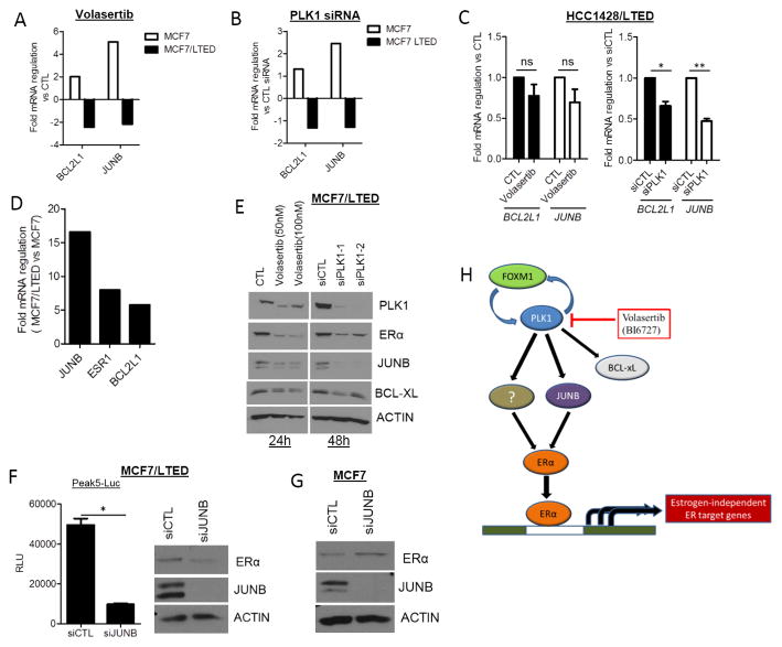
Estrogen receptor (ER) α-positive breast cancers initially respond to antiestrogens but eventually become estrogen independent and recur. ER(+) breast cancer cells resistant to long-term estrogen deprivation (LTED) exhibit hormone-independent ER transcriptional activity and growth. A kinome-wide siRNA screen using a library targeting 720 kinases identified Polo-like kinase 1 (PLK1) as one of the top genes whose downregulation resulted in inhibition of estrogen-independent ER transcriptional activity and growth of LTED cells. High PLK1 mRNA and protein correlated with a high Ki-67 score in primary ER(+) breast cancers after treatment with the aromatase inhibitor letrozole. RNAi-mediated knockdown of PLK1 inhibited ER expression, estrogen-independent growth, and ER transcription in MCF7 and HCC1428 LTED cells. Pharmacologic inhibition of PLK1 with volasertib, a small-molecule ATP-competitive PLK1 inhibitor, decreased LTED cell growth, ER transcriptional activity, and ER expression. Volasertib in combination with the ER antagonist, fulvestrant, decreased MCF7 xenograft growth in ovariectomized mice more potently than each drug alone. JUNB, a component of the AP-1 complex, was expressed 16-fold higher in MCF7/LTED compared with parental MCF7 cells. Furthermore, JUNB and BCL2L1 (which encodes antiapoptotic BCL-xL) mRNA levels were markedly reduced upon volasertib treatment in MCF7/LTED cells, while they were increased in parental MCF7 cells. Finally, JUNB knockdown decreased ER expression and transcriptional activity in MCF7/LTED cells, suggesting that PLK1 drives ER expression and estrogen-independent growth via JUNB. These data support a critical role of PLK1 in acquired hormone-independent growth of ER(+) human breast cancer and is therefore a promising target in tumors that have escaped estrogen deprivation therapy.


Conflict of interest statement

The authors disclose no potential conflicts of interest.

Figures

Figure 1
Figure 1. RNAi screen identifies PLK1 is required for hormone-independent ER transcriptional activity and growth
(A) MCF-7/LTED cells were screened with a siRNA library targeting 720 kinases. Ligand-independent cell growth and ER transcriptional activities were sequentially measured 72 h later using a high-throughput Alamar Blue and Luciferase Reporter assays, respectively. Both cell growth and ER transcriptional activity for each kinase siRNA relative to nonsilencing controls (siCTL) were transformed to a Z-score; the data are presented as median Z-score across 3 independent experiments. (B) Knockdown of 48 and 26 kinases significantly decreased MCF7/LTED cell proliferation and ER transcription, respectively. Knockdown of 10 kinases was found to decrease both cell viability and ER transcription. (C) MCF7/LTED and HCC1428/LTED cells transfected with RNAi targeting PLK1 and cultured for 5 days. Cells were trypsinized and their number determined in a Coulter Counter (*p<0.01). (D) MCF7/LTED and HCC1428/LTED cells were transfected with control (CTL) or PLK1 siRNA and re-seeded in 12-well plates in full growth media. Media was replenished every 3 days and the cells were stained with crystal violet after 7 days (*p<0.0002). (E) RNAi-mediated PLK1 downmodulation was validated by qRT-PCR in MCF7/LTED and HCC1428/LTED cells (*p<0.01). (F) MCF7/LTED cells were transfected with CTL or PLK1 siRNA for 72 h. Cell lysates were prepared and subjected to immunoblot analysis for ER, PLK1 and actin.
Figure 2
Figure 2. High PLK1 expression correlates with antiestrogen resistance in primary ER+ breast cancers
(A) Correlation PLK1 protein levels measured by RPPA with the Ki67 score in 10 post-letrozole breast tumor biopsies (n=10; p=0.007). (B) Correlation of PLK1 and MKI67 expression from RNA-seq analysis of RNA extracted from post-letrozole biopsies of primary ER+ breast cancers (n=47; p<0.001). (C) PLK1 expression was analyzed in patients’ tumors that were categorized as drug-sensitive or -resistant based on the post-letrozole Ki67 mRNA expression as described in Supplementary Methods (*p=0.002; a.u., arbitrary units). (D) Lysates from MCF7, HCC1428 cells and their LTED counterparts were assessed for ER and PLK1 expression by immunoblot analysis. Actin was used as a loading control.
Figure 3
Figure 3. PLK1 downmodulation abrogates estrogen-dependent and independent ER transcriptional activity
(A) MCF7/LTED cells were transfected with CTL, PLK1 or ER siRNA in triplicate wells. After 48 h, cells were transfected with a MERE-Luciferase construct; a Luciferase Assay was performed the following day (*p<0.02). (B) MCF7/LTED and HCC1428/LTED cells were transfected with CTL or PLK1 siRNA for 72 h. RNA was extracted, reverse transcribed and subjected to quantitative PCR with specific primers to PR (*p=0.005), TFF1 (*p=0.04) and GREB1 (*p=0.0007). (C) MCF7/LTED cells transfected with CTL, PLK1 or ER siRNA. After 48 h, cells were transfected with hormone-independent ER reporter (Peak2-Luc and Peak5-Luc) constructs; a Luciferase Assay was performed the following day (*p<0.002; **p 0.03) (D) MCF7/LTED cells were transfected with myristoylated PLK1 or control DNA (pcDNA) for 72 h. Cells were then transfected with Peak5-Luc and TK-Renilla and the Dual Luciferase assay was performed 18 h later (*p<0.0001). MCF7/LTED cells were transfected with CTL, PLK1 or ER siRNA or treated with 1 nM estradiol (E2). After 48 h, cells were transfected with (E) pCAGA-Luc, (F) Ecad-Luc, (G) ErbB3-Luc reporter constructs; a Luciferase assay was performed the following day as described in Methods.
Figure 4
Figure 4. PLK1 inhibitor decreases hormone-independent growth and ER transcriptional activity in LTED cells
MCF7 LTED and HCC1428 LTED cells were treated with increasing concentrations of the PLK1 inhibitors (A) BI2536 or (B) BI6727 (volasertib) for 5 days (*p<0.05). (C) MCF7/LTED and HCC1428/LTED cells transfected with Peak5-luciferase and TK-Renilla luciferase constructs were treated with increasing concentrations of volasertib for 24 h (*p<0.05). Dual luciferase assay was performed and fold luciferase activity to control was calculated as described in Methods. (D, E) MCF7/LTED cells were treated with increasing concentrations of volasertib for 24–72 h. Lysates were collected and after SDS-PAGE, subjected to immunoblot analyses for ER, PARP and phospho-Histone H2AX as described in Methods.
Figure 5
Figure 5. Volasertib enhances the anti-tumor effect of fulvestrant
(A) MCF7 and (B) MCF7/LTED cells were treated with vehicle, fulvestrant (1 μM) or combinations of volasertib and fulvestrant for 72 h. Cell viability was assessed using the MTT assay. (C) Lysates of MCF7 cells treated with vehicle, fulvestrant or combinations of volasertib and fulvestrant for 48 h were prepared and subjected to ER, PARP and p-Histone H2AX immunoblot analyses. (D) MCF7 (5×10⁶) cells were inoculated s.c. in the dorsum of ovariectomized athymic mice supplemented with a 2-week release estradiol pellet. Approximately 4 weeks later, mice with tumors ≥150 mm3 were randomized to treatment with vehicle, volasertib, fulvestrant, and both drugs for 6 weeks. Tumors were measured twice weekly with calipers; tumor volumes in mm3 are shown (*p=0.005). Each data point represents the mean tumor volume in mm3 ± SD (n=9/group). (E) IHC analysis of ER and PR in formalin-fixed paraffin-embedded xenograft sections from each group was performed and scored as described in Methods. Images of ER and PR IHC in representative xenografts are shown. (F) H-scores of ER (*p =0.04) and (G) PR expression in xenografts from all 4 groups are displayed on the right (n=7–8/group).
Figure 6
Figure 6. Inhibition of PLK1 decreases BCL2L1 and JUNB expression in LTED cells
(A) MCF7 and MCF7/LTED cells were treated with vehicle (0.1% DMSO) or 100 nM volasertib for 24 h. An ER Signaling-specific PCR Array was performed with RNA extracted from these cells. The expression of JUNB and BCL2L1 was compared between parental and LTED MCF7 cells. (B) MCF7 and MCF7/LTED cells were transfected with control or PLK1 siRNA for 48 h. RNA was extracted and tested in an ER Signaling-specific PCR Array. The expression of JUNB and BCL2L1 was compared between the parental and LTED cells. (C) HCC1428/LTED cells were treated with vehicle or 100 nM volasertib for 24 h or transfected with control and PLK1 siRNA for 48 h. RNA was extracted and subjected to qRT-PCR analysis for BCL2L1 and JUNB (*p=0.024; **p=0.003). (D) BCL2L1, JUNB and ESR1 (ER) as determined in the Estrogen Signaling PCR Array described in A and B is shown as the ratio of vehicle-treated MCF7/LTED over MCF7 parental cells. (E) MCF7/LTED cells were treated with either vehicle or volasertib (24 h) or transfected with control or PLK1 siRNA (48 h). Cell lysates were prepared and separated by SDS-PAGE followed by immunoblot analysis using the indicated antibodies. (F) Left: MCF7/LTED cells were transfected with control or JUNB siRNA. After 48 h, cells were transfected with the ligand-independent Peak5-Luc reporter construct. A Luciferase Assay was performed 24 h later (*p=0.006). Right: Lysates from cells transfected with Control or JUNB siRNA were assessed for JUNB and ER expression. (G) MCF7 cells transfected with control or JUNB siRNA for 72 h were assessed for ER and JUNB protein levels by immunoblot analysis. (H) Model of PLK1 function in hormone-independent ER+ breast cancer. FOXM1 regulates the expression of one of its transcriptional targets PLK1. In estrogen-deprived cell lines, PLK1 regulates the expression of JUNB and BCL-xL. As shown by the RNAi studies shown herein, JUNB can regulate the expression of ER and ER transcriptional activity.

References

    1. Malvezzi M, Bertuccio P, Levi F, La Vecchia C, Negri E. European cancer mortality predictions for the year 2013. Annals of Oncology. 2013;24:792–800. - PubMed
    1. Goldhirsch A, Wood WC, Coates AS, Gelber RD, Thürlimann B, Senn H-J, et al. Strategies for subtypes—dealing with the diversity of breast cancer: highlights of the St Gallen International Expert Consensus on the Primary Therapy of Early Breast Cancer 2011. Annals of Oncology. 2011;22:1736–47. - PMC - PubMed
    1. El-Tanani MKK, Green CD. Interaction between estradiol and growth factors in the regulation of specific gene expression in MCF-7 human breast cancer cells. The Journal of Steroid Biochemistry and Molecular Biology. 1997;60:269–76. - PubMed
    1. Brenton JD, Carey LA, Ahmed AA, Caldas C. Molecular Classification and Molecular Forecasting of Breast Cancer: Ready for Clinical Application? Journal of Clinical Oncology. 2005;23:7350–60. - PubMed
    1. Mauri D, Pavlidis N, Polyzos NP, Ioannidis JPA. Survival With Aromatase Inhibitors and Inactivators Versus Standard Hormonal Therapy in Advanced Breast Cancer: Meta-analysis. J Natl Cancer Inst. 2006;98:1285–91. - PubMed

Publication types

MeSH terms