Comparative Study
. 2015 Jan 16;427(1):131-45.
doi: 10.1016/j.jmb.2014.11.018. Epub 2014 Dec 4.

Mapping the gating and permeation pathways in the voltage-gated proton channel Hv1

Affiliations
Comparative Study

Mapping the gating and permeation pathways in the voltage-gated proton channel Hv1

Adam Chamberlin et al. J Mol Biol. .

Abstract

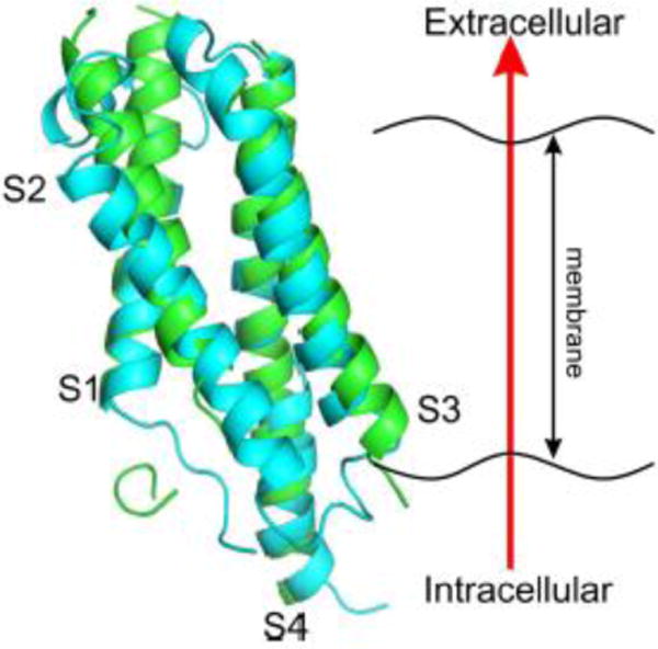
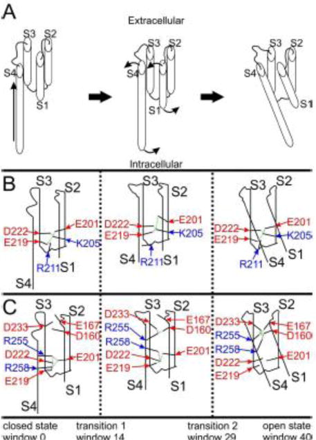
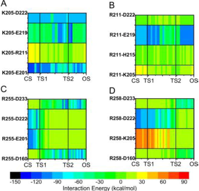
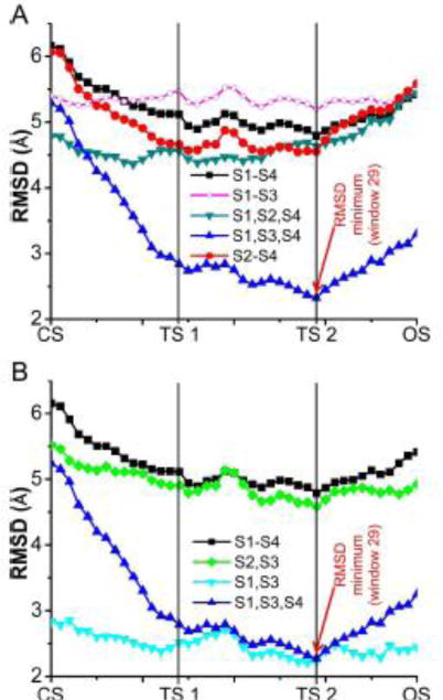
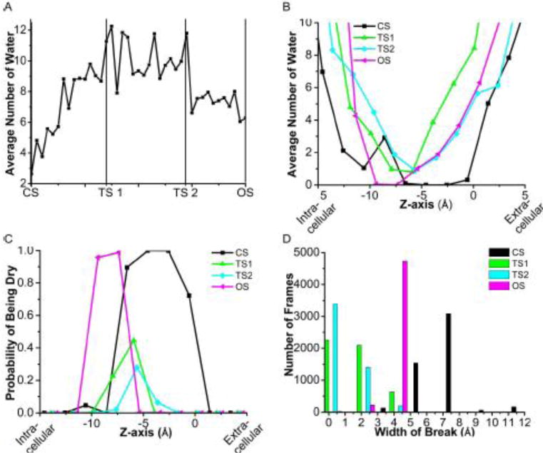
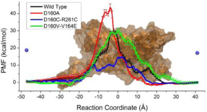
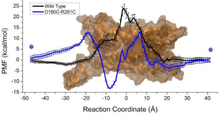
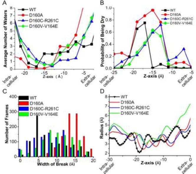
Voltage-gated proton channels (Hv1) are ubiquitous throughout nature and are implicated in numerous physiological processes. The gene encoding for Hv1, however, was only identified in 2006. The lack of sufficient structural information of this channel has hampered the understanding of the molecular mechanism of channel activation and proton permeation. This study uses both simulation and experimental approaches to further develop existing models of the Hv1 channel. Our study provides insights into features of channel gating and proton permeation pathway. We compare open- and closed-state structures developed previously with a recent crystal structure that traps the channel in a presumably closed state. Insights into gating pathways were provided using a combination of all-atom molecular dynamics simulations with a swarm of trajectories with the string method for extensive transition path sampling and evolution. A detailed residue-residue interaction profile and a hydration profile were studied to map the gating pathway in this channel. In particular, it allows us to identify potential intermediate states and compare them to the experimentally observed crystal structure of Takeshita et al. (Takeshita K, Sakata S, Yamashita E, Fujiwara Y, Kawanabe A, Kurokawa T, et al. X-ray crystal structure of voltage-gated proton channel. Nature 2014). The mechanisms governing ion transport in the wild-type and mutant Hv1 channels were studied by a combination of electrophysiological recordings and free energy simulations. With these results, we were able to further refine ideas about the location and function of the selectivity filter. The refined structural models will be essential for future investigations of this channel and the development of new drugs targeting cellular proton transport.

Keywords: gating mechanism; ion transport; voltage-gated proton channels.


Figures

Figure 1
Figure 1. Similarity of Ci-Hv1 model and mHv1 crystal structure
The putative closed-state crystal structure of the mHv1 proton channel (19) (green) is compared to the closest Ci-Hv1 structure taken from our swarm-of-trajectories with the string method (teal). The two structures were aligned using backbone carbons in the S1, S3, and S4 helical domains. For the purposes of this comparison, each domain was defined as: S1 = 150 – 164, S2 = 187 – 205, S3 = 220 – 237, S4 = 250 – 262.
Figure 2
Figure 2. Two transitions during Hv1 channel activation
(A) The large scale motions of the different domains in each part of the transition pathway are shown. (B–C) Salt bridges in closed, intermediate, and open states. (B) K205 in S2 and R211 in S3 are shown in blue, whereas E201 in S2 and E219 and D222 in S3 are shown in red. Weak (green) and strong (purple) attractive interactions are shown. (C) R255 and R258 in S4 are shown in blue, whereas D160 in S1, E201 in S2, and E219, D222, and D233 in S3 domain are shown in red. Weak (green) and strong (purple) attractive interactions are shown.
Figure 3
Figure 3. Salt bridges break and (re-)form during Hv1 channel activation
Color map of interaction energies (color coded in kcal/mol) for selected residues pairs graphed against the window numbers starting from the closed state (CS) at window 0, transition 1 (TS1) at window 14, transition 2 (TS2) at window 29, and open state (OS) at window 40 are shown. The charts are ordered by cationic residues. (A – K205, B – R211, C – R255, D – R258)
Figure 4
Figure 4. mHv1 crystal structure is most similar to an intermediate state of the Ci-Hv1 model
The crystal structure of the mHv1 proton channel from Takeshita et al[1] is compared using the root mean squared difference from the closest Ci-Hv1 model structure taken from each window. The closed state (CS) at window 0, transition 1 (TS1) at window 14, transition 2 (TS2) at window 29, and open state (OS) at window 40 are shown on the z-axis. The RMSD was computed based upon alignment of the two structures and tabulation of the RMSDs using solely the specified domains. For example, the S1, S3, S4 fit shown is for the case where the structures are aligned using the three domains and RMSDs are tabulated only for those three domains. In the graphs, the residues included in each domain are as follows: S1 = 150 – 164, S2 = 187 – 205, S3 = 220 – 237, S4 = 250 – 262.
Figure 5
Figure 5. Intermediate states are more hydrated than the closed and open states
(A) Time averaged number of water molecules within 5 Å of F198. (B) Time averaged number of water molecules present in 1-Å bins along the z-axis, where the 0 point is set to be the position of F198 for selected windows. (C) Fraction of the frames out of 5000 with at least 1 water present in 1-Å bins along the z-axis, where the 0 point is set to be the position of F198 for selected windows. (D) Histogram of the number of frames out of 5000 with a given number of consecutive 1-Å bins without any waters present.
Figure 6
Figure 6. R261C rescues currents in D160C mutation
(A) Currents (top) and Fluorescence (bottom) in response to voltage steps between −40 mV and +120 mV from a holding potential of −60 mV for D160C channels. (B) Normalized steady-state fluorescence (F) and conductance (G) from wild-type (WT) and R261C/S242C Hv1 channels from experiments as in A. (C) Currents from D160C/R261C channels in response to voltage protocol shown above. (D) Normalized conductance (G) versus voltage for D160C/R261C measured as in C.
Figure 7
Figure 7. D160C/R261C retains pH-dependent gating, but not proton selectivity
(A) Normalized conductance (G) versus voltage measured as in Fig 6C in external pH= 7.5 and 8.2. (B) Currents in response to voltage steps between −40 mV and +120 mV from a holding potential of −60 mV in external pH= 7.5 and 8.2. (C–F) Currents in response to voltage steps between −40 mV and +60 mV from a holding potential of −60 mV in external (C) 100 mM NaCl (D) 10 mM NaCl +Sucrose, (E) 100 mM NaCl, (F) 10 mM NaCl + 90 mM CholineCl.
Figure 8
Figure 8. Mutations in Hv1 selectivity filter retain barrier to chloride
PMFs for chloride ion crossing wild-type, D160A, D160C/R261C, and D160V/V164E Ci-Hv1 channels. The starting and ending positions of the chloride ion relative to the channels are shown as blue spheres on the background. The error bars were estimated from block averaging with five blocks.
Figure 9
Figure 9. D160C/R261C mutations create Na+ binding site in Hv1 channel
PMFs for sodium ion crossing wild-type and D160C/R261C Ci-Hv1 channels. The starting and ending positions of the sodium ion relative to the channel are shown as blue spheres. The error bars were estimated from block averaging with five blocks.
Figure 10
Figure 10. Mutations of Ci-Hv1 channels shift position of selectivity filter
(A) Time averaged number of water molecules present in 1-Å bins along the z-axis, where z = 0 is set to be the average position of the backbone atoms of the S1–S4 domains. (B) Fraction of the frames out of 1000 with at least 1 water present in 1-Å bins along the z-axis, where z=0 is set to be the average position of the backbone atoms of the S1–S4 domains. (C) Histogram of the number of frames out of 1000 with a given number of consecutive 1-Å bins without any waters present. (D) The computed radius of the pore along the z-axis.

References

    1. Takeshita K, Sakata S, Yamashita E, Fujiwara Y, Kawanabe A, Kurokawa T, et al. X-ray crystal structure of voltage-gated proton channel. Nature. 2014 - PubMed
    1. Thomas RC, Meech RW. Hydrogen-Ion Currents and Intracellular Ph in Depolarized Voltage-Clamped Snail Neurons. Nature. 1982;299:826–8. - PubMed
    1. Henderson LM, Chappell JB, Jones OTG. Superoxide Generation by the Electrogenic Nadph Oxidase of Human-Neutrophils Is Limited by the Movement of a Compensating Charge. Biochem J. 1988;255:285–90. - PMC - PubMed
    1. Henderson LM, Chappell JB, Jones OTG. The Superoxide-Generating Nadph Oxidase of Human-Neutrophils Is Electrogenic and Associated with an H+ Channel. Biochem J. 1987;246:325–9. - PMC - PubMed
    1. Decoursey TE. Voltage-gated proton channels and other proton transfer pathways. Physiol Rev. 2003;83:475–579. - PubMed

Publication types