. 2014 Dec 7:12:77.
doi: 10.1186/s12964-014-0077-3.

The structure function of the death domain of human IRAK-M

The structure function of the death domain of human IRAK-M

Jiangfeng Du et al. Cell Commun Signal. .

Abstract

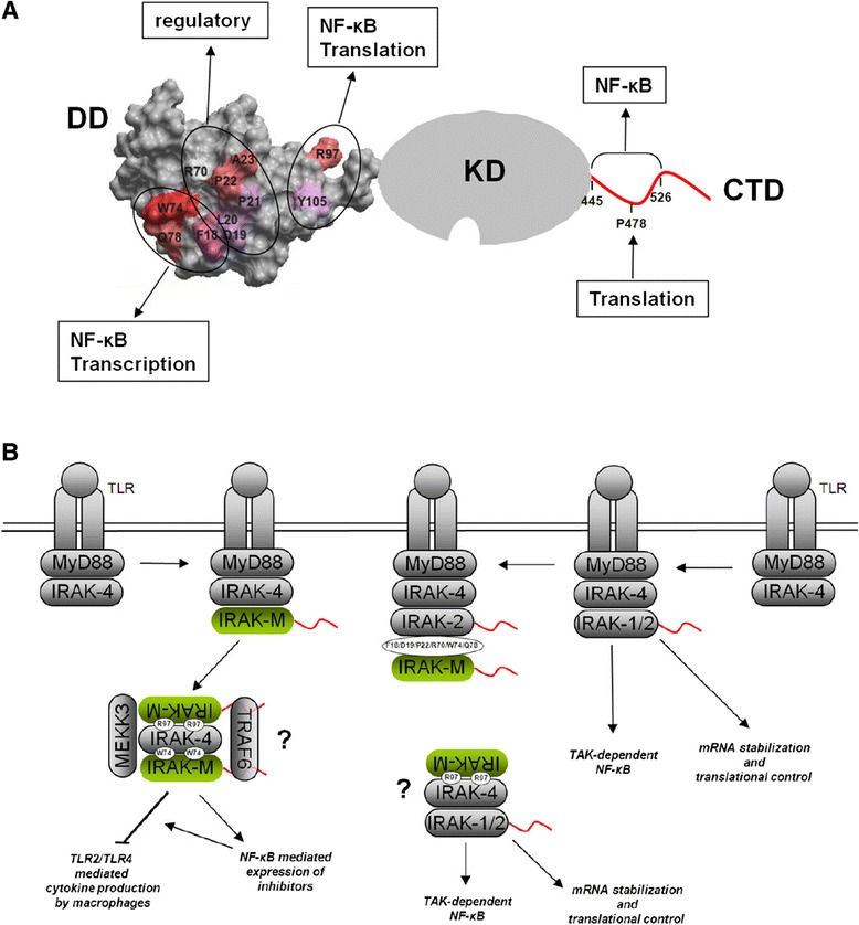
Background: IRAK-M is an inhibitor of Toll-like receptor signaling that acts by re-directing IRAK-4 activity to TAK1 independent NF-κB activation and by inhibition of IRAK-1/IRAK-2 activity. IRAK-M is expressed in monocytes/macrophages and lung epithelial cells. Lack of IRAK-M in mice greatly improves the resistance to nosocomial pneumonia and lung tumors, which entices IRAK-M as a potential therapeutic target. IRAK-M consists of an N-terminal death domain (DD), a dysfunctional kinase domain and unstructured C-terminal domain. Little is known however on IRAK-M's structure-function relationships.

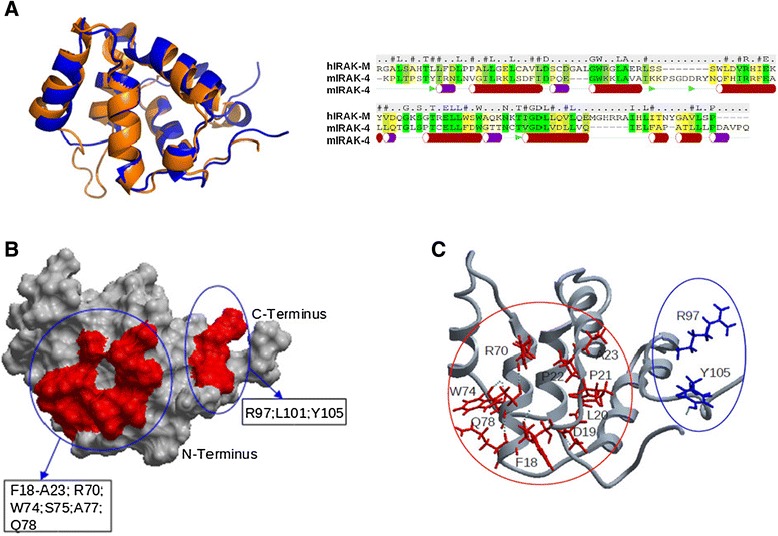
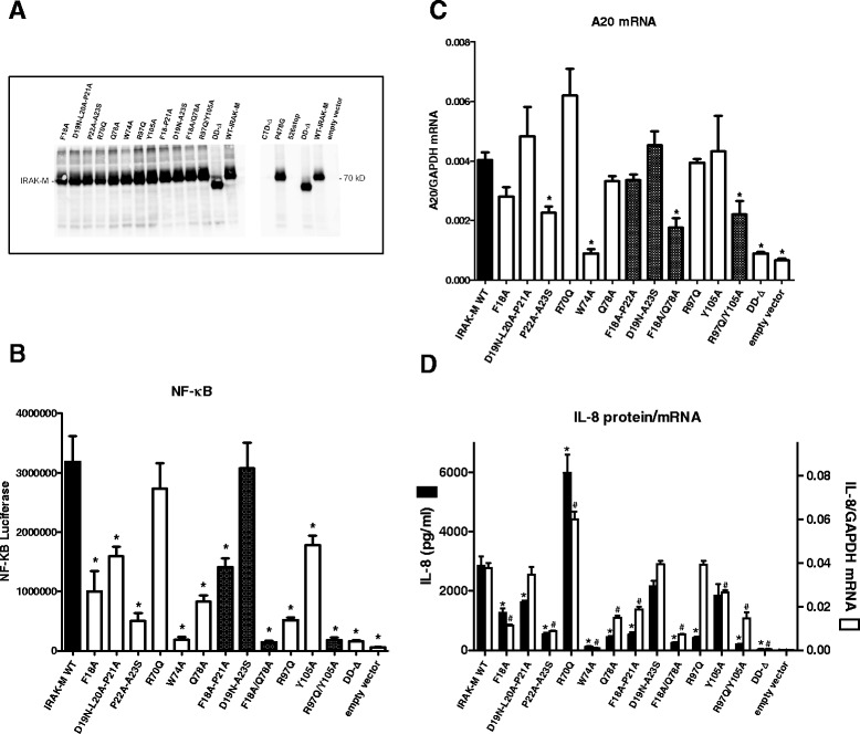
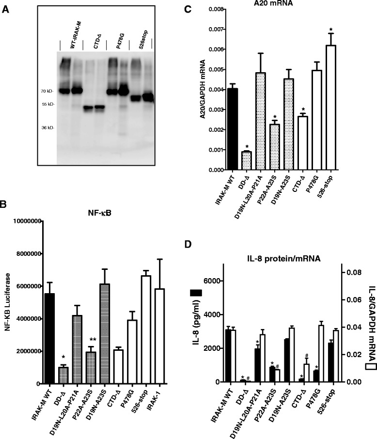
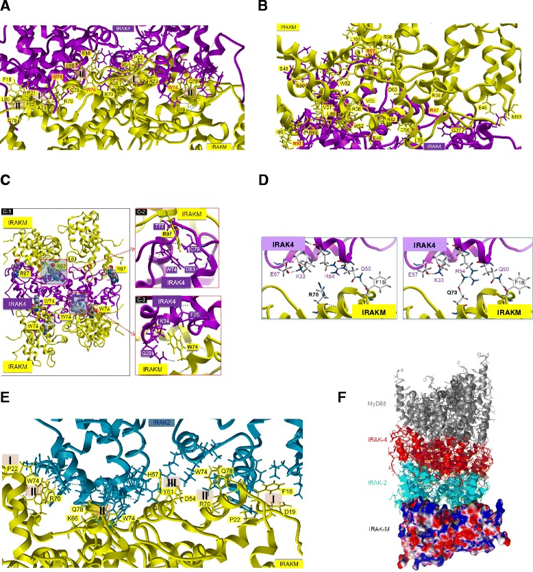
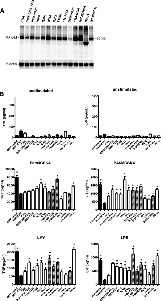
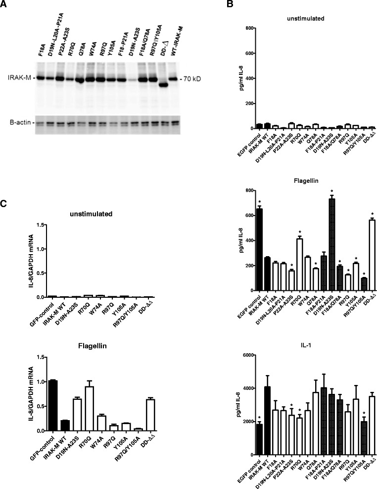
Results: Since death domains provide the important interactions of IRAK-1, IRAK-2 and IRAK-4 molecules, we generated a 3D structure model of the human IRAK-M-DD (residues C5-G119) to guide mutagenesis studies and predict protein-protein interaction points. First we identified the DD residues involved in the endogenous capacity of IRAK-M to activate NF-κB that is displayed upon overexpression in 293T cells. W74 and R97, at distinct interfaces of the IRAK-M-DD, were crucial for this endogenous NF-κB activating capacity, as well as the C-terminal domain (S445-E596) of IRAK-M. Resulting anti-inflammatory A20 and pro-inflammatory IL-8 transcription in 293T cells was W74 dependent, while IL-8 protein expression was dependent on R97 and the TRAF6 binding motif at P478. The IRAK-M-DD W74 and R97 binding interfaces are predicted to interact with opposite sides of IRAK-4-DD's. Secondly we identified DD residues important for the inhibitory action of IRAK-M by stable overexpression of mutants in THP-1 macrophages and H292 lung epithelial cells. IRAK-M inhibited TLR2/4-mediated cytokine production in macrophages in a manner that is largely dependent on W74. R97 was not involved in inhibition of TNF production but was engaged in IL-6 down-regulation by IRAK-M. Protein-interactive residues D19-A23, located in between W74 and R97, were also observed to be crucial for inhibition of TLR2/4 mediated cytokine induction in macrophages. Remarkably, IRAK-M inhibited TLR5 mediated IL-8 production by lung epithelial cells independent of W74 and R97, but dependent on D19-A23 and R70, two surface-exposed regions that harbor predicted IRAK-2-DD interaction points of IRAK-M.

Conclusion: IRAK-M employs alternate residues of its DD to inhibit the different inflammatory mediators induced by varying TLRs and cells.


Figures

Figure 1
Figure 1
3D structure model of the human IRAK-M death domain (DD). (A) Model of the DD of human IRAK-M (Blue) superimposed on the template DD structure 2A9I (Orange) of mIRAK-4 and sequence alignment of hIRAK-M-DD and mIRAK-4-DD. The sequence identity is 28.7%. Secondary structures such as alpha-helices and beta-strands of mIRAK-4-DD (2A9I) are indicated underneath the sequences (Red bar: alpha-helix: Purple bar: unstable helix; Green arrow: beta-strand). (B) Interactive surface prediction of hIRAK-M-DD. Space filling model with predicted interactive residues in red that form two patches. (C) The predicted interactive residues in the two patches which were mutated in this study are shown with side chain and residue number in the back bone model in red and blue.
Figure 2
Figure 2
Expression and functional analysis of human IRAK-M-DD mutants in 293T cells. (A) Analysis of expression levels of IRAK-M variants by transfection in 293T cells by Western blotting performed on cell lysates with an antibody directed to the C-terminal of IRAK-M. (B) Effect of IRAK-M-DD mutations on NF-κB activation by overexpression in 293T cells. (C) Effect of IRAK-M-DD mutations on A20 mRNA induction by overexpression in 293T cells. (D) Effect of IRAK-M-DD mutations on IL-8 protein and mRNA production by overexpression in 293T cells. (B-D) N=4, mean±SEM. *Indicates difference with WT IRAK-M P<0.05. Shaded bars depict results of IRAK-M molecules with combinations of mutated residues/stretches within the same patch.
Figure 3
Figure 3
Expression and functional analysis of IRAK-M C-terminal domain mutants in 293T cells. (A) Analysis of transient expression of IRAK-M and C-terminal domain mutants by transfection in 293T cells by Western blotting performed on cell lysates with an antibody directed to full length IRAK-M. (B) Effect of IRAK-M C-terminal domain mutation on NF-κB activation by overexpression in 293T cells. (C) Effect of IRAK-M C-terminal domain mutation on A20 mRNA induction by overexpressing in 293T cells. (D) Effect of IRAK-M C-terminal domain mutation on IL-8 protein and mRNA production by overexpression in 293T cells. (B-D) Comparison to IRAK-M-DD mutants in the D19-A23 stretch. N=4, mean±SEM. *Indicates difference with WT IRAK-M P<0.05.
Figure 4
Figure 4
Protein:protein docking of IRAK-M-DD tetramers onto IRAK-4-DD tetramers or IRAK-2-DD tetramers. (A) IRAK-M-DD interactions predicted to be important for the contact between IRAK-M and IRAK-4 as obtained by unbiased docking of the IRAK-M tetramer (Yellow) onto the bottom surface of the IRAK-4 tetramer structure (Purple) as is present in the myddosome (3MOP.pdb also F). The residues involved in the interaction are shown with their side chains, of which the residues from IRAK-M are labeled with their respective residue name and number. Two interaction types were involved (type I residues L20, P21, P22, A23, R70, type II residues L16, F18, W74, S75, Q78). This orientation was observed for 63% of the 100 best docked events. (B) IRAK-M-DD interactions predicted to be important for the contact between IRAK-M and IRAK-4 as obtained by unbiased docking of the IRAK-M tetramer (Yellow) on the top surface of IRAK-4 tetramer (Purple). The residues involved in the interaction are shown with their side chains, including R97. This orientation was observed for 37% of the 100 best docked events. (C) Potential sandwich of one IRAK-4-DD tetramer in between 2 IRAK-M-DD tetramers by W74 and R97 mediated interactions. The respective predicted IRAK-4 interactions are shown in detail. (D) Docking of mutant IRAK-M-DD predicts increased IRAK-4 interaction with the IRAK-M R70Q mutant through an extra hydrogen bond formed between Q70 in IRAK-M and R54 in IRAK-4. (E) Part of the composite binding site of the free side of the IRAK-2-DD tetramer in the myddosome is shown with three IRAK-M-DD molecules. The contact residues are shown with their side chain. The residues involved in the interaction from IRAK-M are labeled. Three interaction types were involved. F) A proposed whole myddosome structure with IRAK-M-DD tetramer docked on the free side of the IRAK-2-DD tetramer, based on the myddosome structure, 3MOP.pdb.
Figure 5
Figure 5
Effect of human IRAK-M and death domain mutants in macrophages. WT IRAK-M and DD-mutants were stably expressed in the human monocytic cell line THP-1 by lentiviral transduction and FACS-sorting of EGFP positive cells which is bicistronically expressed. THP-1 cells were matured to macrophages before stimulation as described in Methods. (A) IRAK-M expression evaluated by western blotting. (B) The effect of stable WT IRAK-M expression and IRAK-M mutants was determined on TLR2 (Pam3CSK4) and TLR4 (LPS) mediated TNF and IL-6 production after stimulation for 6 hour. Shaded bars depict results of IRAK-M molecules with combinations of mutated residues/stretches within the same area. N=4, mean±SEM. *Indicates a difference with WT IRAK-M P<0.05.
Figure 6
Figure 6
Effect of human IRAK-M and death domain mutants in lung epithelial cells. WT IRAK-M and DD-mutants were stably expressed in the lung epithelial cell line H292 by lentiviral transduction and FACS-sorting of EGFP positive cells which is bicistronically expressed. (A) IRAK-M expression evaluated by western blotting. (B) The effect of stable IRAK-M expression and IRAK-M mutants was determined on IL-1β and Flagellin (TLR5) mediated IL-8 expression in the supernatant after 6 hour stimulation. Shaded bars depict results of IRAK-M molecules with combinations of mutated residues/stretches within the same area. N=8, mean±SEM. * indicates difference with WT IRAK-M P<0.05. (C) Effect of stable expression of WT and DD domain mutants on IL-8 mRNA levels induced by Flagellin after 3 hours. N=3, mean±SEM.
Figure 7
Figure 7
Schematic representation of the structure-function relationships of IRAK-M based on this study. (A) Functional involvement of the different residues of the Death Domain (DD) of IRAK-M on induction of NF-κB, transcription and translation upon overexpression in 293T cells (data derived from Figures 2 and 3). Graded color code (red->pink->white) indicates the impact of the single mutants on NF-κB in 293T cells (from red important to white no effect). The stretch D19-A23 is not indicated to be essential to NF-κB because mutation of the complete stretch (combined D19-P21 and P22-A23 mutation) results in a mutant with normal NF-κB. (B) Schematic representation of the working mechanism of IRAK-M as inhibitor of MyD88 signaling adapted from Zhou et al. [13], in which IRAK-4/IRAK-M interaction induces transcription of other inhibitors via MEKK3 dependent NF-κB and in which IRAK-2 mediated mRNA stability and translation is inhibited by IRAK-M. Based on our mutagenesis studies both W74 as well as R97 in the IRAK-M-DD are pivotal for IRAK-4/IRAK-M mediated NF-κB activity, but R97 is predicted to bind on the other side of IRAK-4 (see Figure 4). This may induce oligomerization of IRAK-4 and IRAK-1 or IRAK-2 activation, or, since both residues are essential for the unique, previously shown MEKK3 dependent IRAK-M mediated NF-κB activation, IRAK-4 may be sandwiched by IRAK-M to come to this pathway as indicated by W74 and R97 interactions. R97 is not predicted to be involved in the interaction of IRAK-M with IRAK-2 for which F18, D19, P22, R70 and W74 are predicted to be involved.

References

    1. Wesche H, Gao X, Li X, Kirschning CJ, Stark GR, Cao Z. IRAK-M is a novel member of the Pelle/interleukin-1 receptor-associated kinase (IRAK) family. J Biol Chem. 1999;274:19403–19410. doi: 10.1074/jbc.274.27.19403. - DOI - PubMed
    1. Muzio M, Ni J, Feng P, Dixit VM. IRAK (Pelle) family member IRAK-2 and MyD88 as proximal mediators of IL-1 signaling. Science. 1997;278:1612–1615. doi: 10.1126/science.278.5343.1612. - DOI - PubMed
    1. Lin SC, Lo YC, Wu H. Helical assembly in the MyD88-IRAK4-IRAK2 complex in TLR/IL-1R signalling. Nature. 2010;465:885–890. doi: 10.1038/nature09121. - DOI - PMC - PubMed
    1. Nagpal K, Plantinga TS, Sirois CM, Monks BG, Latz E, Netea MG, Golenbock DT. Natural loss-of-function mutation of myeloid differentiation protein 88 disrupts its ability to form Myddosomes. J Biol Chem. 2011;286:11875–11882. doi: 10.1074/jbc.M110.199653. - DOI - PMC - PubMed
    1. Takeuchi O, Akira S. Pattern recognition receptors and inflammation. Cell. 2010;140:805–2. doi: 10.1016/j.cell.2010.01.022. - DOI - PubMed

Publication types

MeSH terms