Review
. 2015 Mar;1852(3):406-20.
doi: 10.1016/j.bbadis.2014.11.020. Epub 2014 Dec 4.

Usher syndrome: Hearing loss, retinal degeneration and associated abnormalities

Affiliations
Review

Usher syndrome: Hearing loss, retinal degeneration and associated abnormalities

Pranav Mathur et al. Biochim Biophys Acta. 2015 Mar.

Abstract

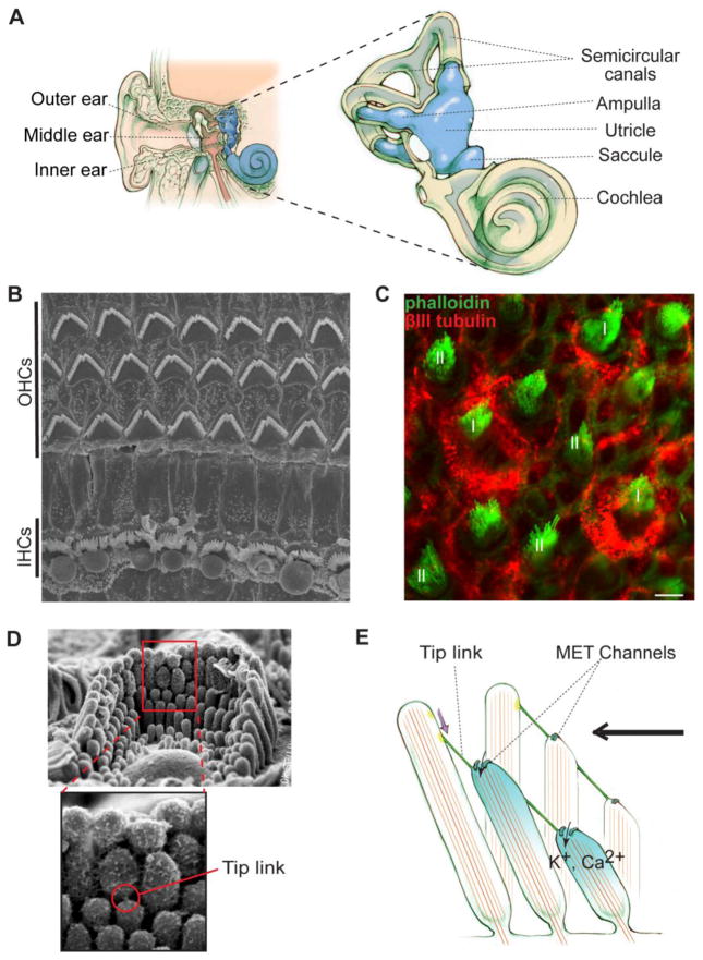
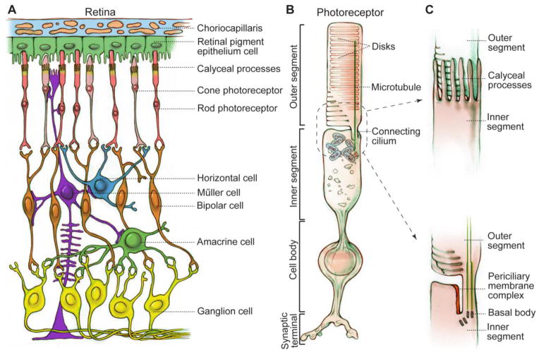
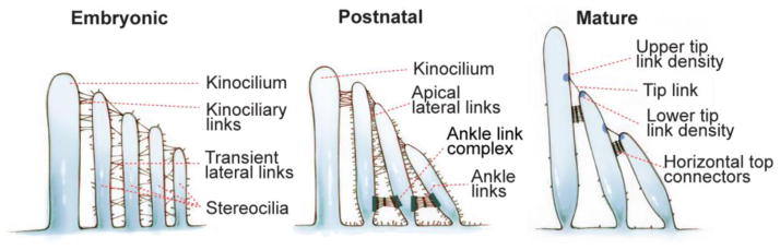
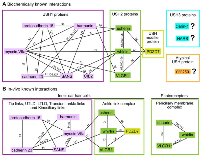
Usher syndrome (USH), clinically and genetically heterogeneous, is the leading genetic cause of combined hearing and vision loss. USH is classified into three types, based on the hearing and vestibular symptoms observed in patients. Sixteen loci have been reported to be involved in the occurrence of USH and atypical USH. Among them, twelve have been identified as causative genes and one as a modifier gene. Studies on the proteins encoded by these USH genes suggest that USH proteins interact among one another and function in multiprotein complexes in vivo. Although their exact functions remain enigmatic in the retina, USH proteins are required for the development, maintenance and function of hair bundles, which are the primary mechanosensitive structure of inner ear hair cells. Despite the unavailability of a cure, progress has been made to develop effective treatments for this disease. In this review, we focus on the most recent discoveries in the field with an emphasis on USH genes, protein complexes and functions in various tissues as well as progress toward therapeutic development for USH.

Keywords: Hair bundle link; Inner ear hair cell; Periciliary membrane complex; Retina photoreceptor; Therapy; USH multiprotein complex.


Figures

Figure 1
Figure 1
Inner ear anatomy and mechanoelectrical transduction (MET). (A) The position and structure of the inner ear. The inner ear is the most inner part of the ear (Left). It contains the cochlea and vestibular system. The vestibular system includes the utricle, saccule and semicircular canal ampullae (Right). (B) Top view of the murine organ of Corti shown by scanning electron microscopy. The organ of Corti has one row of inner hair cells (IHCs) and three rows of outer hair cells (OHCs). (C) Type I (I) and type II (II) hair cells in the murine utricular extrastriola shown by immunofluorescence. Phalloidin signal (green) indicates actin bundles in stereocilia. βIII tubulin (red) labels calyx afferents of type I hair cells. Scale bar, 5 μm. (D) A hair bundle shown by scanning electron microscopy. Top, an entire bundle with a staircase pattern of stereociliary organization. Bottom, an amplified view of the boxed area on the top showing a tip link. (E) MET occurs when the hair bundle is deflected toward the longest stereocilium (excitatory direction, black arrow). Deflection of hair bundles in this direction stretches tip links, which subsequently regulates the gating of MET channels and leads to hair cell depolarization. Note that this is a simplified model and tip links may associate with MET channels indirectly.
Figure 2
Figure 2
Schematic diagrams of the retina and photoreceptor. (A) Cellular organization of the retina. Retinal neurons are arranged into different layers. Photoreceptors and RPE cells are located in the outer retina, while horizontal cells, bipolar cells, amacrine cells and ganglion cells are downstream neurons of photoreceptors in the inner retina. Müller cells are glial cells spanning all cell layers of the retina. (B) Photoreceptor structure exemplified by a rod. Photoreceptors are ciliated cells with several specialized subcellular compartments, consisting of an outer segment having tightly packed membrane disks, a connecting cilium with microtubules, calyceal processes with actin bundles (not shown), an inner segment having all organelles for energy and protein synthesis, a cell body containing the nucleus, and a synaptic terminus for signal transmission to bipolar and horizontal cells. (C) Magnified drawings of calyceal processes (top) and periciliary membrane complex (bottom) around the photoreceptor connecting cilium. In order to show the periciliary membrane complex, calyceal processes are omitted in the bottom drawing.
Figure 3
Figure 3
Dynamic changes of various interstereociliary links in the hair bundle during and after development. During embryogenesis, emerging stereocilia are bundled by transient lateral links and connected with the kinocilium through kinociliary links. Postnatally, the transient lateral links are gradually replaced by tip links, apical lateral links and ankle links. After maturation, the apical lateral links are replaced by horizontal top connectors. Upper tip link density (UTLD) and lower tip link density (LTLD) are electron-dense structures at the insertion sites of tip links in the taller and shorter stereocilia, respectively. In mature rodent cochlear hair cells, the ankle links, kinociliary links and kinocilium disappear.
Figure 4
Figure 4
Known interaction networks among USH proteins. (A) Interactions among USH1 (pink), USH2 (green) and PDZD7 proteins found and confirmed by various in vitro biochemical assays. Interactions among the two USH3 (blue), atypical USH (CEP250) and other USH proteins have not yet been revealed. (B) Interactions of USH proteins confirmed in inner ear hair cells (left) and photoreceptors (right) by genetic studies. Reference numbers are given along each line.

References

    1. LeMasurier M, Gillespie PG. Hair-cell mechanotransduction and cochlear amplification. Neuron. 2005;48:403–415. - PubMed
    1. Eatock RA, Songer JE. Vestibular hair cells and afferents: two channels for head motion signals. Annu Rev Neurosci. 2011;34:501–534. - PubMed
    1. Khan S, Chang R. Anatomy of the vestibular system: a review. NeuroRehabilitation. 2013;32:437–443. - PubMed
    1. Gillespie PG, Muller U. Mechanotransduction by hair cells: models, molecules, and mechanisms. Cell. 2009;139:33–44. - PMC - PubMed
    1. Vollrath MA, Kwan KY, Corey DP. The micromachinery of mechanotransduction in hair cells. Annu Rev Neurosci. 2007;30:339–365. - PMC - PubMed

Publication types

MeSH terms

Substances