. 2015 Feb;14(1):139-47.
doi: 10.1111/acel.12279. Epub 2014 Dec 6.

Reversible cell cycle inhibition and premature aging features imposed by conditional expression of p16Ink4a

Affiliations

Reversible cell cycle inhibition and premature aging features imposed by conditional expression of p16Ink4a

Amelie Boquoi et al. Aging Cell. 2015 Feb.

Abstract

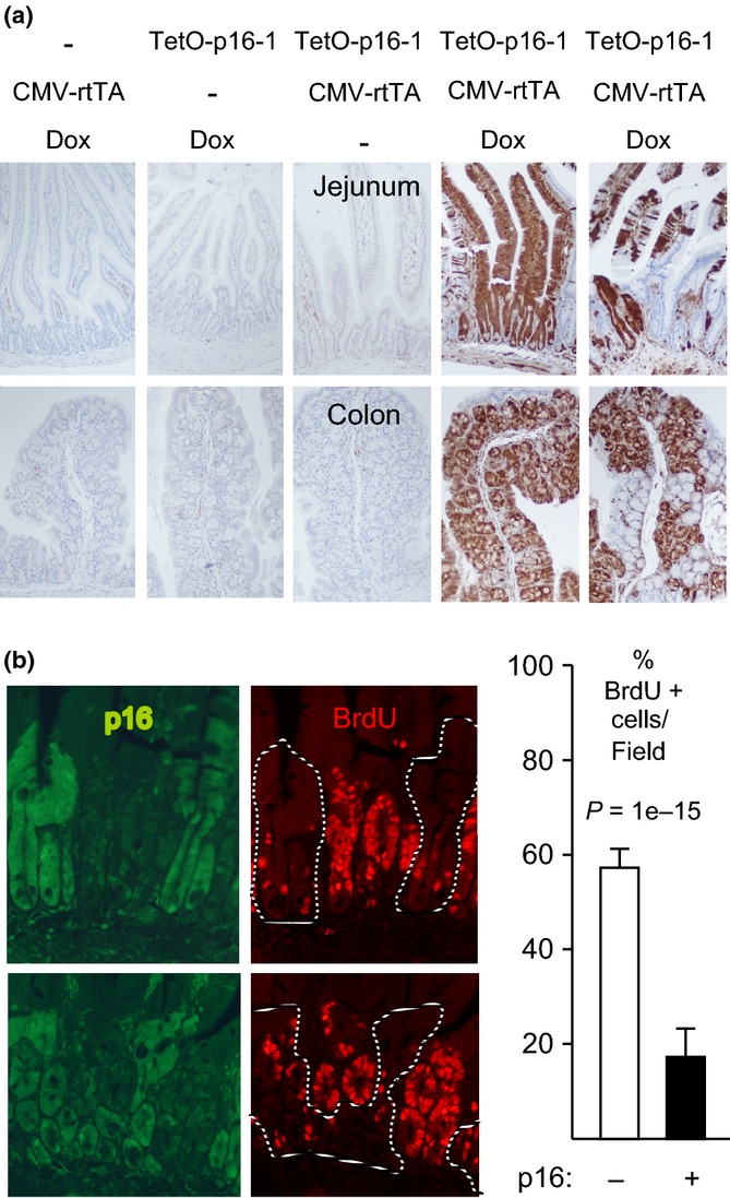
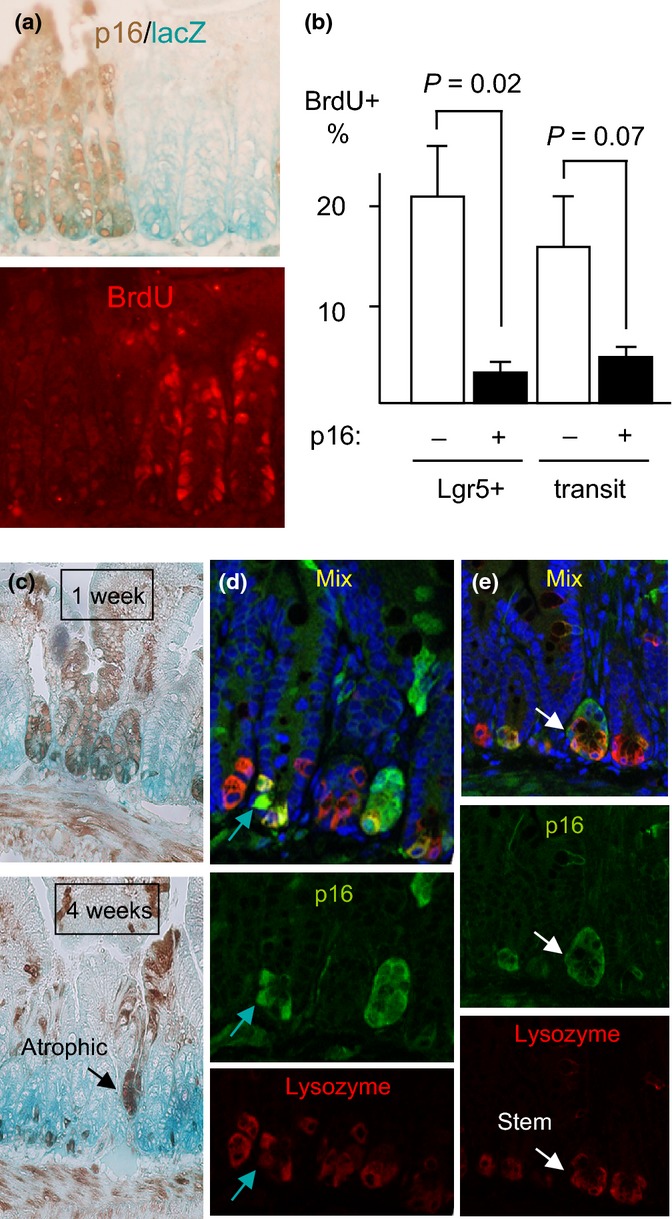
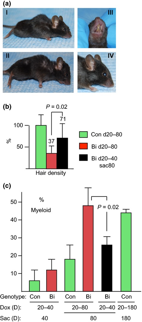
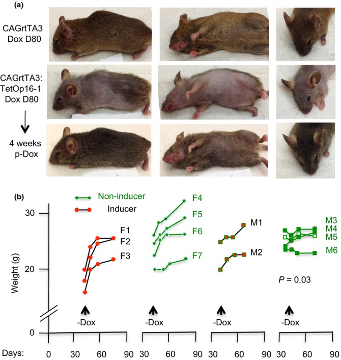
The cyclin-dependent kinase (Cdk) inhibitor p16(Ink4a) (p16) is a canonical mediator of cellular senescence and accumulates in aging tissues, where it constrains proliferation of some progenitor cells. However, whether p16 induction in tissues is sufficient to inhibit cell proliferation, mediate senescence, and/or impose aging features has remained unclear. To address these issues, we generated transgenic mice that permit conditional p16 expression. Broad induction at weaning inhibited proliferation of intestinal transit-amplifying and Lgr5+ stem cells and rapidly imposed features of aging, including hair loss, skin wrinkling, reduced body weight and subcutaneous fat, an increased myeloid fraction in peripheral blood, poor dentition, and cataracts. Aging features were observed with multiple combinations of p16 transgenes and transactivators and were largely abrogated by a germline Cdk4 R24C mutation, confirming that they reflect Cdk inhibition. Senescence markers were not found, and de-induction of p16, even after weeks of sustained expression, allowed rapid recovery of intestinal cell proliferation and reversal of aging features in most mice. These results suggest that p16-mediated inhibition of Cdk activity is sufficient to inhibit cell proliferation and impose aging features in somatic tissues of mammals and that at least some of these aging features are reversible.

Keywords: Cdk; Ink4a; aging; p16; senescence; stem cell.


Figures

Figure 1
Figure 1
p16 induction inhibits intestinal epithelial cell proliferation. (a) Mice of different genotypes were or were not treated with Dox for 1 week, as designated. IHC for exogenous p16 (brown, 10× fields). Note strong mosaic p16 induction in the presence of both transgenes and Dox. (b) CMV-rtTA:TetO-p16-1 mice treated with Dox for 1 week and injected with BrdU. Panels (left): Two 20× fields (above, below) with co-IF for p16 (left, green; right, dashed lines) and BrdU (right, red). Graph (right): Among 1122 crypt cells scored, 19% of p16+ cells were BrdU+ vs. 58% of p16- cells. Modest BrdU signal was visible in the green filter.
Figure 2
Figure 2
Induced p16 inhibits proliferation of intestinal stem cells. (a) CMV-rtTA:TetO-p16:Lgr5-lacZ mice were treated with Dox for 1 week and injected with BrdU. LacZ was detected by activity stain (left, aqua), p16 by IHC (left, brown), and BrdU in the same section by IF (right, red). Note paucity of BrdU staining in the p16+ Lgr5-lacZ+ cells (left 3 crypts). Across 8 20× fields, p16+ Lgr5+ cells (N = 331) showed no BrdU staining vs. 15% of the p16- Lgr5+ cells (N = 461, P = 0.02) (b) In the same mice, p16 and BrdU were detected by IHC in serial 4- to 5-micrometer sections and the % BrdU+ cells were scored in Lgr5+ and transit-amplifying cells, respectively. P values by two-sided t-tests. The one-sided t-test for transit cells = 0.03. N = 3 mice; 5803 cells counted. (c) Reduced p16 expression in small intestines of mice with continuous p16 induction. CMV-rtTA:TetO-p16:Lgr5-lacZ mice were treated with Dox near 2 mos of age for 1 or 4 weeks. IHC for p16 (brown) and activity assay for lacZ (aqua). Fields 10x. N = 3 mice per time point. Note the gradual loss of p16 expression, with preferential retention at the crypt base (P = 0.02 vs. transit-amplifying zones, P = 0.007 vs. villi). An atrophic, p16+ crypt is marked. (d, e) Partial costaining for p16 and lysozyme in crypt base cells, detected by confocal co-IF. Mice treated with Dox d20-40 were stained for p16 (green), lysozyme (red), and DNA (DAPI, blue; top). Green arrows (d) mark a p16+ cell that stains weakly for lysozyme, reflecting partial paneth cell differentiation. White arrows (e) identify a p16+ lysozyme- cell that is surrounded by lysozyme+ cells and, hence, is likely a stem cell.
Figure 3
Figure 3
Progression vs. recovery of aging features. CMV-rtTA:TetO-p16 littermates treated with Dox d20-40 were divided into 2 groups. Dox was sustained in one and withdrawn in the other. Mice were sacrificed at d80. (a) Sustained Dox treatment d20-80 results in persistent defects. Note the thin hair, hunched posture, thin body, and sunken eyes in representative mice (a1, all); malocclusion of the incisors, with resulting overgrowth (alll, 5/17 mice); and cataracts (alV, bilateral, confirmed by histology; 2/17 mice). (b) Back hair density (mg cm−2) in bitransgenic mice (Bi) treated with Dox d20-80 (N = 14) and Bi mice treated with Dox d20-40 and sacrificed at d80 (N = 6) were normalized to that of their wild-type and singly transgenic littermate controls (Con) treated with Dox d20-80 (N = 12) and depicted as mean ± SD (percentages relative to controls listed). Hair density of Bi d20-40sac80 mice was significantly greater than in Bi d20-80 mice (P = 0.02) and did not differ significantly from control mice. (c) Increase in myeloid to lymphoid ratio with sustained Dox treatment d20-80 in bitransgenic mice (P = 0.01 compared to control d20-80 mice), ameliorated by discontinuing Dox at d40 (P = 0.02 compared to Bi d20-80 mice). Results shown are means ± SD from 3 to 5 mice, except the control d20-180 results, which are mean ± range from two mice.
Figure 4
Figure 4
Rapid reversal of aging features in CAG-rtTA3 mice following p16 de-induction. (a) Top row: a noninducing littermate mouse at D80 of Dox treatment (different angles). Middle row: a CAG-rtTA3:TetOp16 mouse at D80 of Dox treatment. Lower row: the same CAG-rtTA3:TetOp16 mouse following 4 weeks of Dox withdrawal. Note dramatic recovery in body hair and skin wrinkling, filling out of face and eyes, and straightening of whisker hairs. Only the hair on the ventral trunk lagged (center), and the eyes remained sunken. (b) Left: littermate female CAG-rtTA3:TetOp16 mice (‘inducers’ F1-3) and wild-type or singly transgenic mice (‘noninducers’ F4-7) were treated with Dox from D20-40. Mice were weighed weekly for 2 weeks and then 4 weeks after Dox withdrawal (X-axis: days). Inducers showed a dramatic rebound in weight (Y-axis: g) within the first week. Right: littermate male inducers (M1-2) and noninducers (M3-6) were treated with Dox D20-80, and then Dox was withdrawn. Inducers showed a prompt recovery in weight within 2–4 weeks. The slope of weight gain was greater in the mice with prior p16 induction (P = 0.03).

References

    1. Alcorta DA, Xiong Y, Phelps D, Hannon G, Beach D, Barret JC. Involvement of the cyclin-dependent kinase inhibitor p16(INK4a) in replicative senescence of normal human fibroblasts. PNAS. 1996;93:13742–13747. - PMC - PubMed
    1. Barker N, van Es JH, Kuipers J, Kujala P, van den Born M, Cozijnsen M, Haegebarth A, Korving J, Begthel H, Peters PJ, Clevers H. Identification of stem cells in small intestine and colon by marker gene Lgr5. Nature. 2007;449:1003–1007. - PubMed
    1. Beausejour CM, Krtolica A, Galimi F, Narita M, Lowe SW, Yaswen P, Campisi J. Reversal of human cellular senescence: roles of the p53 and p16 pathways. EMBO J. 2003;22:4212–4222. - PMC - PubMed
    1. Chong JL, Wenzel PL, Saenz-Robles MT, Nair V, Ferrey A, Hagan JP, Gomez YM, Sharma N, Chen HZ, Ouseph M, Wang SH, Trikha P, Culp B, Mezache L, Winton DJ, Sansom OJ, Chen D, Bremner R, Cantalupo PG, Robinson ML, Pipas JM, Leone G. E2f1-3 switch from activators in progenitor cells to repressors in differentiating cells. Nature. 2009;462:930–934. - PMC - PubMed
    1. Collado M, Gil J, Efeyan A, Guerra C, Schuhmacher AJ, Barradas M, Benguria A, Zaballos A, Flores JM, Barbacid M, et al. Tumour biology: senescence in premalignant tumours. Nature. 2005;436:642. - PubMed

Publication types

Substances