

Human mesenchymal stromal cells enhance the immunomodulatory function of CD8(+)CD28(-) regulatory T cells
- PMID: 25482073
- PMCID: PMC4716622
- DOI: 10.1038/cmi.2014.118
Human mesenchymal stromal cells enhance the immunomodulatory function of CD8(+)CD28(-) regulatory T cells
Abstract
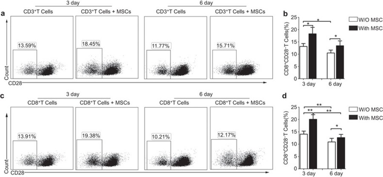
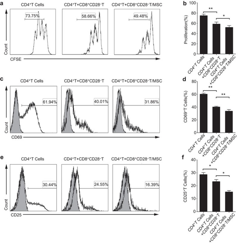
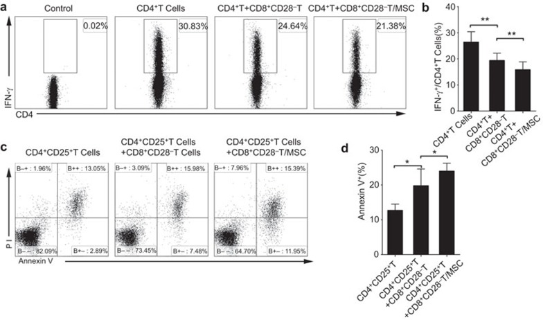
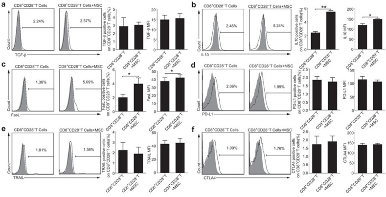
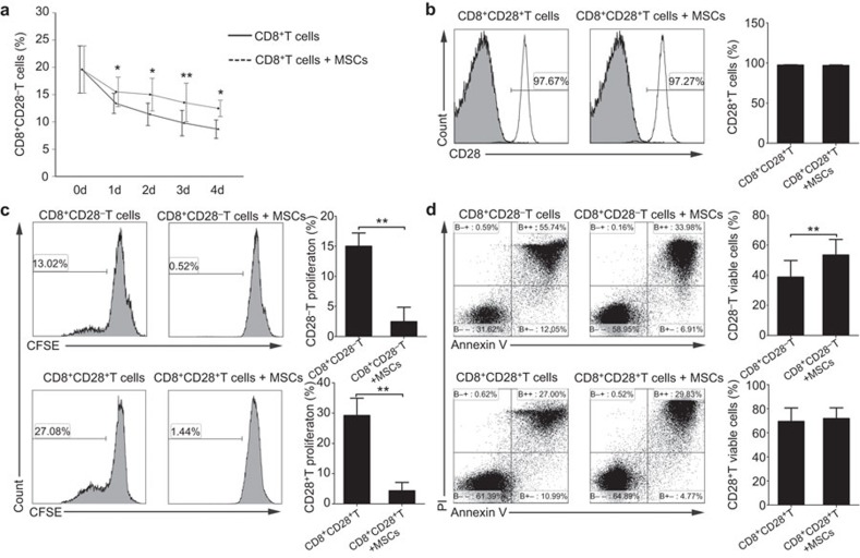
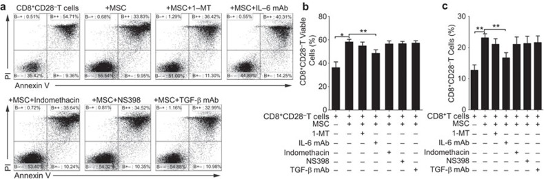
One important aspect of mesenchymal stromal cells (MSCs)-mediated immunomodulation is the recruitment and induction of regulatory T (Treg) cells. However, we do not yet know whether MSCs have similar effects on the other subsets of Treg cells. Herein, we studied the effects of MSCs on CD8(+)CD28(-) Treg cells and found that the MSCs could not only increase the proportion of CD8(+)CD28(-) T cells, but also enhance CD8(+)CD28(-)T cells' ability of hampering naive CD4(+) T-cell proliferation and activation, decreasing the production of IFN-γ by activated CD4(+) T cells and inducing the apoptosis of activated CD4(+) T cells. Mechanistically, the MSCs affected the functions of the CD8(+)CD28(-) T cells partially through moderate upregulating the expression of IL-10 and FasL. The MSCs had no distinct effect on the shift from CD8(+)CD28(+) T cells to CD8(+)CD28(-) T cells, but did increase the proportion of CD8(+)CD28(-) T cells by reducing their rate of apoptosis. In summary, this study shows that MSCs can enhance the regulatory function of CD8(+)CD28(-) Treg cells, shedding new light on MSCs-mediated immune regulation.
Figures
Similar articles
-
An increase in CD3+CD4+CD25+ regulatory T cells after administration of umbilical cord-derived mesenchymal stem cells during sepsis.PLoS One. 2014 Oct 22;9(10):e110338. doi: 10.1371/journal.pone.0110338. eCollection 2014. PLoS One. 2014. PMID: 25337817 Free PMC article.
-
Interferon-γ and tumor necrosis factor-α promote the ability of human placenta-derived mesenchymal stromal cells to express programmed death ligand-2 and induce the differentiation of CD4(+)interleukin-10(+) and CD8(+)interleukin-10(+)Treg subsets.Cytotherapy. 2015 Nov;17(11):1560-71. doi: 10.1016/j.jcyt.2015.07.018. Cytotherapy. 2015. PMID: 26432559
-
Combination cell therapy using mesenchymal stem cells and regulatory T-cells provides a synergistic immunomodulatory effect associated with reciprocal regulation of TH1/TH2 and th17/treg cells in a murine acute graft-versus-host disease model.Cell Transplant. 2014 Apr;23(6):703-14. doi: 10.3727/096368913X664577. Epub 2013 Feb 26. Cell Transplant. 2014. PMID: 23452894
-
Mesenchymal stromal cells and immunomodulation: A gathering of regulatory immune cells.Cytotherapy. 2016 Feb;18(2):160-71. doi: 10.1016/j.jcyt.2015.10.011. Cytotherapy. 2016. PMID: 26794710 Review.
-
[Recent advance in research on immunomodulatory function of mesenchymal stem cells].Zhongguo Shi Yan Xue Ye Xue Za Zhi. 2007 Oct;15(5):1117-20. Zhongguo Shi Yan Xue Ye Xue Za Zhi. 2007. PMID: 17956703 Review. Chinese.
Cited by
-
Antitumor activity of pluripotent cell-engineered vaccines and their potential to treat lung cancer in relation to different levels of irradiation.Onco Targets Ther. 2016 Mar 11;9:1425-36. doi: 10.2147/OTT.S97587. eCollection 2016. Onco Targets Ther. 2016. PMID: 27042111 Free PMC article.
-
Autologous Bone Marrow-Derived Mesenchymal Stem Cells Modulate Molecular Markers of Inflammation in Dogs with Cruciate Ligament Rupture.PLoS One. 2016 Aug 30;11(8):e0159095. doi: 10.1371/journal.pone.0159095. eCollection 2016. PLoS One. 2016. PMID: 27575050 Free PMC article.
-
Immune modulation by mesenchymal stem cells.Cell Prolif. 2020 Jan;53(1):e12712. doi: 10.1111/cpr.12712. Epub 2019 Nov 15. Cell Prolif. 2020. PMID: 31730279 Free PMC article. Review.
-
Immunomodulatory Activity of Human Bone Marrow and Adipose-Derived Mesenchymal Stem Cells Prolongs Allogenic Skin Graft Survival in Nonhuman Primates.Cell J. 2021 Apr;23(1):1-13. doi: 10.22074/cellj.2021.6895. Epub 2021 Mar 1. Cell J. 2021. PMID: 33650815 Free PMC article.
-
CD8+ T Lymphocytes: Crucial Players in Sjögren's Syndrome.Front Immunol. 2021 Jan 28;11:602823. doi: 10.3389/fimmu.2020.602823. eCollection 2020. Front Immunol. 2021. PMID: 33584670 Free PMC article. Review.
References
-
- 1da Silva Meirelles L, Chagastelles PC, Nardi NB. Mesenchymal stem cells reside in virtually all post-natal organs and tissues. J Cell Sci 2006; 119: 2204–2213. - PubMed
-
- 2Shi Y, Hu G, Su J, Li W, Chen Q, Shou P, et al. Mesenchymal stem cells: a new strategy for immunosuppression and tissue repair. Cell Res 2010; 20: 510–518. - PubMed
-
- 4Sensebe L, Krampera M, Schrezenmeier H, Bourin P, Giordano R. Mesenchymal stem cells for clinical application. Vox Sang 2010; 98: 93–107. - PubMed
Publication types
MeSH terms
Substances
LinkOut - more resources
Full Text Sources
Other Literature Sources
Research Materials
