Review
. 2014 Sep-Oct;5(5):402-26.
doi: 10.4161/nucl.32235.

Nucleolar stress with and without p53

Affiliations
Review

Nucleolar stress with and without p53

Allison James et al. Nucleus. 2014 Sep-Oct.

Abstract

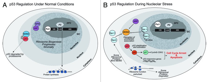
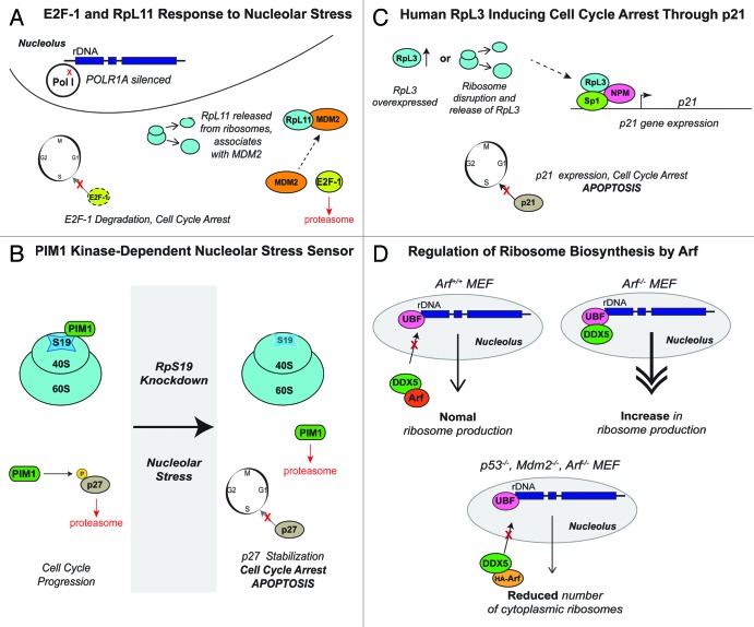
A veritable explosion of primary research papers within the past 10 years focuses on nucleolar and ribosomal stress, and for good reason: with ribosome biosynthesis consuming ~80% of a cell's energy, nearly all metabolic and signaling pathways lead ultimately to or from the nucleolus. We begin by describing p53 activation upon nucleolar stress resulting in cell cycle arrest or apoptosis. The significance of this mechanism cannot be understated, as oncologists are now inducing nucleolar stress strategically in cancer cells as a potential anti-cancer therapy. We also summarize the human ribosomopathies, syndromes in which ribosome biogenesis or function are impaired leading to birth defects or bone narrow failures; the perplexing problem in the ribosomopathies is why only certain cells are affected despite the fact that the causative mutation is systemic. We then describe p53-independent nucleolar stress, first in yeast which lacks p53, and then in other model metazoans that lack MDM2, the critical E3 ubiquitin ligase that normally inactivates p53. Do these presumably ancient p53-independent nucleolar stress pathways remain latent in human cells? If they still exist, can we use them to target >50% of known human cancers that lack functional p53?

Keywords: cell cycle; nucleolar stress; p53; ribosomal proteins; ribosomopathies.


Figures

None
Figure 1. Ribosome biogenesis in eukaryotic cells occurs in the nucleolus, a nuclear compartment that displays the fibrillar center (FC), the dense fibrillar component (DFC), and the granular component (GC) at the ultra-structural level. Transcription of pre-rRNA (47S in mammals) by RNA polymerase I (Pol I) is generally thought to occur at the boarders of the FC and DFC on tandem gene repeats emanating as radial loops from more condensed rDNA within the FCs. The 5S rRNA is transcribed by RNA Pol III in all eukaryotes; in yeast the 5S genes reside within the nucleolus within intergenic sequences that lie between the larger tandem rDNA gene repeats, but in metazoans, the 5S genes reside as tandem repeats outside the nucleolus. Processing and cleavage of the 47S pre-rRNA occurs in the DFC as ribosomal proteins synthesized in the cytoplasm enter the nucleolus and assemble with the 18S rRNA to form the small ribosomal subunit (SSU) and with the 5.8S, 28S, and 5S rRNAs to form the large ribosomal subunit (LSU). Large and small ribosomal subunits continue to assemble and mature as they enter the GC which contains GTPases (e.g., GNL3) that likely function in subunit maturation or nucleolar release. The GC is drawn here as a heterogeneous nucleolar compartment consisting of RNA-containing and RNA-deficient zones. Another GTPase, Nucleostemin (NS), resides in the RNA-deficient zones of the GC. Rather than participating in ribosome biogenesis directly, NS functions in maintaining stem cell homeostasis and in responding to nucleolar stress. Export of the ribosomal subunits is finally mediated by nuclear export factors, NMD3 and CRM1. See text for more details.
None
Figure 2. Regulation of p53 during normal and nucleolar stress conditions. (A) During normal, non-stressed conditions, the E3 ubiquitin ligase MDM2 associates with p53, promoting p53’s degradation. Nucleophosmin (NPM) and ARF are located in the nucleolus. (B) During nucleolar stress, normal ribosome biogenesis and function are perturbed. The association between MDM2 and p53 is disrupted; additional proteins such as ribosomal proteins (RpL5, RpL11) with the 5S rRNA, and Arf can associate with MDM2. p53 is stabilized and activates the cell cycle inhibitor p21 and other p53-responsive genes. These events lead to cell cycle arrest and apoptosis.
None
Figure 3. MDM2 and p53 Regulation. MDM2 has three main functional domains: a N-terminal p53-binding domain, a central acidic region, and a carboxy RING finger, which has E3 ubiquitin ligase activity. (A) MDM2 folds such that the carboxy RING finger domain interacts with p53 bound at MDM2’s N-terminus. p53 is then ubiquitinylated and targeted for degradation., (B) Protein folding is prevented when certain proteins such as ARF, Nucleostemin (NS), or other ribosomal proteins bind to the phosphorylated central domain of MDM2. This results in stabilization and accumulation of p53 levels.,
None
Figure 4. p53-Independent Responses to Nucleolar Stress. (A) Donati et al. expressed RNAi to silence POLR1A, a subunit of Pol I, in order to mimic nucleolar stress in p53-deficient HCT-110 human cancer cells. Donati et al. proposes a model in which, during perturbations in ribosome biogenesis, modeled by silencing of POLR1A, RpL11 is released from ribosomes and subsequently associates with MDM2. The E2F-1-MDM2 interaction is severed, leading to proteasomal degradation of E2F-1 and cell cycle arrest. (B) In p53−/− erythroid cells, PIM1 kinase normally associates with RpS19. PIM1 kinase also phosphorylates the cell cycle inhibitor p27Kip1 at Thr157, thus marking it for degradation, allowing normal cell cycle progression. Iadevaia et al. disrupted the RpS19/PIM1 kinase interaction, either by expressing RNAi against RpS19 or applying treatments known to induce nucleolar stress in human erythroleukemic TF-1 (p53+/+) and K562 (p53−/−) cell lines. PIM1 kinase, being unable to associate with the small subunit of the ribosome, is degraded, allowing p27Kip1 stabilization, cell cycle arrest, and apoptosis, even in K562 (p53−/−) cells. (C) Russo et al. overexpressed RpL3 in order to mimic a disruption of ribosome biogenesis. Upon RpL3 overexpression, a multi-protein complex containing RpL3, Sp1, and NPM formed at the p21 gene promoter, activating its expression, which resulted in cell cycle arrest or apoptosis. (D) Arf functions as a negative regulator of ribosome biogenesis. In Arf+/+ murine embryonic fibroblasts (MEFs), Arf associates with DDX5, a DEAD-box RNA helicase, sequestering it in the nucleoplasm, allowing normal ribosome production. In Arf−/− MEFs, DDX5 localizes to the rDNA promoters in the nucleolus, resulting in increased ribosome production. In p53−/−, Mdm2−/−, Arf−/− MEFs, exogenous expression of HA-Arf resulted in reduced ribosome production once again.

References

    1. Pederson T. The plurifunctional nucleolus. Nucleic Acids Res. 1998;26:3871–6. doi: 10.1093/nar/26.17.3871. - DOI - PMC - PubMed
    1. Boisvert F-M, van Koningsbruggen S, Navascués J, Lamond AI. The multifunctional nucleolus. Nat Rev Mol Cell Biol. 2007;8:574–85. doi: 10.1038/nrm2184. - DOI - PubMed
    1. Olson MOJ. Sensing cellular stress: another new function for the nucleolus? Sci STKE. 2004;2004:pe10. - PubMed
    1. Hein N, Hannan KM, George AJ, Sanij E, Hannan RD. The nucleolus: an emerging target for cancer therapy. Trends Mol Med. 2013;19:643–54. doi: 10.1016/j.molmed.2013.07.005. - DOI - PubMed
    1. Drygin D, O’Brien SE, Hannan RD, McArthur GA, Von Hoff DD. Targeting the nucleolus for cancer-specific activation of p53. Drug Discov Today. 2014;19:259–65. doi: 10.1016/j.drudis.2013.08.012. - DOI - PubMed

Publication types

MeSH terms

Substances