. 2015 Aug;62(2):615-26.
doi: 10.1002/hep.27645. Epub 2015 Mar 23.

Liver X receptors balance lipid stores in hepatic stellate cells through Rab18, a retinoid responsive lipid droplet protein

Affiliations

Liver X receptors balance lipid stores in hepatic stellate cells through Rab18, a retinoid responsive lipid droplet protein

Fiona O'Mahony et al. Hepatology. 2015 Aug.

Abstract

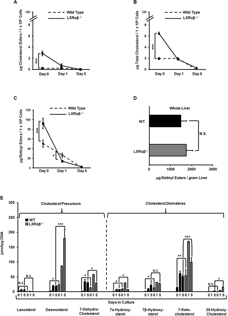
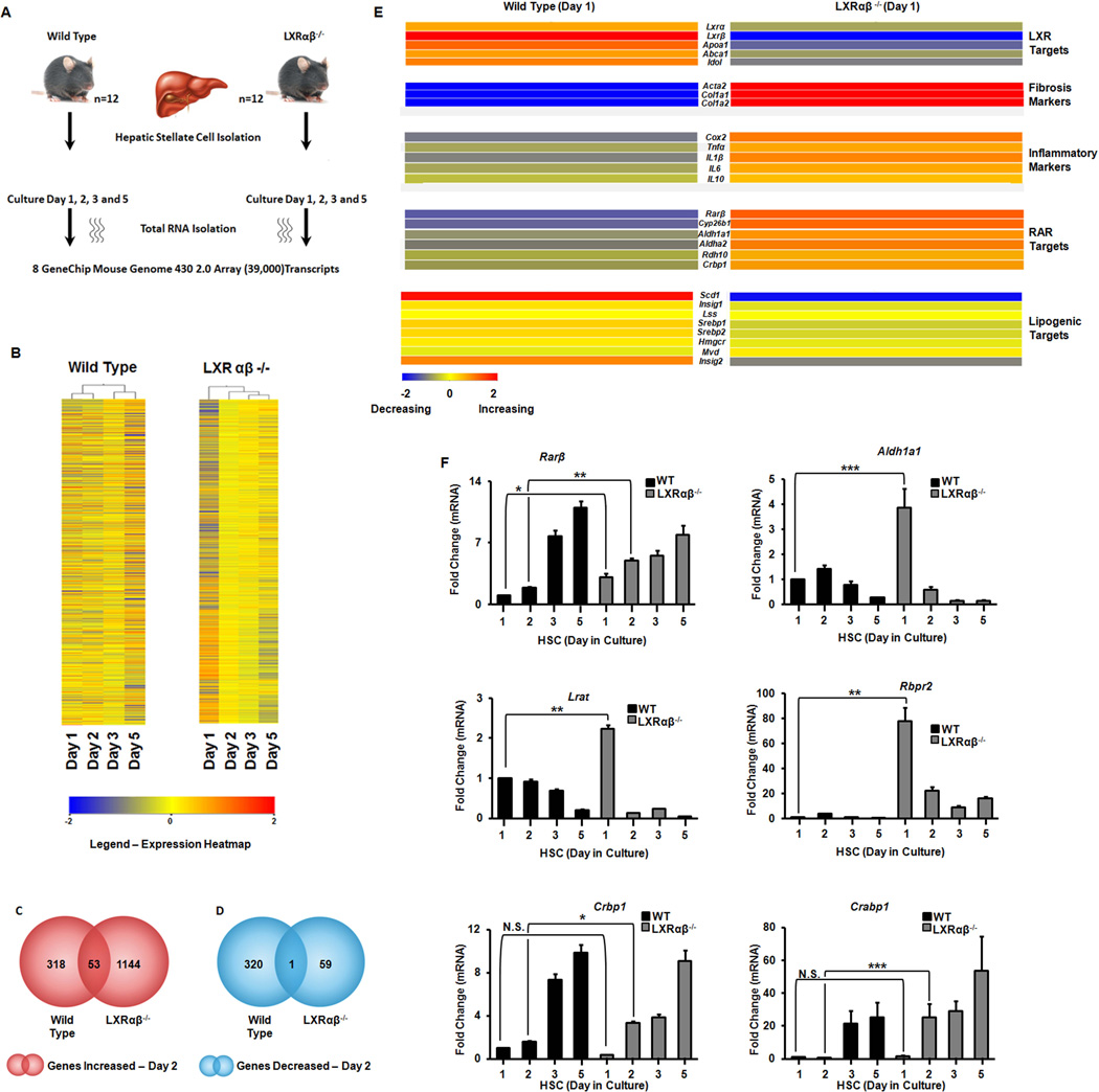
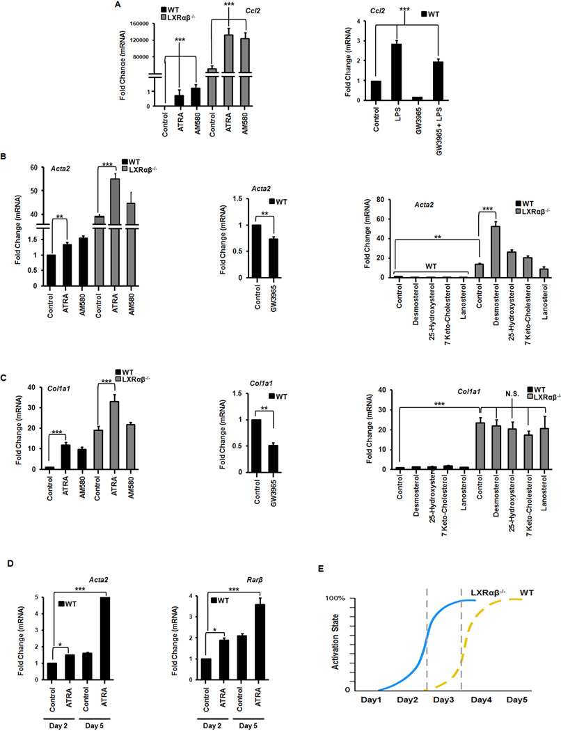
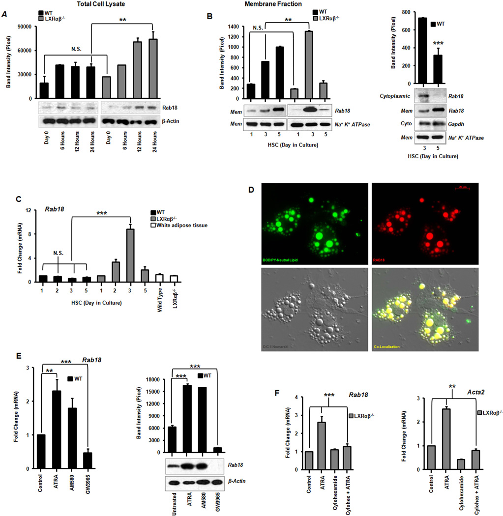
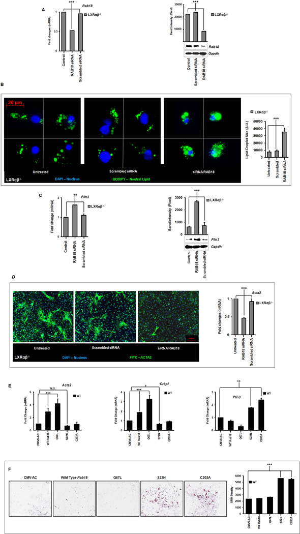
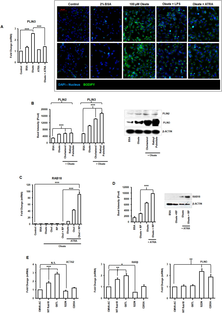
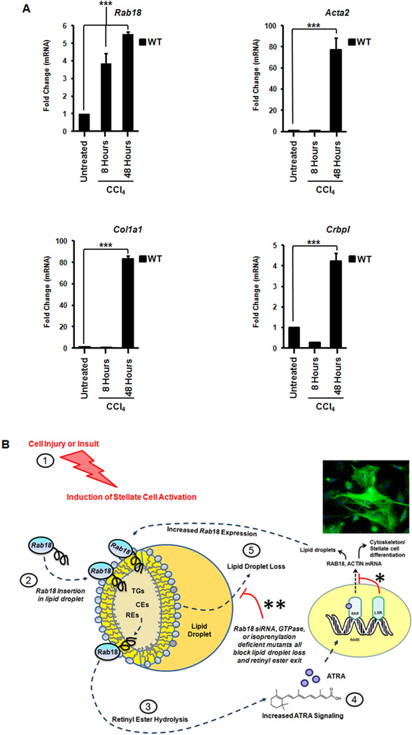
Liver X receptors (LXRs) are determinants of hepatic stellate cell (HSC) activation and liver fibrosis. Freshly isolated HSCs from Lxrαβ(-/-) mice have increased lipid droplet (LD) size, but the functional consequences of this are unknown. Our aim was to determine whether LXRs link cholesterol to retinoid storage in HSCs and how this impacts activation. Primary HSCs from Lxrαβ(-/-) and wild-type mice were profiled by gene array during in vitro activation. Lipid content was quantified by high-performance liquid chromatography and mass spectroscopy. Primary HSCs were treated with nuclear receptor ligands, transfected with small interfering RNA and plasmid constructs, and analyzed by immunocytochemistry. Lxrαβ(-/-) HSCs have increased cholesterol and retinyl esters. The retinoid increase drives intrinsic retinoic acid receptor signaling, and activation occurs more rapidly in Lxrαβ(-/-) HSCs. We identify Rab18 as a novel retinoic acid-responsive, LD-associated protein that helps mediate stellate cell activation. Rab18 mRNA, protein, and membrane insertion increase during activation. Both Rab18 guanosine triphosphatase activity and isoprenylation are required for stellate cell LD loss and induction of activation markers. These phenomena are accelerated in Lxrαβ(-/-) HSCs, where there is greater retinoic acid flux. Conversely, Rab18 knockdown retards LD loss in culture and blocks activation, just like the functional mutants. Rab18 is also induced with acute liver injury in vivo.

Conclusion: Retinoid and cholesterol metabolism are linked in stellate cells by the LD-associated protein Rab18. Retinoid overload helps explain the profibrotic phenotype of Lxrαβ(-/-) mice, and we establish a pivotal role for Rab18 GTPase activity and membrane insertion in wild-type stellate cell activation. Interference with Rab18 may have significant therapeutic benefit in ameliorating liver fibrosis.


Figures

Figure 1
Figure 1. LXR null hepatic stellate cells have increased retinyl ester stores and retinoic acid signaling
Primary HSCs were isolated from WT and Lxrαβ−/− mice and cultured on plastic for 1–5 days. (A–C) Measurements of CEs, cellular cholesterol and REs; N=8–10 mice/genotype. (D) Measurements of REs from whole liver samples. N=5 mice/genotype. (E) LC/MS analysis of sterol species. N=8–10 mice/genotype. All data are mean ± SEM, analyzed on matched days by two-tailed t test (A–D) or 1-way ANOVA (E) with post-hoc tests: *, P < .05; **, P < .01; ***, P < .001; NS, P > .05.
Figure 2
Figure 2. Gene array analysis of activating hepatic stellate cells
Primary HSCs from WT and Lxrαβ−/− mice (N=12 mice/genotype) were cultured on plastic. (A) Schematic of the work-flow. (B) Hierarchical clustering analysis of array data. K-means analysis is shown. (C–D) Total transcripts increasing (red) or decreasing (blue) by >2× difference. (E) Heat map of LXR, RAR, fibrotic, and inflammatory genes in primary HSCs after one day of culture activation. (F) Validation of RAR responsive genes by qRT-PCR from the array. Data are normalized to 36b4 expression and day 1 WT. Fold changes are mean ± SEM and differences between multiple groups compared by 1-way ANOVA with post-hoc tests: *, P < .05; **, P < .01; ***, P < .001.
Figure 3
Figure 3. Retinoic acid has a pro-inflammatory and pro-fibrotic effect on activating hepatic stellate cells
Gene expression of inflammatory (A) and fibrotic (B–C) gene expression in HSCs on day 2 of culture activation exposed to LPS (1 ng/mL), ATRA (100 nM), AM580 (100 nM), GW3965 (1 µM), or indicated cholesterol intermediates (100 µg/mL). N=3–5 mice/genotype. (D) WT stellate cells become more responsive to ATRA, as measured by the expression of fibrotic and RAR target genes, as culture activation progresses (day 2 in culture versus day 5). (E) Lxrαβ−/− stellate cells are ‘frame shifted’ in regards to the timing of fibrotic gene expression. They do not express higher absolute levels of these genes, but reach maximal expression sooner than WT cells. All data are mean ± SEM, analyzed by 1-way ANOVA with post-hoc tests. *, P < .05; **, P < .01; ***, P < .001; NS, P > .05.
Figure 4
Figure 4. Identification of Rab18, a retinoid responsive lipid droplet associated protein
(A) Rab18 protein expression by immunoblotting from total cell lysates and corresponding band densitometry in the first 24 hours of primary stellate cell culture activation. (B) Immunoblot analysis of Rab18 in HSC membrane and cytosolic fractions showing a shift from cytoplasm to membrane inserted Rab18 as activation proceeds in wild type stellate cells. (C) Rab18 gene expression in culture activated primary HSCs and compared to white adipose tissue (N=12 mice/genotype). (D) Immunofluorescence microscopy demonstrates Rab18 localization to LD surfaces. BODIPY (green), Rab18 (red), Nomarski DIC imaging, and the merged image bottom right; magnification 63×. (E) Rab18 mRNA and protein expression in day 2 culture activated primary WT HSCs treated with ATRA (100 nM), AM580 (100 nM), or GW3965 (1 µM). (F) Cycloheximide (10 µM) abrogates ATRA-induced Rab18 expression. All data are mean ± SEM, analyzed by 1-way ANOVA with post-hoc tests: *, P < .05; **, P < .01; ***, P < .001; NS, P > .05.
Figure 5
Figure 5. Rab18 knockdown retards lipid droplet loss and induction of α-smooth muscle actin
Primary HSCs were transfected on day 2 of culture activation with a Rab18 siRNA or scrambled control. (A) Gene and protein expression from whole cell lysates following 24 hours of Rab18 knockdown. (B) Immunofluorescence microscopy shows retention of neutral LDs (BODIPY, green). Nuclei are blue (DAPI), magnification 63×. Quantitation of lipid droplet surface area using densitometry (36 cells represented) is shown at right. (C) Plin3 gene and protein expression after Rab18 knockdown. (D) Immunofluorescence microscopy of α-smooth muscle actin (anti-actin, green; nuclei, blue (DAPI); magnification 10×) and gene expression following Rab18 knockdown. (E) Gene expression of Acta2, Crbp1 and Plin3 following overexpression of native Rab18, GTPase (S22N – “off”; C67L – “on”), or isoprenylation (C203A) mutants. (F) Lipid retention in wild type stellate cells on day 5 of primary cell culture by Oil red O staining occurs when either Rab18 GTPase activity is quenched (S22N) or Rab 18 membrane insertion is abrogated (C203A). Quantification by total pixel counts is shown at right. N=3–5 mice/genotype. All data are mean ± SEM, analyzed by 1-way ANOVA with post-hoc tests: *, P < .05; **, P < .01; ***, P < .001; NS, P > .05.
Figure 6
Figure 6. Regulation of RAB18 in an immortalized human stellate cell line
(A) PLIN3 gene expression and fluorescence microscopy of neutral lipid storage (BODIPY, green) in LX-2 cells, oleate-loaded and treated with LPS (1 ng/mL) or ATRA (100 nM). Magnification 200×. (B) PLIN2 and PLIN3 protein expression in LX-2 cells reloaded with combinations of oleate (100 µM), cholesterol (0.2 µg) or retinyl palmitate (RP) (50.6 µg)). (C) Gene expression of RAB18 in lipid loaded LX-2s treated with ATRA (100 nM). (D) Protein expression of RAB18 in lipid loaded cells treated with or without ATRA. Changes quantified by densitometry. (E) Overexpression of RAB18 mutants (S22N, Q67L, C203A) in LX-2 cells coordinately alters expression of fibrotic (ACTA2), retinoid (RARβ) and lipid droplet (PLIN3) genes. All data are mean ± SEM, analyzed by 1-way ANOVA with post-hoc tests: *, P < .05; **, P < .01; ***, P < .001; NS, P > .05.
Figure 7
Figure 7. Rab18 is specifically induced in vivo after acute liver injury in LXR null mice
Wild-type mice were treated with a single dose of carbon tetrachloride: 5 microliters/gram of 10% CCl4 in sterile olive oil injected intraperitoneally, for 8–48 hours. (A) Rab18 mRNA is induced in WT livers eight hours after acute injury. Fibrotic (Acta2 and Col1a1) and retinoid trafficking genes (Crbp1)are also increased, but Rab18 induction precedes these genes. All data are mean ± SEM, analyzed by two-tailed t tests: *, P < .05; **, P < .01; ***, P < .001; NS, P > .05. (B) ATRA-dependent regulation of Rab18. Stellate cell activation begins in response to external cell injury/insult (1). This leads to pre-existing Rab18 insertion into lipid droplet membranes (2) with subsequent lipid hydrolysis (3). A positive feedback loop is established as progressive hydrolysis of retinyl esters produces more ATRA and ATRA signaling (4), thereby inducing more Rab18 and lipid droplet loss (5). In WT mice, LXRs dampen this ATRA-dependent Rab18 response (*). Higher basal levels of retinyl esters in Lxrαβ−/− HSCs amplify the Rab18 response throughout this cycle. Rab18 expression is attenuated when lipid droplets are fully lost, ending the cycle. Knockdown of Rab18 by siRNA or expression of Rab18 mutants causes retention of auto-fluorescent lipid droplets (** and Fig. 5B,F), indicating a block on retinoid loss from the droplet. This correlates with diminished expression of actin and other markers of stellate cell activation. Abbreviations: TG = triglycerides, CE = cholesterol esters, RE = retinyl esters.

References

    1. Mederacke I, Hsu CC, Troeger JS, Huebener P, Mu X, Dapito DH, Pradere JP, et al. Fate tracing reveals hepatic stellate cells as dominant contributors to liver fibrosis independent of its aetiology. Nat Commun. 2013;4:2823. - PMC - PubMed
    1. Friedman SL. Hepatic stellate cells: protean, multifunctional, and enigmatic cells of the liver. Physiol Rev. 2008;88:125–172. - PMC - PubMed
    1. Blaner WS, O'Byrne SM, Wongsiriroj N, Kluwe J, D'Ambrosio DM, Jiang H, Schwabe RF, et al. Hepatic stellate cell lipid droplets: A specialized lipid droplet for retinoid storage. Biochim Biophys Acta. 2009;1791:467–473. - PMC - PubMed
    1. Friedman SL, Roll FJ, Boyles J, Bissell DM. Hepatic lipocytes: the principal collagen-producing cells of normal rat liver. Proc Natl Acad Sci U S A. 1985;82:8681–8685. - PMC - PubMed
    1. Kluwe J, Wongsiriroj N, Troeger JS, Gwak GY, Dapito DH, Pradere JP, Jiang H, et al. Absence of hepatic stellate cell retinoid lipid droplets does not enhance hepatic fibrosis but decreases hepatic carcinogenesis. Gut. 2011;60:1260–1268. - PubMed

Publication types

MeSH terms