. 2015 Jan 8;57(1):69-82.
doi: 10.1016/j.molcel.2014.10.028. Epub 2014 Dec 4.

Mitochondrial shape governs BAX-induced membrane permeabilization and apoptosis

Affiliations

Mitochondrial shape governs BAX-induced membrane permeabilization and apoptosis

Thibaud T Renault et al. Mol Cell. .

Abstract

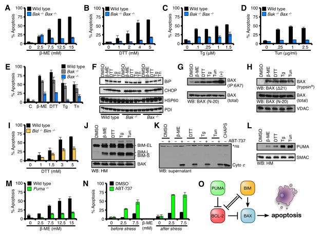
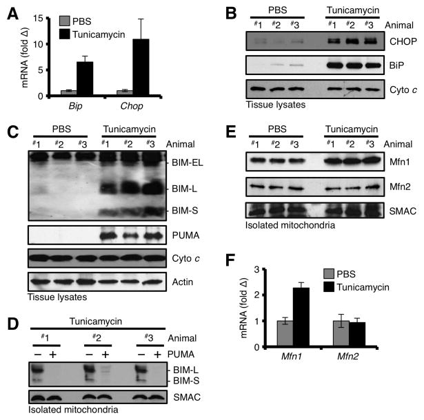
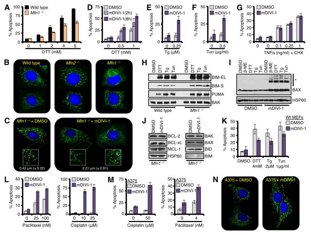
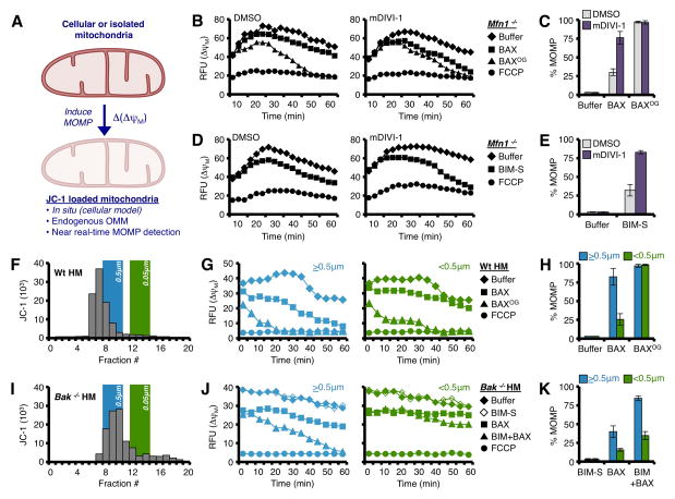
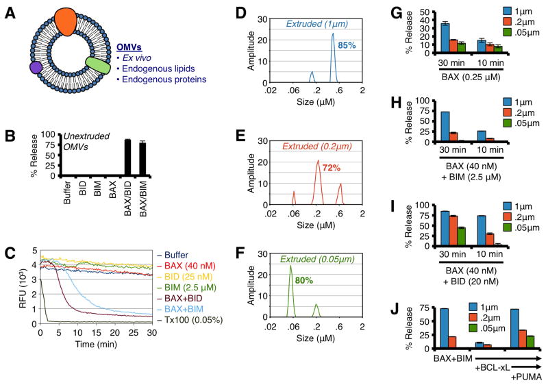
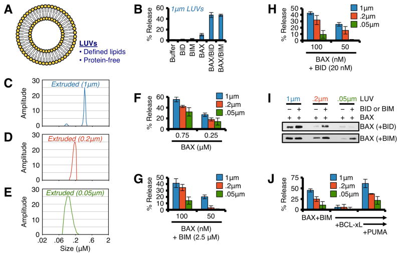
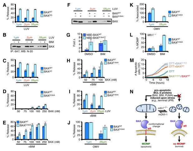
Proapoptotic BCL-2 proteins converge upon the outer mitochondrial membrane (OMM) to promote mitochondrial outer membrane permeabilization (MOMP) and apoptosis. Here we investigated the mechanistic relationship between mitochondrial shape and MOMP and provide evidence that BAX requires a distinct mitochondrial size to induce MOMP. We utilized the terminal unfolded protein response pathway to systematically define proapoptotic BCL-2 protein composition after stress and then directly interrogated their requirement for a productive mitochondrial size. Complementary biochemical, cellular, in vivo, and ex vivo studies reveal that Mfn1, a GTPase involved in mitochondrial fusion, establishes a mitochondrial size that is permissive for proapoptotic BCL-2 family function. Cells with hyperfragmented mitochondria, along with size-restricted OMM model systems, fail to support BAX-dependent membrane association and permeabilization due to an inability to stabilize BAXα9·membrane interactions. This work identifies a mechanistic contribution of mitochondrial size in dictating BAX activation, MOMP, and apoptosis.


Figures

Figure 1
Figure 1. Terminal UPR requires BAX
(A–D) Wt and Bak−/−Bax−/− MEFs were treated with β-ME, DTT, Tg, or Tun for 18 h. (E) Wt, Bak−/−, and Bax−/− MEFs were treated with β-ME (15 mM), DTT (5 mM), Tg (1.5 μM), or Tun (2.5 μg/ml) for 18 h. (F) Lysates from ER stress treated Wt, Bak−/−, and Bax−/− MEFs in E were analyzed by western blot. (G) CHAPS lysates from ER stress treated Wt MEFs (highest doses at 18 h) were subjected to 6A7 IP and western blot. Total cell lysates (5%) were analyzed as a loading control. (H) HM fractions isolated from ER stress treated Wt MEFs (highest doses at 18 h) were subjected to trypsinization, and analyzed by western blot. Total cell lysates (5%) were analyzed as a loading control for BAX, VDAC is a pre-trypsinization mitochondrial loading control. (I) Wt and Bid−/−Bim−/− MEFs were treated with Tg for 18 h. (J) HM fractions isolated from ER stress treated Wt MEFs (highest doses at 18 h) were analyzed by western blot. (K) HM fractions from ER stress treated Wt MEFs (highest doses at 18 h) were incubated with ABT-737 (1 μM) for 30 min at 37°C, centrifuged, and the supernatants were analyzed by western blot. CHAPS (0.25%) lysed mitochondria indicates total cyto c within each lane. *Indicates a non-specific band. (L) Same as J, but probed for PUMA and SMAC. (M) Wt and Puma−/− MEFs were treated with Tg for 18 h. (N) Puma−/− MEFs were pre-treated with ABT-737 (1 μM) for 1 h, then β-ME for 18 h; or β-ME for 18 h, then ABT-737 for an additional 6 h. (O) A summary schematic of BCL-2 family interactions required for apoptosis to proceed. All data are representative of at least triplicate experiments, and reported as ± S.D., as required. See also Figures S1–S3.
Figure 2
Figure 2. In vivo induction of the UPR promotes BIM, PUMA, and Mfn1 accumulation at the OMM
(A) Mice were injected with 2 mg/kg Tun or PBS, sacrificed 24 h later, and liver total RNA was analyzed for Bip and Chop. Expression was normalized to 18S. (B) Tissue lysates from livers in A were analyzed for CHOP and BIP. (C) Tissue lysates from livers in A were analyzed for BIM and PUMA. (D) Mitochondria from livers in A were isolated, incubated with PUMA (100 nM) or PBS for 30 min at 37°C, pelleted, and analyzed by western blot. (E) Mitochondria from livers in A were isolated and analyzed by western blot. (F) Same as A, but total RNA was analyzed for Mfn1 and Mfn2 by qPCR. All data are representative of at least triplicate experiments, and reported as ± S.D., as required.
Figure 3
Figure 3. Mitochondrial network shape regulates tUPR
(A) Wt and Mfn1−/− MEFs were treated with DTT for 18 h. (B) Wt, Mfn2−/−, and Mfn1−/− MEFs were loaded with MitoTracker Green® (50 nM) and Hoechst 33342 (20 μM) before imaging (400×). (C) Mfn1−/− MEFs were treated with mDIVI-1 (25 μM) for 2 h before imaging (400×). Further magnified regions (2.5×) are shown in white boxes. The average length of ~ 200 mitochondria is shown. (D) Mfn1−/− MEFs were pre-treated with mDIVI-1 (25 μM) for 2 or 8 h, and then DTT for 18 h. (E-F) Mfn1−/− MEFs were pre-treated with mDIVI-1 (25 μM) for 8 h, then Tg (0.25 μM) or Tun (0.5 μg/ml) for 18 h. (G) Mfn1−/− MEFs were pre-treated with mDIVI-1 (25 μM) for 8 h, then TNFα and CHX (10 μg/ml) for 18 h. (H) HM fractions from ER stress treated Mfn1−/− MEFs were analyzed by western blot. (I) Mfn1−/− MEFs were pre-treated with mDIVI-1 (25 μM) for 2 h, ER stress agents for 18 h, and mitochondria were isolated and analyzed by western blot. High molecular weight complexes of BAX are indicated (*). VDAC is a loading control. (J) Mfn1−/− MEFs were treated with mDIVI-1 (25 μM) for 8 h, and lysates were analyzed by western blot. (K) Wt MEFs were pre-treated with mDIVI-1 (25 μM) for 8 h, and ER stress agents for 18 h. (L) Mfn1−/− MEFs were pre-treated with mDIVI-1 (25 μM) for 8 h, and Paclitaxel or Cisplatin for 18 h. (M) Same as L, but A375. (N) Same as C, but A375. All data are representative of at least triplicate experiments, and reported as ± S.D., as required. See also Figure S4.
Figure 4
Figure 4. Mitochondrial size dictates sensitivity to BAX-dependent MOMP
(A) Schematic representation of measuring Δ(ΔψM) to detect MOMP. (B–C) Digitonin-permeabilized, JC-1 loaded Mfn1−/−MEFs (pre-treated with 25 μM mDIVI-1, or DMSO, for 8 h) were incubated with BAX (0.25 μM) or OG-BAX (0.25 μM), and mitochondrial depolarization (ΔψM) was determined. Kinetic and endpoint measurements are shown in B and C, respectively. (D–E) Same as B, but with BIM-S (25 nM). Kinetic and endpoint measurements are shown in D and E, respectively (F) JC-1 loaded Wt liver mitochondria were fractionated by size, and the relationships between 0.5 and 0.05 μm LUVs are indicated on the same graph. (G–H) Larger (>0.5 μm; fractions 6–8) and smaller (<0.5 μm; fractions 11–15) Wt mitochondria were treated with BAX (100 nM) or OG-BAX (100 nM) for 1 h at 37°C. Kinetic and endpoint measurements are shown in G and H, respectively. (I) JC-1 loaded Bak−/− liver mitochondria were fractionated by size. (J–K) Larger (>0.5 μm; fractions 8–10) and smaller (<0.5 μm; fractions 12–16) Bak−/− mitochondria were treated with BAX (20 nM) ± BIM-S (20 nM) for 1 h. Kinetic and endpoint measurements are shown in J and K, respectively. All data are representative of at least triplicate experiments, and reported as ± S.D., as required. See also Figure S5.
Figure 5
Figure 5. BAX preferentially permeabilizes OMVs with diameters similar to Wt mitochondria
(A) Schematic representation of OMVs. (B) Unextruded OMVs were combined with BAX (40 nM), and N/C-BID (20 nM) or BIM BH3 peptide (2.5 μM) for 30 min at 37°C. (C) Kinetic traces of unextruded OMV permeabilization with BAX (40 nM) and BID (25 nM) or BIM BH3 (2.5 μM) for 30 min at 37°C. Triton X-100 solubilizes OMVs and establishes 100% release. An anti-FITC antibody is used to quench the FITC-dextran released during permeabilization. (D–F) DLS analyses of extruded (1, 0.2 & 0.05 μm) OMVs. The major peak was calculated as the area under the curve and is reported as a %. (G) OMVs were combined with BAX (0.25 μM) for 10 or 30 min. (H) OMVs were combined with BAX (40 nM) and BIM BH3 (2.5 μM) for 10 or 30 min. (I) Same as H, but with N/C-BID (20 nM). (J) OMVs were combined with BAX (40 nM), BIM BH3 (2.5 μM), BCL-xLΔC (300 nM), and PUMA BH3 (5 μM) for 30 min. All data are representative of at least triplicate experiments, and reported as ± S.D., as required. See also Figure S6.
Figure 6
Figure 6. BAX preferentially permeabilizes LUVs with diameters similar to Wt mitochondria
(A) Schematic representation of LUVs. (B) Standard LUVs (1 μm) were combined with BAX (100 nM), and N/C-BID (20 nM) or BIM BH3 (2.5 μM) for 1 h at 37°C. (C–E) DLS analyses of extruded (1, 0.2 & 0.05 μm) LUVs. (F) LUVs were combined with BAX (0.25 & 0.75 μM) for 1 h. (G) LUVs were combined with BAX (75 & 100 nM) and BIM BH3 (2.5 μM) for 1 h. (H) Same as G, but with N/C-BID (20 nM). (I) LUVs were combined with BAX (100 nM), and N/C-BID (20 nM) or BIM BH3 (2.5 μM) for 30 min at 37°C prior to centrifugation, solubilization, and western blot for associated BAX. (J) LUVs were combined with BAX (100 nM), BIM BH3 (2.5 μM), BCL-xLΔC (300 nM), and PUMA BH3 (5 μM) for 1 h. All data are representative of at least triplicate experiments, and reported as ± S.D., as required. See also Figure S7.
Figure 7
Figure 7. BAX α9 displays requirements for membrane shape
(A) LUVs were combined with BAX or BAXOG (0.25 μM) for 15 min at 37°C. (B) BAX (100 ng) was incubated in the presence of BIM BH3 (2.5 μM) and LUVs for 30 min prior to 6A7 IP and western blot. (C) LUVs were combined with BAXWT or BAXΔC (0.25 μM) for 1 h at 37°C. The required incubation time is longer for BAXΔC compared to BAXWT, which increases BAXWT activity. (D) LUVs were combined with BAXWT or BAXS184A for 30 min at 37°C. (E) Same as D, but with BIM BH3 (2.5 μM). (F) LUVs were combined with BAXWT or BAXS184A (100 nM) for 30 min at 37°C prior to centrifugation, solubilization, and western blot for associated BAX. (G) NBD-BAXWT or NBD-BAXS184A was incubated with 1 μm LUVs for 5 min, ± BIM BH3 (2.5 μM). An increase in NBD fluorescence indicates BAX·LUV interactions, and is reported as fold increase compared to NBD-BAXWT + LUVs. (H) LUVs (1 μm) were combined with BAXWT or BAXS184A (50, 75, 100 nM) with BIM BH3 (2.5 μM) for 30 min at 37°C. (I) LUVs were combined with BAXWT or BAXS184A (50 nM) with BIM BH3 (2.5 μM) for 30 min at 37°C. (J) OMVs were combined with BAXWT or BAXS184A (50 nM) and BIM BH3 (2.5 μM) for 30 min at 37°C. (K) NBD-BAXWT or NBD-BAXS184A ± BIM BH3 (2.5 μM) was incubated with OMVs for 30 min at 37°C. The interaction between NBD-BAXWT + BIM BH3 with 1 μm OMVs is reported as 100%. (L) Digitonin-permeabilized, JC-1 loaded Mfn1−/− MEFs were incubated with BIM BH3 (0.1 μM), BAXWT (50 nM), and BAXS184A (50 nM), and ΔΔψM was determined. (M) Mfn1−/− MEFs expressing shBax were reconstituted with human BAXWT or BAXS184A, treated with DTT (1.5 mM), and the kinetics of tUPR was evaluated by IncuCyte. (N) A schematic summarizing the relationship between BAX, mitochondrial shape, and apoptosis. All data are representative of at least triplicate experiments, and reported as ± S.D., as required. See also Figure S7.

Similar articles

Cited by

References

    1. Asciolla JJ, Renault TT, Chipuk JE. Examining BCL-2 family function with large unilamellar vesicles. J Vis Exp. 2012 Oct;5(68) pii: 4291. - PMC - PubMed
    1. Beverly LJ, Howell LA, Hernandez-Corbacho M, Casson L, Chipuk JE, Siskind LJ. BAK activation is necessary and sufficient to drive ceramide synthase-dependent ceramide accumulation following inhibition of BCL2-like proteins. Biochem J. 2013;452:111–119. - PMC - PubMed
    1. Blobel G. Protein targeting. Biosci Rep. 2000;20:303–344. - PubMed
    1. Cassidy-Stone A, Chipuk JE, Ingerman E, Song C, Yoo C, Kuwana T, Kurth MJ, Shaw JT, Hinshaw JE, Green DR, et al. Chemical inhibition of the mitochondrial division dynamin reveals its role in Bax/Bak-dependent mitochondrial outer membrane permeabilization. Dev Cell. 2008;14:193–204. - PMC - PubMed
    1. Certo M, Del Gaizo Moore V, Nishino M, Wei G, Korsmeyer S, Armstrong SA, Letai A. Mitochondria primed by death signals determine cellular addiction to antiapoptotic BCL-2 family members. Cancer Cell. 2006;9:351–365. - PubMed

Publication types

MeSH terms