. 2015 Jan 6;23(1):68-79.
doi: 10.1016/j.str.2014.10.014. Epub 2014 Dec 4.

Electrostatic interactions between the Bni1p Formin FH2 domain and actin influence actin filament nucleation

Affiliations

Electrostatic interactions between the Bni1p Formin FH2 domain and actin influence actin filament nucleation

Joseph L Baker et al. Structure. .

Abstract

Formins catalyze nucleation and growth of actin filaments. Here, we study the structure and interactions of actin with the FH2 domain of budding yeast formin Bni1p. We built an all-atom model of the formin dimer on an Oda actin filament 7-mer and studied structural relaxation and interprotein interactions by molecular dynamics simulations. These simulations produced a refined model for the FH2 dimer associated with the barbed end of the filament and showed electrostatic interactions between the formin knob and actin target-binding cleft. Mutations of two formin residues contributing to these interactions (R1423N, K1467L, or both) reduced the interaction energies between the proteins, and in coarse-grained simulations, the formin lost more interprotein contacts with an actin dimer than with an actin 7-mer. Biochemical experiments confirmed a strong influence of these mutations on Bni1p-mediated actin filament nucleation, but not elongation, suggesting that different interactions contribute to these two functions of formins.


Figures

Figure 1
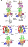
Figure 1. Comparison of the initial model and the refined model from all atom molecular dynamics simulation of a dimer of Bni1p FH2 domains associated with the barbed end of an actin filament
The initial model was constructed by aligning the FH2 and actins from the Otomo crystal structure (Otomo et al., 2005) on the end of the Oda actin filament structure (Oda et al., 2009). This structure relaxed during 160 ns of all atom molecular dynamics simulation. Snapshots are from the 160 ns WT simulation. (A) Schematic ribbon diagrams for addition of an actin subunit (grey) to an actin filament barbed end with a bound dimer of FH2 domains (red and blue). On the left an incoming actin subunit is shown approaching the “open” conformation of the actin filament. On the right, the newly added subunit is incorporated onto the barbed end of the filament, ready for the trailing FH2 domain to take a step along the filament. We simulated the state on the right in panel (A), prior to the step of the trailing FH2 domain. (B-E) Ribbon diagrams of (blue) the leading FH2 domain and (red) and trailing FH2 domain with four of the seven actin subunits from the actin 7-mer. (B) and (C) are initial and final views down the central filament axis, and (D) and (E) are initial and final views of the system from the side. Also see Figure S1, S2, S3 and Table S1, S2, and S3 for additional structural information.
Figure 2
Figure 2. Contacts between formin residues and actin in the refined structure of the Bni1pFH2/actin filament seven-mer
Number of average contacts between each residue in (A) FH2L and (B) FH2T and each actin subunit calculated as described in the main text. A contact is defined as 12 Å between alpha-carbons of two residues. The regions of Bni1p are labeled with horizontal lines. See also Table S1 for the label definitions, and also Figure S4.
Figure 3
Figure 3. Salt bridges and H-bonds of Bni1p R1423 and K1467 with actin
Ribbon diagrams of actin filament subunits A1 to A3 are shown in blue, gold and yellow, along with formin helices KnA (residues 1422-1440, cyan and pink in FH2L and FH2T respectively) and KnB (residues 1457-1479, blue and red in FH2L and FH2T respectively) The backbone of the actin TBC is shown in purple (defined as residues 22-26, 139-149, 167-169, 338-355). The insets show close-up views of the FH2T interactions with A3 (upper right) and the FH2L interactions with A2 (lower right). Salt-bridges/H-bonds are highlighted with light blue, transparent ovals. The residues comprising TBC, KnA and KnB were used to calculate the electrostatic interaction energies in Table 2 and Table S2. See also Table S5 for information about the evolutionary conservation of these residues.
Figure 4
Figure 4. Coarse-grained (CG) model of formin and actin
(A) Ribbon diagrams of the backbones of all-atom models of actin and Bni1p next to their CG hENM models. In the actin CG model, the color scale represents the residue number (dark blue is the N-terminal end, and dark red is the C-terminal end). In the formin CG model, FH2L is colored blue and FH2T is colored red. Lines connect CG sites within 10 Å, as in the hENM model. The varying CG site sizes represent the radii used for the LAMMPS excluded volume terms. (B and C) Models with the CG beads superimposed on the all-atom ribbon diagrams of the protein backbones. Bead sizes represent the excluded volume radii, as in (A). (B) Model of the CG dimer system, consisting of the formin dimer and two actin subunits extracted from the filament model. (C) Model of the formin dimer with the actin 7-mer filament.
Figure 5
Figure 5. Contacts between formin residues and actin in CG structures of the wild type and mutant Bni1p/actin filament heptamer systems
Number of average contacts between each formin residue and each actin subunit in the CG heptamer systems: (A) WT FH2L; (B) WT FH2T; (C) double-mutant FH2L; and (D) double-mutant FH2T.
Figure 6
Figure 6. Contacts between formin residues and actin in CG structures of WT and double mutant Bni1p/actin dimer systems
Number of average contacts between each formin residue and each actin subunit in the CG dimer systems: (A) WT FH2L; (B) WT FH2T; (C) double- mutant FH2L; and (D) double-mutant FH2T. (E-G) CG models of Bni1p FH2 domains (FH2L blue, FH2T red) on a dimer of actin subunits (orange) A2 and (yellow) A3. The model has one site per amino acid, and every site has a charge assigned from the charge fitting procedure described in the Experimental Methods. Lines represent the elastic network bonds between CG sites. (E) Side view. (F, G) View looking from the barbed end towards the pointed end of actin. Purple spheres are the Bni1p knob helix KnB, and green spheres represent actin residues within 12 Å of the KnB residues. (F) WT system. (G) Double-mutant system.
Figure 7
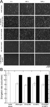
Figure 7. Substitutions R1423N and K1467L do not affect the elongation of actin filament barbed ends mediated by Bni1(pP1FH2)p
Conditions: 10 mM imidazole (pH 7.0), 50 mM KCl, 1 mM MgCl2, 1 mM EGTA, 50 mM DTT, 0.3 mM ATP, 0.02 mM CaCl2, 15 mM glucose, 0.02 mg/ml catalase, 0.1 mg/ml glucose oxidase and 0.5% methylcellulose (4,000 cP at 2% (w/v)). Data were collected with TIRFM. (A) Time series of images of 1.5 μM actin (33% Oregon green-labeled) filaments growing in the presence of wild-type and mutant Bni1(pP1FH2)p constructs. The concentration of each formin is indicated. (B) Rates of barbed end elongation mediated by wild-type and mutant Bni1(pP1FH2)p in the absence (open bars) or presence (filled bars) of 5 μM profilin. For each sample, we measured the elongation of 10-20 filaments typically over a span of at least 300 s. The error bars are standard errors of the mean. See also Figures S5 and S6 for additional information about the experimental characterization of the actin/formin system.
Figure 8
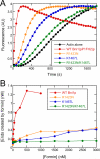
Figure 8. Substitutions R1423N and K1467L of Bni1(pP1FH2)p strongly impair actin filament nucleation
Conditions: 10 mM imidazole (pH 7.0), 50 mM KCl, 1 mM MgCl2, 1 mM EGTA, 0.17 mM ATP, 0.5 mM DTT, 0.03 mM CaCl2, 1.7 mM Tris (pH 8.0), 1 part per 105 (w/v) Antifoam 204. (A) Representative time courses of spontaneous polymerization of 4 μM actin (20% pyrene-labeled) alone or in presence of 1 μM wild-type or mutant Bni1(pP1FH2)p constructs. For clarity, every 5th data point is shown. (B) Dependence of the concentration of formin-nucleated barbed ends on the concentration of Bni1(pP1FH2)p constructs. Barbed end concentrations were calculated from the elongation rates in bulk samples when half of the total actin was polymerized and the elongation rates measured in the TIRF microscopy experiments. See also Figures S5 and S6 for additional information about the experimental characterization of the actin/formin system.

Similar articles

Cited by

References

    1. Brown WM, Kohlmeyer A, Plimpton SJ, Tharrington AN. Implementing molecular dynamics on hybrid high performance computers - Particle-particle particle-mesh. Comput Phys Commun. 2012;183:449–459.
    1. Brown WM, Wang P, Plimpton SJ, Tharrington AN. Implementing molecular dynamics on hybrid high performance computers - short range forces. Comput Phys Commun. 2011;182:898–911.
    1. Chang F, Drubin D, Nurse P. cdc12p, a protein required for cytokinesis in fission yeast, is a component of the cell division ring and interacts with profilin. J Cell Biol. 1997;137:169–182. - PMC - PubMed
    1. Chu JW, Voth GA. Allostery of actin filaments: Molecular dynamics simulations and coarse-grained analysis. P Natl Acad Sci USA. 2005;102:13111–13116. - PMC - PubMed
    1. Courtemanche N, Pollard TD. Determinants of Formin Homology 1 (FH1) domain function in actin filament elongation by formins. J Biol Chem. 2012;287:7812–7820. - PMC - PubMed

Publication types

MeSH terms